













![Extracellular vesicles as a tool for diagnostics and therapeutics: insights and challenges [WEBINAR]](https://i.ytimg.com/vi_webp/g2aeCH40mfE/maxresdefault.webp)


Codiak BioSciences developed a versatile platform for generating engineered extracellular vesicles with defined therapeutic properties by identifying novel scaffolds (EWI subfamily proteins and MARX family proteins) through proteomic analysis of high-purity EV isolates, enabling efficient surface display and lumenal packaging of diverse therapeutic cargoes including proteins, RNAs, and small molecules for potential clinical applications in cancer immunotherapy and vaccines.
Engineering Extracellular Vesicles: Scaffolds for Therapeutic Cargo
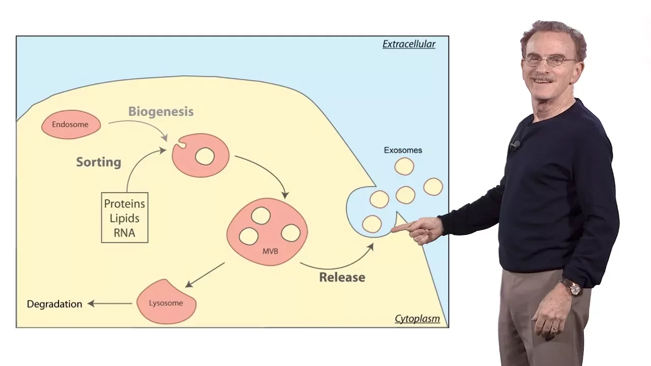
Added:Fundamental biology and biogenesis of extracellular vesicles (EVs), such as exosomes and microvesicles, and their natural roles in intercellular communication.
Principles of molecular biology and genetic engineering, specifically the design of fusion proteins and recombinant DNA technology.
Basic concepts of membrane biology, including protein-lipid interactions and membrane-anchoring domains (e.g., GPI anchors, transmembrane domains).
Core principles of drug delivery systems, including targeting strategies, physiological barriers, and biocompatibility.
Advanced active and passive cargo loading methodologies for non-protein molecules, such as therapeutic nucleic acids (mRNA, siRNA, and CRISPR/Cas9 systems).
Strategies for active targeting of engineered EVs to specific tissues or organs using homing peptides or monoclonal antibodies.
Bioprocessing, scalability, and purification techniques required for the clinical-grade manufacturing of therapeutic extracellular vesicles.
Regulatory frameworks and current clinical trials evaluating engineered EV-based therapies for oncology, immunology, and regenerative medicine.
916 views15likes1:01:52Original Release: 2021-05-12
welcome everyone to this week's edition of the extracellular vesicle club this is a special edition of the club uh because we have the isev annual meeting coming up but we wanted to squeeze this this one in before the meeting starts so i'm just going to say a few words about the annual meeting we have an education day that will be on wednesday may 12th this week and then there are going to be several satellite events over the next few days finally we will have the main meeting from the 18th to the 21st of may and you can still register for this meeting so this is the biggest and most comprehensive ev meeting in the world and it's possible to attend just the education day if you if you prefer if you're new to ev studies want to find out what's going on in the field what some of the basics are the education day is a great way to get introduced to that if you're not ready for the full meeting we also have some some some satellite events that are going to be free so for example there's a a collaboration of the michael j fox foundation and isev on neuronal markers of evs specifically l1 cam and how it can be used or not used as the case may be to pull down extracellular vesicles from biofluids to understand the state of the brain so there's a bit of i guess different different interpretations and different findings in that area and we're going to address those head-on in a satellite meeting so today it's my pleasure to introduce russell mcconnell and kevin dooley they're from from kodiak kodiak biosciences and they're going to tell us a little bit about kodiak and then they're going to go into their their recently published paper in molecular therapies i want to stress here that this is a research presentation so this is not a sponsor presentation rather they are describing some of the research that they've done at kodiak in engineering extracellular vesicles and in indeed their their constructs as they'll probably tell us are available to the community and they'd be glad to share those with you for your research um so russell and and kevin thank you so much for joining today and for presenting your work to the extracellular vesicle club i'm going to turn the screen sharing over to you now wonderful well uh thank you very much for having us ken really appreciate the opportunity to share some of our work thanks ken for having us we yeah really appreciate the opportunity to share some of our work with the community and also for hosting this uh you know really wonderful theories over the past year plus it's been a great way to stay in touch with you know what's going on in uh eevee research so before we jump into the data i just wanted to give a one slide overview of the history of kodiak um so the company was actually founded towards the end of 2015 and lab operations started in the beginning of 2016 which is when myself and russell joined the company we've completed a couple of venture raises over the years our first headquarters is actually in tech square but we have since moved to our current location in cambridge in february 2020 just in time for the pandemic and completed our ipo in october of 2020 we also initiated two phase one clinical trials in september of last year and have recently opened a clinical manufacturing facility to make our engineer dds and from the beginning of the company we were always interested in using engineered extracellular vesicles with very defined therapeutic proper posed to relying on any intrinsic therapeutic property of the ev so in order to do that we needed to develop new systems for engineering both the surface and the lumen of evs to display or package different molecules of interest and this is the work that was recently published in molecular therapy and it's broken down into two sections which is highlighted in the correct direction so in the first part of the talk which is what i'm going to be covering goes over the purification protocol that we use to isolate ev as well as the proteomic analysis that we used to identify different scaffold candidates in the second part of the talk which my colleague russell mcconnell will be taking on and describes how we then use those scaffolds that we identified by proteomics to engineer these with different therapeutic cargoes and ultimately how we tested those both in vitro and in vivo and so the real backbone of the work is really predicated on both the cell line that we use for ev production as well as the purification protocol that we use to ultimately isolate the population of evs that we use and so um this slide here just details the purification protocol all of the work that was reported in the paper was done with a suspension adapted heck 293 cell line and so this is something that we can grow and chemically define medium so there's no serum or anything like that and we can grow these to very very high cell densities and we typically do this anywhere from the 200 ml scale up to 25 liters in wave bioreactors and at the termination of cell culture the cell density is typically between 10 and 15 million cells per ml so there's a lot more biomass here compared to conventional 2d plastic culture and so after the cell culture is completed we remove the cells by centrifugation filter the condition medium and then treat with a broad spectrum nuclease to digest any extra vesicular dna or rna species that are in the media the media is then concentrated by tangential flow filtration depending on the size of the production run and then pelleted by ultracentrifugation to prepare a crude ed pellet this is then loaded into the bottom of an iodicinol density gradient and fractionated overnight by spinning at 150 000 times t and when you're working with such high vcds you can begin to use visual cues to actually see the different subpopulations that are fractionated on the gradient and that's shown in the image sort of in the middle of the slide here where we can see four distinct populations after the gradient is completed and so what we've done in the manuscript is to isolate these different fractions and then characterize them both biochemically and ultra-structurally to get a better understanding of where the evs are actually fractionating here and typically what we've done in the past is pull the fractions ultra centrifuge them again and then analyze them but what i wanted to call the group's attention to is this additional 20 000 times g centrifugation step that we needed to add to our purification protocol and the reason for that is when we imaged that top fraction of the gradient fraction one by two different modes uh two different imaging modalities we saw two different very different pictures and so on the left is a conventional tem grid of fraction one and on the right is imaging a similar population just by cryotem what we saw was an abundance of this web-like material that coded the entire grid that really wasn't detectable by conventional tem so we implemented this 20k spin which pellets out this web-like material and what we're left with is a clean fraction of evs you know without any cellular debris and we characterized both the pellet and the supernatant from that low speed spin and found the pellet to be primarily composed of actin and actin binding proteins which is shown in the proteomics data on the right hand side of the slide so in addition to imaging we also used a number of other techniques to get a better understanding of the different populations that were fractionating on the iodicinol gradient so we have conventional tem grids here in the middle of the slide for fractions one through four and we can see the prevalence of vesicles in f1 and f2 and in f3 and f4 there are detectable vesicles but they are coded in a blanket of protein and we can see that more clearly when we um analyze these fractions for total protein you can see in the plots on the right that the fraction four contains um the majority of the protein as well as the majority of the dna which was resistant to the broad spectrum nuclease treatment we also looked at cholesterol levels which have been shown previously to be enriched in ev membranes and we see the majority of the cholesterol associated with fraction one interestingly when you take these four fractions and analyze them on the nano site all four will register particles and all four will give you size distributions that fall within the described dv range but we know from the other characterization that we've done that you know the particles that are registering fraction four are probably very different from those in fraction one um which just you know goes to show that you do need to extensively characterize um the material that you're working with in order to very clearly distinguish evs from non-uv species we also used sds page and immunoblot analysis to get a better understanding of the different proteins that are enriched in the different fractions so in the middle is an sds page gel loaded by equal total protein from the producer cell lysate the crude ultra centrifuge pellet that we used to load the idicts and all gradient and then the four fractions and just by looking at the total protein profile it's very clear that the material in fraction one and two is distinct from that in fractions three and fractions four and when we use immunoblot analysis to look for particular markers we can see proteins that are involved in ev biogenesis and vesicle trafficking enriched in fractions one and two things like centennial one um then the tetraspanning cd9 81 and 63 whereas proteins that are you know bound up in chromatin things like high mobility group box protein one or histone h3 are enriched in the higher density fraction four and there's one particular protein i want to call the group's attention to just because it was really useful for us as we were developing our purification protocol and that's galectin-3 binding protein or algal s3bp here we can see that it's very enriched in the uc pellet and predominantly fractionates in fraction four and this was something that we used as a marker for contamination essentially for evs derived from hec293 producer cells and we looked at a bunch of different methods for isolating eb's things like peg precipitation or peg coupled with size exclusion chromatography which is shown in the protein gel here here as well as ultracentrifugation and you can see by the blots down below for galectin-3 binding protein all of these methods peg peg plus sec and ultracentrifugation alone result in an enrichment of collecting three binding protein and we really needed to use the iodicinal fractionation in order to deplete that contaminant from our eb population and interestingly um there's a bit of literature on this protein which is also called mac2 binding protein and it forms oligomeric protein structures i form these these rings which can be imaged by tem and i know there's no scale bar on here but these are about 35 to 40 nanometers which i think can be easily confused with evs by just looking at tem grids now we also used lcms to and proteomic analysis to get a more granular picture of the proteins that are enriched in these different fractions that's shown in the heat map on the left so we're showing here a subset of that analysis and highlighting proteins that are enriched in fraction one in the top portion of the heat map and those enriched in fraction four on the bottom portion and if you look at the specific proteins you can see they're very different fraction four is primarily composed of secreted extracellular matrix constituents and histones which you know are probably responsible for the high amounts of dna that are still present in fraction four after nuclease treatment and if we look at what's enriched in fraction one we can see proteins that are again involved in vesicle biogenesis things like alex zentennant one lactic hearing but we also found a number of proteins that hadn't been previously well characterized as being specific to evs and there are two families of proteins here one is the ewi subfamily of the immunoglobulin super family and the two members are ptgfrn and igsf8 shown with the pink arrows here and then we also saw a number of proteins from the marx protein family enriched in fraction one including marx mark cell one and best one and so these were the candidates that we had identified from the proteomic analysis i have a schematic in the middle of the slide here which just shows the differences in topology of the scaffolds that we identified as well as uh tetraspanins which have probably been used most widely as a mechanism for engineering evs so the the marx protein family falls into this lipid anchor uh engineering scaffold um all three of these proteins contain an n-terminal meristemation modification which is anchored into the inner leaflet of the ep membrane and then ptgfrn and igsf8 are single pass transmembrane proteins which are similar to sequences like lamp2b or p display and now to specifically assess the capacity of each of these candidate scaffolds to direct fusion proteins into evs we used flag tag gfp as a surrogate cargo so we made genetic fusions of each scaffold to gfp and then stably transfected an exosome producer cells this is the heck 293 suspension cell that we described earlier and all the work that's in the manuscript is from stably selected cell pools we found that there's a lot of variability associated with transient transfection as well as some other potential artifacts which can make downstream analysis uh quite challenging so everything is from a stably selected producer cell and everything is also purified using the idicts and all gradient that we just walked through and now as a way of directly comparing the efficiency by which these scaffolds can direct gfp into evs we first looked at the producer cell and we wanted to make sure that all of the producer cells could indeed make the fusion protein at the cellular level and we're looking at flow cytometry data on the left-hand portion of the slide i'm looking at all the different scaffolds as well as a cytosolic gfp control which has no membrane targeting sequence you can see the cytosolic tfp results in about 10-fold higher gfp than the rest of the scaffolds but for the most part they're all um you know about the same with you know the exception of lamp2b and p display are a little bit lower but then when we look at the levels of gfp in the evs from these producer cells we see a pretty wide spread and so this data was generated using quantitative elisa coupled with a nanoparticle tracking analysis to generate um you know to quantitate the number of gfp molecules per ev and what we're showing here is that the scaffolds that we identified by proteomic analysis resulted in the highest levels of gfp per exosome compared to previously described scaffolds and the last piece of data i wanted to share with the group is actually a bit of deeper characterization on those evs themselves using nano fcm to look at the single vesicle level we were curious if we were isolating different subpopulations some of which would be highly enriched and some not and so we looked here i'm looking at gfp expression on the x-axis and normalized count on the y-axis we see really nice monodisperse populations of gfp-positive eds for the four scaffolds that we identified by proteomics so now i'm going to pass it over to my colleague russell mcconnell who's going to walk us through how we use these scaffolds to actually engineer eds all right great thanks kevin um so when we were looking at these um we first took an approach where we wanted to look at over-expression of the full-length proteins and then really dive into you know which portions of the proteins are required for a very high level exosome expression and so on the left here is just a schematic of the ptgfrn truncations that we tested um so ptgfrn has six ig domains on the outside of the uh on its extracellular portion and basically we just did a serial truncation uh essentially just removing one uh ig domain at a time and uh what you can see is that uh there's uh something a little funny going on where as you truncate off these you actually lose quite a bit of expression but then the very shortest um truncations um so the 687 here actually expresses at a pretty decent level um so some work that i'm not going to go into now but we go into in the in the paper quite a bit um is actually looking at what's going on with these longer truncations and it's actually destabilization of the protein but the shorter truncation actually loads quite well and really i think highlighting uh just how well pt grfm localizes to exosomes if you look and this is just a sds page protein gel so this is a wild-type exosome and this is the ptgr for an x band here if you overexpress ptgfrn you can actually get this massive band of protein here where it's by far the most abundant protein that you can see in the protein gel and if you look at cryo-oem images of a wild type exosome and compare that to the overexpression you can actually see this like protein corona kind of surrounding uh the vesicle in the ptgfrm over expressing exosome and we believe these are actually the the ptgfrn molecules sticking out from the exosome surface so you can express ptgfrn um at extremely high levels uh on on these vesicles so what can you do with this protein so we actually use this system to display a number of different types of proteins on the surface of exosomes um so this ranged from a secreted molecule in the case of il-7 all the way up to trimeric protein like cb40 ligands single chain fabs so this is an anti cd3 single chain fab that's displayed on the n terminus here um as well as il-12 so i'll go into a little bit more detail uh in the in the next slide on on what we saw with these constructs uh so the interesting thing here is that uh you're actually able to confer biologic biological activity onto these evs by generating these fusion proteins um but we didn't see that all of these sort of worked equally well uh when you compare them with some with some other controls um so this data is highlighting these these four different examples uh and they're using different assays so generally speaking we're either looking at cells isolated from human blood or cells isolated from mouse spleens so these are all assays that we're running using primary cells so in the case of il-7 if you look and in all of these we actually use two different versions of pt grfrn either the full length shown in blue or that very short deletion construct that still worked quite well and that's shown in red so if we compare this versus p display which is the pdgfr transmembrane region which we use as a control for a lot of these experiments if you look in the case of il-7 you can see that the ptgfram fusions actually did quite a bit better than the p-display version but if you compare it to recombinant protein shown here uh actually all of the uh exosome uh the vesicle um versions of il-7 uh didn't really work as well as just the the purified recombinant protein um so this actually contrasted quite quite a bit with what we saw with some of the other constructs so in the case of cd40 ligands again the ptgfrn fusions worked quite well relative to the p display version but in contrast to il-7 the cd40 ligand uh actually worked quite a bit better so the they had lower ec 50s uh in the case of the um the short fusion as well as the full-length ptgfrn fusion uh when you compare it to the either the recombinant protein or the p display version of this uh and again you see something similar when you look at the activity of the cd anti cd3 single chain fab um so the the full length version of pt giraffron actually did a better job of displaying this than the short version and they they both seem to be pretty active um you know relative to the recombinant protein and in the case of il-12 fusion we saw similar activity between the two ptjfram fusions and we also saw pretty comparable activity to recombinant il-12 and this is really just looking at the interference gamma secreted from from pbmc's following stimulation with an anti-cd3 antibody so i'd like to jump dive into this il-12 data a little bit better a little bit deeper and what you can see is that when we treated mice that it had tumor tumors implanted in them um when we looked at the response so here we're just looking at the full length idol 12 ptgr for infusions we saw that at the 100 nanogram dose you know these actually did a lot better so the tumors were much smaller than either with the recombinant protein or pbs control group and when you increase this to 200 nanograms the recombinant protein kind of caught up to the il-12 the exo il-12 but you know and in both cases they they were doing better than the pbs treated controls and if you look at the survival curves associated with these you can see that um you know in in both cases the exo-il-12 and the blue group here did better than either the recombinant protein or the the pbs controls so all of this data together was showing us that this il-12 ptgfr infusion protein that was displayed on the evs was really having a potent anti-tumor effect in these mice um and i just wanted to plug uh this this paper that uh was recently published from some folks here at kodiak uh that goes into this result in in a lot more detail but they're using here il-12 fused the ptgr friend molecule that that we're describing so that was all work where we were displaying proteins on the surface of the evs so we next wanted to look and see what was happening with the marx family proteins and what was required in those to localize proteins and protein fusions to to evs so the marx family proteins that we identified so marx marxel one and bass blunt um share a number of common structural features so um they're meristelated on their immunoterminus and in the case of marks and marks l1 have a polybasic region that sits sort of in the middle of the protein bas1 has a slightly different organization and has a polybasic region that's proximal to the amino terminus so we basically expressed either full-length versions of these or just the uh immunoterminal 30 amino acids of these proteins so in the case of mark's mark's l1 uh this construct just has the mercylation site whereas in the case of aspan it has the restylation site and the polybasic sequence uh so similar to what we did with the ptgrf infusions we looked at the producing cells by flow cytometry to examine gfp expression just to see if they were all making the protein and we saw extremely similar levels across the board for all of these constructs when we looked at the purified exosomes and again use a quantitative elisa to determine gfp levels and normalize that to the particle counts what you can see is the full-length proteins all three of them express pretty well and they localized exosomes um decently uh we saw something very different when we looked at the immunoterminal truncations of these proteins however so both marks and marks l1 um when you're using just the first 30 amino acids of these two proteins had much lower expression than basp1 which had uh you know a very a very high level comparable to what we saw with the the full-length proteins so we did a little bit further analysis and truncated this down further and shown on the left here is actually a commodity stain protein gel as well as the corresponding anti-gfp western blot and what you can see is we did a serial truncation from the 1 through 30 sequence all the way down to the first three amino acids and what you'll notice is that um you know you have very strong localization for all of these constructs up through one through nine and then when you chop off and you just have the first six amino acids you lose this pretty strongly and if we dive in a little bit deeper we were able to trim this down through the first eight amino acids and you start to see some loss when you get down to just the first seven um so the consensus sequence that um seemed to work uh the best was this one through eight um containing the sequence mgg kls kkk i mean you can see there's a couple additional lysine residues here in the positions nine and ten um so for most of the work that i'm showing from now on we'll actually be looking at the first 10 amino acids on this um so in addition to this polybasic region which seems to be required for the localization we also wanted to see if the meristeallation was required um so uh in work that's been long established uh the way that proteins get meristelated is the initiator methionine gets cleaved off by a methionine aminopeptidase and then the exposed alpha carbon on the glycine in position two uh has the meristeal group attached to it um so what we did was mutated either this second glycine which is the mercilation acceptor site or the third the glycine in position three which shouldn't have any effect on restylation and what we see is that the glycine to aluminium mutation in position two basically eliminates ev localization whereas uh mutating the glycine position three has no effect and if you make the double mutation um you know you you of course uh abrogate binding here as well um so this is the uh you know the the protein gel and the the western blot data for all this localization um but you could actually kind of tell what was working it wasn't working just uh by looking at the gradients so at the right here is actually an image of the idixanal gradients and if you look at the band for the ev proteins you can actually see that the one through nine sequence and the g3a sequences are are actually green so you can actually see the gfp by eye for these whereas the other bands where you don't have a strong localization of the gfp construct uh have a more whitish color to them so this is a really um very pronounced effect that's readily apparent when you're when you're looking at the samples themselves so what can you do with this um so we've actually used uh this construct to load all sorts of different things in into uh evs so um you know the work i was just showing was gfp but we've also used this to load um you know chemically induced dimerization systems uh we've actually used it to load transmembrane proteins as well affinity reagents so camelot nanobodies rna binding proteins cas9 and this is all pretty impressive for this short little one through ten peptide sequence so all the crystal structures here roughly to scale um so this tiny little peptide here is capable of redirecting all these different proteins into evs just by fusing it to their immunoterminus so i'm going to talk a little bit more about this particular protein ovalbumin which we actually use as an is a classic antigen for vaccine formulations so what we're doing here is actually loading the vesicles with with the ovalbumin as well as a cyclic dinucleotide sting agonist so again i'm going to plug this another paper that recently came out from the group here that was looking at using these cyclic dinucleotides to basically activate the immune system and here they're using them as a way of basically melting tumors so if you'd like more details about the sting agonist and how it works you can you can see this this recently published paper but in this context here we're actually using the sting agonist as an adjuvant so what we're going to be showing in the next couple slides is um ovolbumin either as a free protein or loaded inside a vesicle and we administered this in a couple of different ways so one was that we call exo-vac is actually the antigen plus the cyclic dinucleotide loaded in the same population of vesicles we also tested uh if you just have the antigen inside a vesicle and you co-administer the cyclic nucleotide not associated with the vesicles um and then finally we looked at free protein and free protein plus the cyclic dinucleotide so what you can see from uh the the data presented in the graphs on the right so we looked at this a couple of different ways um so in this panel we're looking actually at the um tetramer standing for mhc class ones so this is basically detecting uh effectory effector memory t cells that are loaded uh with the ova peptide synthecal on mhc class one and we're texting this by flow cytometry and what you can see very clearly is that um the exo-vac formulation was really the only thing that elicited a very strong immune response so actually if you give the ovalbumin inside of an exosome with a sting agonist but the sting agonist isn't actually associated with the vesicles you fail to elicit this response and you also don't see a very strong response if you administer either free protein free protein plus the adjuvant or just the antigen inside the vesicle alone and i should mention here also that we're looking at both effector memory t cells from the lung so these were all administered intra-nasally so this is looking at a local immune response as well as looking at t cells from the spleen so this would be you know both local and a systemic immune response that we're able to illicit with the exo-vac formulation so you also see something very similar when you look um by ali spots so here we're looking at interferon gamma secretion from splenocytes and uh we're looking both for the um cd8 and cd4 uh peptides uh from the obelbumin so in both cases we're seeing a very strong response uh in the exo-vac administer administration but much less response from the other conditions so the other question that we had is um all those experiments were done with uh intranasal administration or other routes of administration also viable so basically we took the exo-vac formulation and administered either intravenously intra-nasally or subcutaneously and in all cases saw a really nice robust response and for comparison we're looking at a subcutaneous administration of ova plus a more traditional adjuvant attivax which is used in vaccines for a number of viruses so compared to the sort of traditional formulation of protein plus adjuvant we're actually getting a much better response with the exo-vac so just to summarize um we're looking here at some new scaffolds that we identified by using the high purity ev isolation that kevin outlined at the beginning of the talk and this was really critical to be able to separate things that are truly vesicle associated from other things that were co-purifying with the vesicles using some of the prep preparation methods that we initially looked at so these new scaffolds can be used to load diverse types of proteins and you can also load multiple cargos simultaneously either by just expressing two different things fused to ptgfrm for instance um or you can display things on the outside of the exosomes uh the vesicles using ptgfrn or on the inside using uh bass one and uh as we showed with the vaccine data you can actually uh combine these protein engineering methods with other loading methods so in the case that we presented here is a small molecule so cyclic dynucleotide sting agonist that was added to these engineered vesicles so we think that the scaffold discovery workflow that we've outlined here you know of course is readily adaptable to other types of vesicles so you can look at vesicles from other cell lines from tissues from body fluids lots of different sources but really you know keeping the emphasis on a high purity vesicle purification and really rigorous validation of your capability of isolating the vesicles away from other co-purification co-purifying molecules so we've tested the particular ptgfrn and basp1 fusion proteins um in some other solid lines and they've they've worked in the cell lines that we've tested but we you know really like to see these uh test in other ev systems and uh it is worth mentioning that we have isolated vesicles from a number of different cell lines and we've detected uh these fam protein families in pretty much every vesicle type that we've examined so far so in this table you can see there's a number of different cell lines and they all have um you know at least one member of the ewi protein family um and at least one member of the marx protein family and in most cases they actually have quite a few of these proteins of interest and then last but certainly not least i will mention that there are currently two clinical trials underway that utilize these engineered vesicles so exo-il-12 is currently being evaluated for early-stage cutaneous t-cell lymphoma and exo-sting which is the ptgfrn over-expressing exosome loaded with the small molecule sting agonist is currently being investigated for use in solid tumors and with that i would just like to thank the many many folks who have contributed to this work and i would be happy to take any questions well thank you very much russell and thank you kevin for presenting this exciting work to us and uh telling us about you know some of the things that you're doing with these engineered evs as well uh so we will now open this up for discussion and i'm going to allow i'm going to ask for specific people to please unmute yourselves so when i call your name please go ahead and do that and you can ask your question so we will start with uh raja gopal ramesh who has a question about the galectin-3 protein please go ahead roger gopal thanks for the excellent talk my question is so the collecting three expression is it unique only to the 293 cells or or have you ever tested in any other cell line because at least in the precursor studies other cell lines are also used as producer cells so for evs do you have any idea about it yeah thanks for the question unfortunately we haven't really tested for the presence of collecting three binding protein and other ev production cell lines i'm justin heck 293 thanks great so we next have a question from guido david and i it appears that you're not on but i think i think i did see your name still here so guido are you there i think i'll go ahead and just ask this question then it's about gamma secretase so does the truncation of ptgfrn cause it to become a constitutive substrate of the gamma secretases you know which would include the presidents i don't think that's actually what's going on here so some of the data we weren't able to present in the talk is actually looking at the producer cell lines that we use to make those truncations and if you look at the gfp fluorescence it's pretty constant across all of them so what i actually think might be happening is they're getting hung up either in the er or some organelle and not actually making it to the ev membrane would be my guess but that would require you know further further investigation but i don't think they're getting uh chewed up by gamma secretase great thank you next is john spurdudo so john has several questions about about the processing procedures uh hi yeah uh first really good presentation uh really welcome to see the thorough characterization that what you're using is actually evs definitely welcome um so yeah my questions were uh foremost so um something that people are starting to notice and i've noticed myself having done uc and uh uf or excuse me tff is uh uc seems to deflate the evs in a sense whereas tff if you look at the tem they tend to be much more rounded um do you think that that is what's happening here or do you have plans to switch to tff or perhaps another isolation method because obviously the iodicinal gradient is not a scalable bioprocessing method like others are and then the second question um is just an idea what do you think that fibrillar structure is do you think that that's artificial as a function of the uc or do you think it's something inherent to how evs interact with soluble uh fibrillar ecm um and then the third one which is more just a curiosity do you think that any of this ptgf grfn loading would vary between adherent and suspension cell lines um i think maybe i'll take the first two and uh kevin can take the last question there um so uh one um you know the the the deflating uh vesicle question i think it really depends on how you're looking at these um the sample so if you're looking by negative stain em um you know of course you're you're putting them into a vacuum and you know some of them deflate and then you get sort of like a flattened football shape right um but if you look by cryo-em uh where you're just you know freezing the sample in place and then examining it um you know we haven't really seen any uh effects like you're describing uh with the uc purified materials so they you know the the vesicles appear to be um intact and and roughly spherical um so i i don't think that that's a um necessarily an issue with the purification maybe maybe more of an issue with the visualization method um and uh yeah we you know we have uh published a i think uh in a few places now but for large-scale applications we're not using um the ultracentrifugation for ev purification so we have come up with a scalable chromatographic method to purify evs and that's what's being used for um you know clinical scale production um so the fibrillar material um we're not exactly sure where that's coming from but it may be a consequence of using a high density suspension cell culture so it's definitely you know all the characterization that we've done has been showing that it's basically an actin based structure so it's an actin protein and a lot of actin binding proteins that are associated with it um and it's not really associated with the the vesicles because you can actually separate it from the vesicles quite easily using the slow speed spin that um that kevin outlined um so you know we're not exactly sure where it's um where it's coming from but it's probably coming from um you know dead cells that are uh basically releasing their contents out into the medium and it's not exactly clear why you only see it um why it's making it through the purification process because there is a lower speed spin prior to the um you know ultra speed spin that should theoretically get rid of it um but it makes it through that step somehow so um you know we we see it uh very reproducibly uh in the you know that fraction one material when we pull the iodicional gradient uh fractions um but we're not exactly sure where it's coming from and then for the the last question you asked here about differences in loading between suspension versus adherent unfortunately i don't have a great answer for you i think this is something that is going to need to be tested empirically and as ken mentioned at the top of the hour we're working on getting these plasmas available just so you know people in the community can can try this out in different systems my suspicion is it is going to work we tried a number of different heck 293 cell lines uh granted they are grown in suspension but it's not specific to the one cell line that we've tested here awesome no yeah all makes sense awesome presentation and awesome work looking forward to what's coming thanks for the questions john and um and and again just to emphasize as kevin said uh these plasmids are are being made available so feel free to reach out um to kevin and russell if you're interested in in trying this system or these systems i should say so the next uh we have a couple of questions here about the mouse model uh from maria elisa vasquez and then also a follow-up from patrick james so if the two of you want to go ahead and ask your questions in sequence please so perhaps maria is having a problem with her microphone so i'll just go ahead and ask this question about the dose so 100 versus 200 nanograms in tumor therapy is that total protein in ebs or is that a specific protein that you measured that is the il-12 dose so that's the that's the answer then that's the that's the dose related to il-12 and patrick go ahead yes i was following on from that um so with the il-12 pdg rfn how was the the mice dosed was it intra-tumor iv yep so all of them were dosed intra-tumor early in those studies thank you and patrick you had another question too so why don't you go ahead and ask that awesome yeah so you mentioned at the start that you you did a lot of your work with cell pools i was wondering whether have you i assume you've done some limiting delusions are you going for some monoclonality have you noticed so what kind of variation on eeveels have you observed that's a good question i don't think i have a great answer for you unfortunately i think things are pretty consistent from cell pools to individual clones but again can't comment directly unfortunately thank you all right so next uh next questions come from kyle schuler uh yeah thank you for the the great talk i just had uh two questions um regarding the ptgfrm platform uh and if you if you've tried to utilize this uh to adhere specific targeting proteins or proteins with known targeting properties for specific cells or tissues to the outside of the evs my second question was just regarding your transfection methods i think in the paper you mentioned uh neon electroporation and a transporter 5 reagent i was just wondering if you if you've seen any particular method to be the most effective and maybe how that compares to other methods like lentivirus stable transfections things of that nature yeah i think i can hop in here so yes absolutely up to your first question we have thought about and are currently working on appending all different sorts of targeting proteins to the eb surface using pt geoffren it's a it's a large body of work that we're currently focused on and in a sense i mean appending like the anti-cd3 single chain fab is in in a sense a targeting ligand so yep that's something we're actively pursuing and then in terms of the transfection method we haven't noticed really any difference in terms of transfection efficiency or protein expression using either the neon electroporation or transporter 5 method but we haven't compared that directly to lentiviral transduction we stuck with you know transporter 5 and electroporation really just for ease of use it's it's very quick to do but again you haven't haven't noticed any differences between the two that we routinely use okay yeah that's great to know yeah thank you so much and i'll just add quickly too that um you know as kevin mentioned we're also putting these through uh selection so you know it's it's not a transient transfection so small differences in transfection efficiency kind of come out in the wash after the um the selection right yeah it makes sense thank you so our next question which i will just ask myself i think i'll ask the next two myself but um the questioners are welcome to come on too if they have follow-ups um how do you inactivate benzenes or do you you don't uh yeah so the benzenes activity is removed through the um sequential steps including density gradient fractionation it's something that we've tested and we don't see any residual benzenes activity in fraction one after we pull that band and put it through the subsequent purification great okay so that was a question from carla pereira so thank you carla for the question um and next from artem's giveaway uh have you tested the ev samples for negative controls or depleted controls like april lipoproteins g130 histones i think you had some some of those in there didn't you yeah so we blotted specifically for a couple of different histones and we don't see any of that in fraction one we see an accumulation of those types of proteins in fraction four which was also shown in the proteomic table we didn't specifically look for gm-130 or a-lipo proteins by western blotting but we don't see any of those proteins by by proteomic analysis great okay thanks for the question artem we will next go to jean-vivard when you do you want to ask your question about the basque one well actually it's uh hello very interesting um it's not exactly just about bus one but i was just wondering um if the expression of the protein in dvs is correlated with expression in the cells because you showed in your last table that some of the cells you tested didn't express i just wrote bus one for instance that was not expressed in the evs from k562 cells but is it expressed in the cells and is the level similar to what you see in hex cells yeah thanks for the question um so you're absolutely right um the case is where we see um where we don't see these proteins localizing into the vesicles the cells aren't expressing the protein um so i think it's more of an issue with uh protein expression and not really with localizing these proteins to the the vesicles themselves okay so you can print at least for those ones you can predict if the cells express them the evs will have them yes uh in the limited number of cases that we have that that that has been true thank you uh next is marcelo clarici hey hi yeah i'm not sure if you have partially answered this in a previous question but your um acting related proteins you seem to get rid of them uh in the 20 000 g centrifugation step uh but don't you expect somehow to get rid of them also in your flotation step so i just want to know if you have some data about this or if maybe you you suspect they are somehow associated to the evs thank you yeah that's a that's a great great question um uh yeah if if it was just actin and actin binding proteins you would expect them to be very dense and uh to not wind up in that in that fraction one um and so the you know the way that we're um running the gradient as well is that we're um you know mixing our sample with the iodicinol and loading it at the bottom of the gradient and then floating the evs up to fraction one so uh you would expect very little uh material in that fraction one that doesn't have a significant membrane component to it um so we actually don't know if there's cellular membranes associated with that the actin and actin uh binding proteins that are present in that sort of what we call web material um but that would be my guess as to why it's um floating up to the top and when you look at it higher magnification in the cryo-em images um you you don't necessarily see just actin fibers so it's probably cellular membranes associated with the actin fibers and that's what's causing it to float to the top of the gradient okay we have um i see three more questions so we'll try to get to all of these um and jay hold on because i see your question but the other two are sort of related to each other so i'm going to go first to jagdeep standu you have a question about micrornas hello excellent talk i really learned a lot um i have a very simple question actually so these um evs they're not just nothing right not inert so they have a lot of micro rnas proteins and other things so what could be the effect of these on the recipient cells yeah do you want to take it russell go for it kevin no yeah that's uh that's a great question i guess there was a spirited debate at the uh previous session of the ev journal club on the microrna content of the of evs but i'll save my personal commentary on that and but just for your your question in all of the activity data that we presented in the manuscript we always include an unengineered ev control at the same dose that we're giving the engineered version and we haven't measured any detectable phenotype of the unengineered hek293ev whether that's mediated by a protein a lipid or a microrna we can't say because there's really no no measurable impact of treatment with evs alone but when you say no measurable in impact so what are we measuring like how do we know what kind of uh side effect it might have right so if we don't measure we don't know right but if we measure what do we measure sure so in the case of the different engineered constructs that we tested we you know we looked for particular activation markers associated with those those proteins but you know we've also looked at things like cell growth cell viability things like that and haven't been able to really detect any any sort of changes in biology associated with treatment with the unengineered dbs it's kind of hard to look for something when you don't know what it is you're exactly looking for okay thank you at least it's something you're looking at they're not causing a lot of proliferation by themselves they're not becoming too neurogenic or something like that right right okay thank you so much that is indeed the the next question that we had from uh yoon jae jong so so you and jay do you want to ask this too yeah thank you for your um exciting presentation but my question is following with objective questions but yeah actually because of this safety issue um the tumor geneticity or immunogenesis is very important for clinical trials so and agk-293 cells are known to tumor genetics so i think their evils also have traumatogenic hemogenic potential so did you remove did there are proteins or microorganisms from agk these are maybe you think their effects are marginal right so what do you think of this you want to take russell you want me to jump in sure um yeah so you know as kevin mentioned you know we haven't seen any effects of these these vesicles when we administer them to you know either cells in vitro we've done a number of animal studies um where we're also not seeing any tumorogenic effects of these vesicles and that's in everything from um you know mice up up through a number of different um non-human primate models that we've looked at um so you know we're not overly concerned with uh tumor genetic potential of these but of course it's something that um you know we we're of course keeping an eye out for but we haven't seen any signs of that in any of our either pre-clinical data um and i don't think we've released any of our uh clinical data uh with these just yet but um you know that's of course something that we're going to be paying close attention to in the phase 1 trials so i think i would actually recommend that these be tested in some ipsc derived cells because at least those are human as we know like many things we don't observe in rat and mice we do see some effects in the human cells so maybe it would be nice to actually do in some kind of ipsc cells yes it's a great suggestion very good all right and we'll come back then to um to to to jay so um migare and jaya singh um you you've uh i think that that uh kevin and russell have shown us that they're they're able to put a lot of proteins in but jay has a question about the the cargo right uh thanks for the great talk it was an amazing work stuff here um i mean i don't i read your papers on the sting against and il-12 and the data is amazing i was just wondering if i think a couple years back at aser you all presented some work on the mrna vaccine values showed data or i'm not sure if this is just a schematic by mrna do you have any data or any direction where you all have tried to load the like nucleic acid cargo's either peripheral cell transfection or exogenously or yeah either cool opinion station or any other methods yeah absolutely and so russell actually briefly touched on this but using the bass1 sequence to load rna binding proteins is a method that we've explored for specifically loading rnas that contain a cognate hairpin sequence into the lumen of exosomes and we've also done quite a bit of quite a bit of work exogenously loading uh different olga nucleotide sequences onto the surface of the ev using things like lipid anchors i think thank you very much so you do see like five significant function like enough to get a functional effect right depending on which avenue we've used for loading the activity is a you know a bit variable but that's something that we're continuing to uh to develop and work on here all right so thank you very much so i have a brief follow-up on that um you you mentioned kevin that uh you you have some personal ideas on her personal views on micro rnas and evs uh i i'm wondering if there's any um if you can say anything about what would happen if you had let's say an argonaut protein that was now engineered to be attached to one of these scaffolds it would obviously be in very close proximity to the membrane and i'm wondering if the activity or the cargo of that of that specific rna binding protein might might be different from one that is essentially cytoplasmic not necessarily in in proximity to the uh to the plasma membrane or any membrane absolutely i think that's a that's a really good idea and i think the uh you know the jefferson work that came out and sell in 2019 did a very nice job i think in my opinion debunking the whole uh argonaut loading um hypothesis but specifically tagging them with sequences that would localize them to ev membranes might completely you know turn that on its head that's a great idea okay everybody i hope you heard that everyone get out there and do the work uh uh and we i see we have one final question uh from rupadhya go ahead and ask your question and i think this will be the last one for our session all right let me just ask this then is it possible to release the protein of interest that's attached to best one so you load it but then maybe you don't want it right there you want it you want to release it um is that is that something that can be done so i you know i think um either bass 1 or pc gfrn can be used very nicely as sort of platform so if you have an you know inducible loading system you can certainly employ that and there's been a number of those used in the literature um but i i i'd like also to mention and and highlight there's actually a whole body of work around bass one so it was originally identified as a cofactor for a transcriptional regulator wilms tumor one wt1 and there's this beautiful body of work showing that actually that one can shuttle between the membrane and the nucleus uh in a in a calmodulin regulated fashion so in a very highly unusual binding mode calmodulin actually uh binds to the myristal group uh on bass one sort of covering up that hydrophobic sequence and that allows the whole protein to translocate into the nucleus uh so this is something that we haven't uh you know really examined in in depth um but it's possible that this um system would be you know sort of allow proteins to come on and off of the membrane um you know and this whole system is regulated through pkc phosphorylation of the serine in position six and in best one so like i said there's an extensive literature on this already but not in the context of vesicles so you know if if somebody wants to look at that please please do i think it's fascinating but it's not something we followed up on very cool well i think we've uh we've raised some questions today too um in addition to hearing about the exciting work that you've already done um so so that's i think that's a a very nice use of our time you know to come up with some ideas too so um kevin and russell thank you so much for joining us today uh we really appreciate your taking the time to uh to share your research with us uh we wish you the best as you uh as you continue um the the work that you're doing and um and thanks to everybody for joining today thanks for the questions thanks for the participation i hope to see many of you at the ice 2021 meeting starting on wednesday this week um and we'll be back to regular journal club sessions then at the end of may so if anybody would like to sign up to give a talk or to moderate a session just feel free to reach out to me and let me know so thanks again everybody take care stay well and we'll talk to you soon bye now thanks ken take care
Up Next

25:13
AAV Production in HEK293 Cells: A Lab Protocol | NIIMBL
@innovatebio
2.2K views•2021-12-04
14:12
Quantitative Real-Time PCR (qPCR): Principle, Method & Data Analysis
@animatedbiologywitharpan
129.2K views•2023-09-06
23:33
Microbial Degradation of Plastics: Biodegradation Pathways & Sustainability
@majeedhammad
2.9K views•2021-04-11
16:04
CRISPR and Genetic Engineering: How Gene Editing Works and Why It Matters
@kurzgesagt
30.5M views•2016-08-10
Related Study Plans & Knowledge Roadmaps
Structured learning paths in Biotechnology
biotechnology
AAV Production: Transfection, Prep & qPCR
01Introduction to Gene Therapy and AAV Biology
02Mammalian Cell Culture: Suspension HEK293 Cells
03Upstream Bioprocessing: Triple-Transfection Optimization
+ 2 more learning modules
18h5 modules
Explore Roadmap
biotechnology
Building Gut-on-a-Chip: PDMS, Cells & TEER
01Foundations of Microfluidics and Gut Anatomy
02Cleanroom Fabrication & PDMS Soft Lithography
03Epithelial Cell Culture and On-Chip Seeding
+ 2 more learning modules
26h5 modules
Explore Roadmap
biotechnology
Optimizing CFPS: E. coli Extract & Yield
01Fundamentals of Molecular Biology and Translation
02Introduction to Cell-Free Protein Synthesis (CFPS)
03Preparing E. coli Crude Lysate (The Extract)
+ 2 more learning modules
19h5 modules
Explore Roadmap
biotechnology
Haematococcus PBR: Design, Stress & Astaxanthin
01Biology and Life Cycle of Haematococcus pluvialis
02Bench-Scale Photobioreactor (PBR) Design and Engineering
03Culture Media Formulation and Inoculation
+ 2 more learning modules
15h5 modules
Explore Roadmap