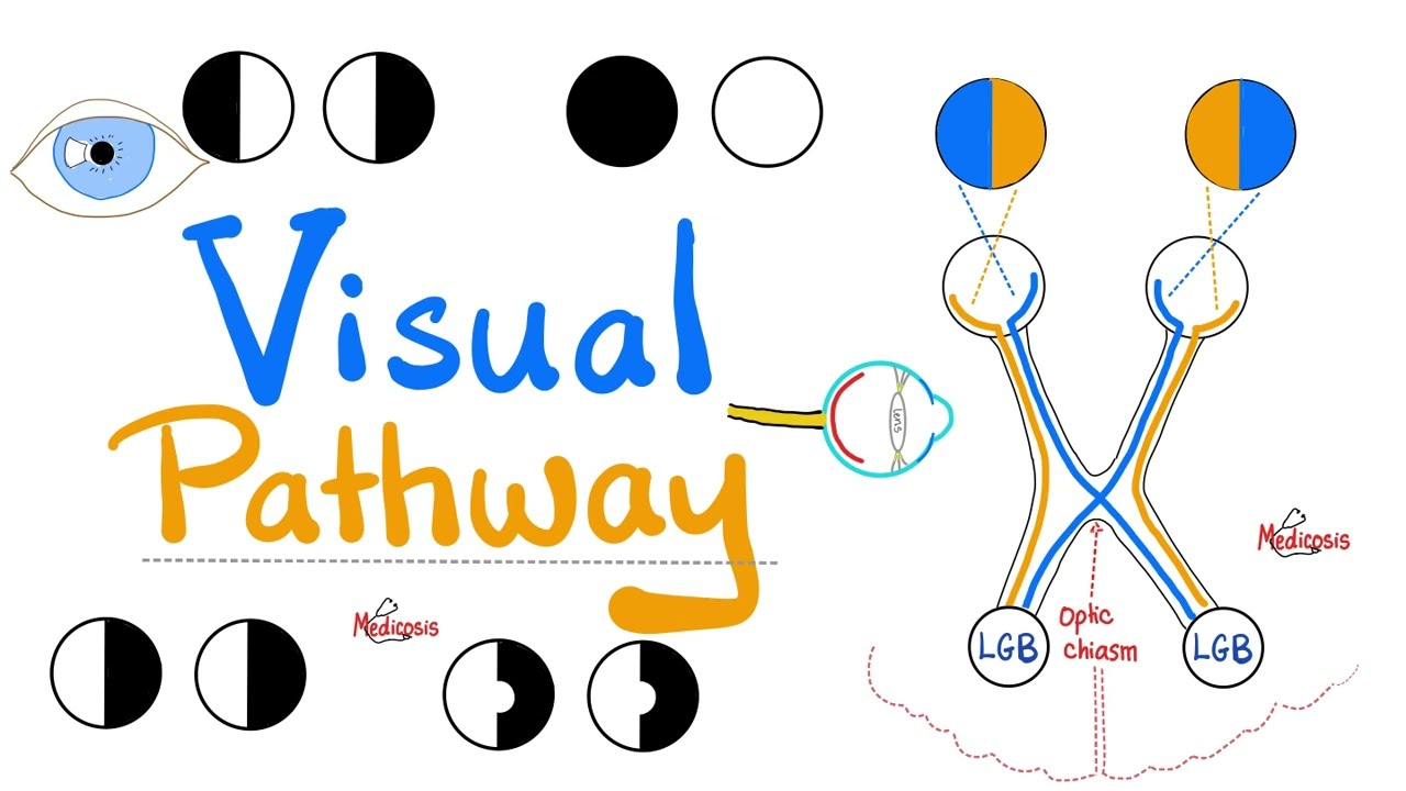






















Visual fields represent the total area visible to each eye, extending approximately 60 degrees upward, 75 degrees downward, 60 degrees nasally, and 110 degrees temporally; the visual pathway transmits signals from retinal photoreceptors through bipolar and ganglion cells to the optic nerve, optic chiasm (where nasal fibers cross), optic tract, lateral geniculate nucleus, and finally to the striate cortex in the occipital lobe, with damage at different locations producing characteristic field defects such as central scotomas from macular degeneration, arcuate scotomas from glaucoma, altitudinal defects from optic nerve damage, bitemporal hemianopia from chiasmal compression, and homonymous defects from retrochiasmal lesions, and visual field assessment methods include confrontation testing for screening, Humphrey field analyzer for static threshold perimetry (particularly for glaucoma monitoring), and Goldmann visual field for kinetic threshold perimetry (for neurological field changes).
Visual Fields Examination: Confrontation & Perimetry Guide
Added:hi our field of vision is the total area we're able to see and it's a combination of the individual visual fields of our two eyes when looking straight ahead a normal field extends about 60 degrees up 75 degrees down 60 degrees nasally and an impressive 110 degrees temporally the shape of the field of each eye is rather like the shape of a pair of sunglasses for simplicity the field of each sight can be documented as an oval shape with the visual field of the right eye drawn on the right side of the page and the left eye on the left side. This is as if the patient is looking out through the page you have in front of you. For example a patient who could not see below the horizon from his right eye would be documented like this with the dark area representing the area the patient is unable to see. You should notice that this is different from how you would record a scar on the right side of a patient's face where the drawing would be as if the patient was sitting in front of you. This reversal of sides when recording visual fields can cause confusion so remember that fields have drawn the wrong way round compared to the rest of the notes and make sure that you label them accordingly. This is a right inferior altitudinal defect.
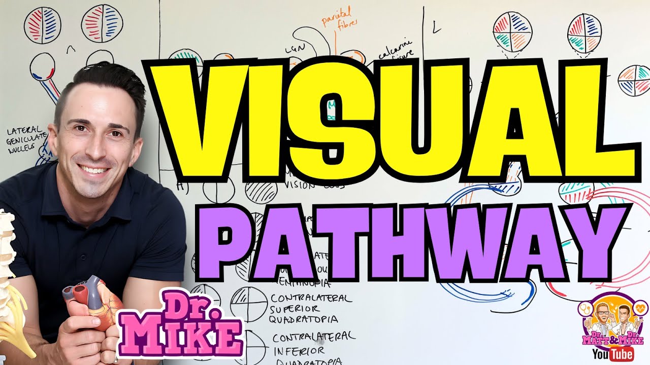
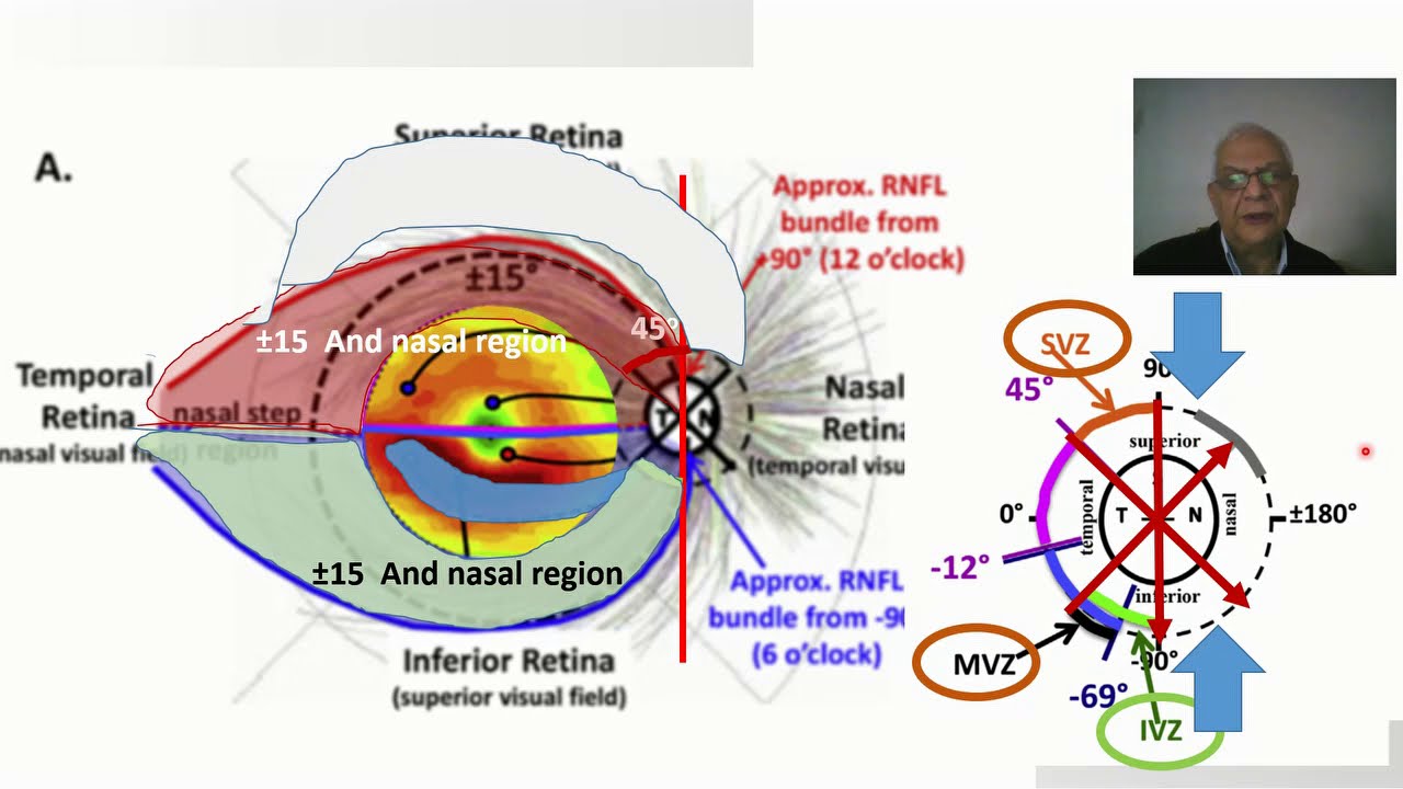
The visual pathway is the wiring from eye to brain and the destination being the primary visual cortex in the occipital lobes also known as the striate cortex. Damage along this pathway causes characteristic field defects. The pathway starts in the eye with rod and cone photoreceptors in the retina. These synapse via bipolar cells with the ganglion cells of the retinal nerve fiber layer these unmyelinated fibers pass into the optic disk and become myelinated as they become the optic nerves. We all have a small blind spot or scotoma corresponding to the optic disk which lacks photoreceptors this physiological scotoma is slightly to the side or temporal to the center of our vision damage to the macula area of retina causes loss of central vision or central scotoma seen most commonly in age-related macular degeneration or AMD an Amsler grid is a simple 5 millimeter grid pattern where the patient attempts to fix at the central dot from about 30 centimeters with each eye in turn. These can be useful to identify and monitor central scotoma. A new or changing central scotoma or indeed distortion of central vision may indicate treatable wet AMD and should be seen promptly by an eye specialist damage to nerve fibers as they pass through the optic disc is seen in glaucoma these defects cause an arcuate scotoma which follows the pattern of the nerve fiber layer here an upper nerve fiber layer defect causes a lower arcuate scotoma later this can extend to an upper or lower altitude neural defect then complete loss of peripheral field and finally blindness identification of early glaucoma field defects can allow treatment to delay or prevent this progression and preserve vision as well as the visual field, assessment of glaucoma features the appearance of the optic disc, the intraocular pressure and the medical history. The optic nerve function can be affected in many ways for example in thyroid eye disease it can be compressed in optic neuritis it can be inflamed and in giant cell arteritis it can be ischemic these may cause various shapes of field defect such as an altitudinal defect or a mixed field defect which does not respect the horizontal or vertical midline.These causes mentioned can affect one or both eyes at the same time. The optic chiasm is named after the Greek letter Chi or X. At this crossing the signal from each eye is split into two with the nasal retinal fibers crossing sides while the temporal fibers stay on the same side. The effect of this selective crossing is that the vision corresponding to the left hand field of view is supplied to the right hand side of the brain with the right visual field going to the left brain compression of the chiasm most commonly by a pituitary tumor can cause a bitemporal hemianopia with only the crossing fibers affected in the early stages these field defects may be missed sometimes for years in practice these defects are neither complete nor symmetrical. Beyond the chiasm is the optic tract which terminates in the lateral geniculate nucleus in the thalamus from here fibers fan out into the optic radiations which synapse in the striate cortex. Defects beyond the chasm the so-called retro chiasmal pathway cause homonymous defects meaning a similar area of field is affected in both the eyes. It is important to note that the vertical midline is never crossed. The optic radiations are also called the geniculo-calcarine tract. Damage to this tract occurs most commonly due to stroke and can cause quadrantanopia or hemianopia depending upon the lesion size. Temporal lobe lesions cause superior quadrantanopia or pie-in-the-sky and may have associated complex partial seizures, memory problems or wernickes aphasia if dominant hemisphere. Parietal lobe lesions cause inferior quadrantic defects or pie on the floor with possible associated sensory disturbances and Gerstmanns and aphasic syndromes if dominant hemisphere. Homonymous hemianopia and quadrantanopia most commonly arise from occipital strokes affecting the striate cortex. Posterior cerebral artery infarcts with intact middle cerebral collateral perfusion may cause so-called macular sparing where the hemianopia spares the central vision visual inattention is common in stroke patients and may mimic or accompany homonymous hemianopia with visual inattention the visual pathway may be intact but the patient fails to notice the vision on the affected side this can be demonstrated by offering the patient bilateral simultaneous stimuli so how should fields be assessed well this depends upon the context of the examination so far we have looked at field defects as being absolute in practice the visual field is like an island of vision with the center of the vision being most sensitive this means a small or dim target can be detected centrally just as easily as a bright or large target is detected peripherally accurately mapping the shape of this island of field allows for early detection of problems and to demonstrate changes over time. Two machines are in common use in the UK to assist with this. First is a Humphry field analyzer second is a Goldmann visual field. Both machines set the person's face inside a bowl of uniform light the Humphry machine typically only measures the central 24 or 30 degrees of field one eye is covered while the other fixates upon a central target lights of various brightness are shown across a grid pattern within the bowl until the threshold for detection of the light is established at each point when the person sees the flash of light they press a button the results are printed as a numeric value for the sensitivity at each point and then represented as a grayscale picture to aid interpretation this is typically used for screening and monitoring of glaucoma here is a normal Humphrey Field and here is an arcuate defect in glaucoma this is termed static threshold perimetry as the stimuli are shown in fixed grid positions to establish a threshold at each point in contrast the Goldmann field is kinetic threshold perimetry where the light stimulus is moved in towards the center of the vision until it is detected the patient again fixes the tested eye upon the central light target well this time an examiner manually brings lights towards the center of vision until it is detected these points are then plotted into isopter, like contours on a map by repeating with different light brightness or size the shape of the visual field is plotted the Goldmann field can be measured out to 90 degrees and is most commonly used to monitor neurological field changes for example before and after treatment of a pituitary tumor finally we have visual field testing - confrontation in an asymptomatic individual I think this should be a brief screening examination taking a few seconds I check central vision for quadrants and temporal field if there are possible stroke patient begin by checking fit in attention first ask the patient to cover one eye and look at your face then ask them to tell you if any part of the vision is missing if the patient thinks it is all present then normally they have no gross field defect if they cannot see part of your face then use an anslee grid to check their central vision for distortion and scotoma patients may use this grid to monitor the central vision at home next the four quadrants again with one eye covered ask a patient to look you in the eye and tell you if they can see your finger wiggling in each quadrant if they cannot see part then it can be mapped out more carefully with a red hat pin particularly noticing whether it crosses a horizontal or vertical midline finally for peripheral field I ask the patient whether they can see a finger wiggle at 90 degrees to each eye about five centimeters temporal of course a more detailed examination is possible but I think the most likely reason to miss an unsuspected field problem is not because of a brief examination but because the visual fields are not examined at all so let's wrap up central loss of field is most common in macular degeneration and should be assessed by an eye specialist urgently if recentl in onset. Field defects which respect the horizontal midline usually arise from the eye or optic nerve. Causes include glaucoma, branch retinal vein occlusion, optic neuritis and optic nerve ischaemia. They should be assessed by an eye specialist. Field defects which respect the vertical midline may arise from the optic chiasm and beyond causes include stroke and brain tumors Humphry field testing is routinely used to screen and monitor glaucoma while Goldmann testing may be used in neurological field defects examination to confrontation can be done quickly so that it need not be missed out of any routine neurological or ophthalmological assessment. To criticize comment or share your knowledge with others please go to eyevideos.blogspot.com where you will find transcripts links and more videos Thanks
Up Next

Orbital Elements Explained: Orbiter Tutorial for Beginners Part 1
@TexFilms
67.5K views•2012-06-20

Triumph of Orthodoxy Icon: Byzantine Art & History Explained
@BenCallan
2.1K views•2024-08-06

FastAPI vs Flask vs Django: Choosing the Right Python Web Framework
@TechWithTim
302.5K views•2024-05-26

Game of Thrones Opening Credits: A Cinematic Analysis
@gameofthrones
46.3M views•2011-04-18
Related Study Plans & Knowledge Roadmaps
Structured learning paths in General & Interdisciplinary Studies