














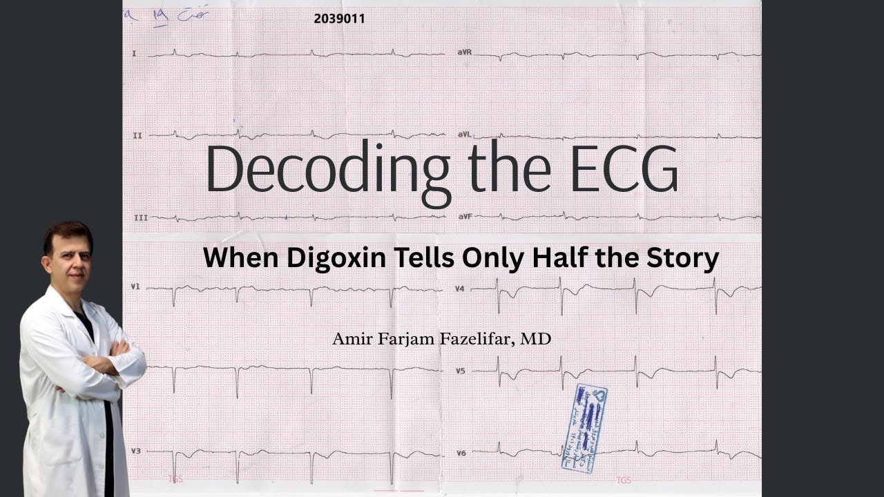

An electrocardiogram (ECG/EKG) records the heart's electrical activity through three main waves: the P-wave represents atrial depolarization initiated by the SA node, the QRS complex represents ventricular depolarization with hidden atrial repolarization occurring simultaneously, and the T-wave represents ventricular repolarization; common abnormalities include ST-segment elevation indicating myocardial infarction, T-wave inversion suggesting ischemia, first-degree heart block showing prolonged but consistent PR interval, second-degree heart blocks (Mobitz I with progressively lengthening PR intervals and Mobitz II with normal PR intervals but dropped beats), third-degree heart block with complete AV dissociation requiring pacemaker intervention, Wolf-Parkinson-White syndrome featuring characteristic delta waves from accessory pathways, atrial flutter with sawtooth F-waves, atrial fibrillation with irregular fibrillatory f-waves, wandering atrial pacemaker showing multiple P-wave morphologies, premature ventricular contractions, and torsades de pointes representing a life-threatening polymorphic VT associated with prolonged QT syndrome.
ECG Basics: Waveforms, Intervals, and Heart Blocks
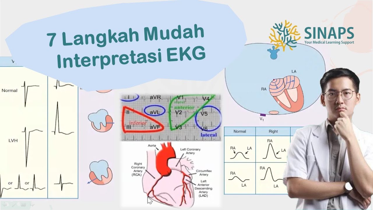
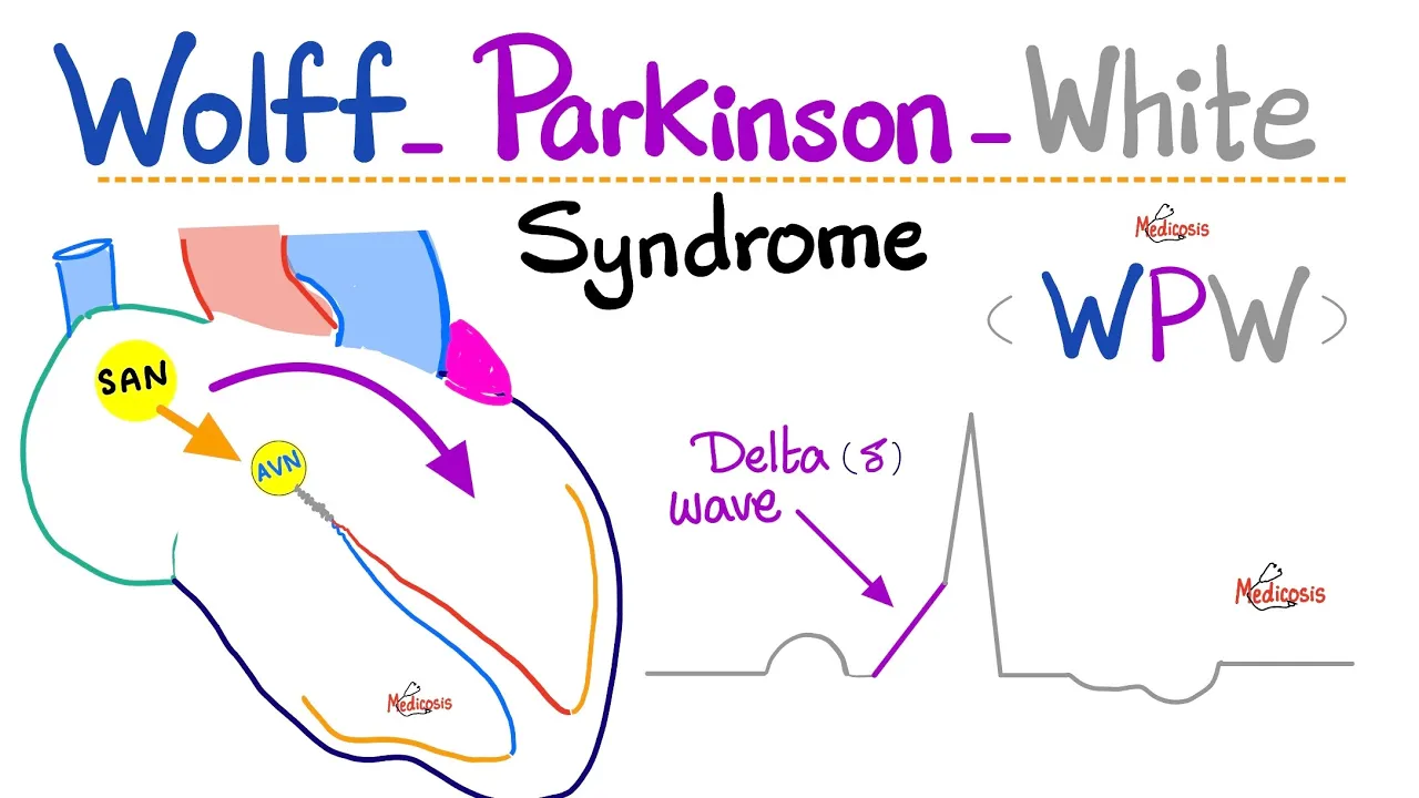
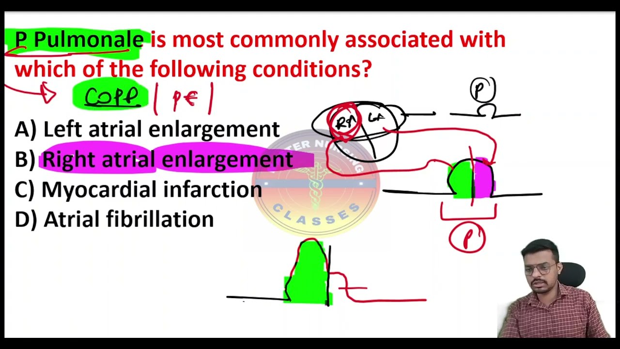
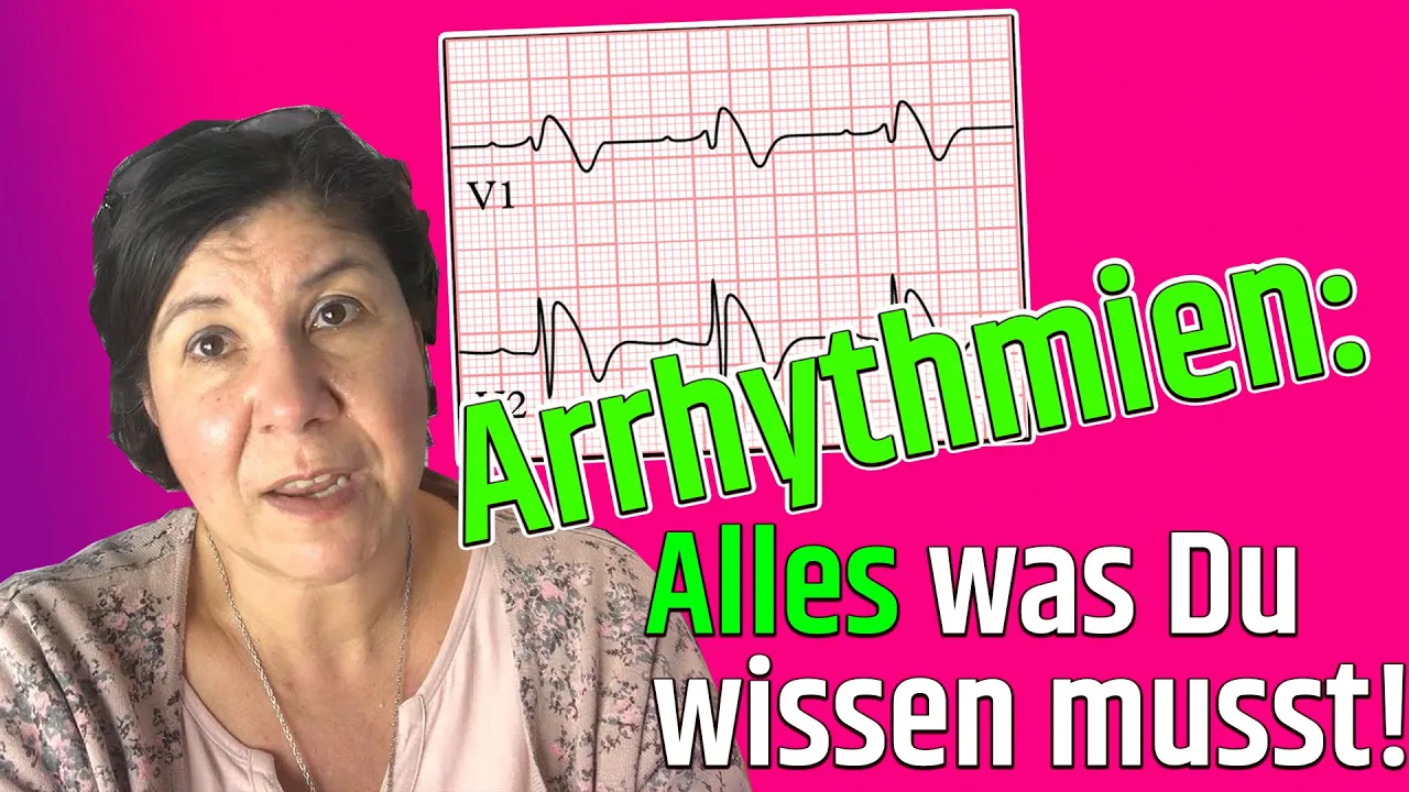
Added:all right Engineers we're going to talk about EKGs so before I get into all of these abnormal EKGs because we're going to talk about each one of these in a little bit of detail we'll go over them in much much more detail whenever we get into pathophysiology for right now I want to take a look at just the basic eekg another thing that we'll look at in future videos is we'll go over the 12 lead system so we'll go over the three bipolar limb leads the three augmented unipolar limb leads and the six chest leads but for right now we're going to just take a basic look at an EKG and then some common abnormalities first things first if we look at here at the EKG we have three distinguishable waves right this first one right here we refer to this wave as the p-wave all right so this is our p-wave so p-wave is this positive deflection from that isoelectric line right so we have that positive deflection then you might see this little negative deflection right there that's your QRS wave so we have our QRS wave and then this last wave over here with this other positive deflection is the t-wave now let's talk about each one of these waves what they are designed to what they specify so first off let's go over here to p-wave p-wave so the p-wave tells us that the Atria are polarizing in other words whenever the Atria so you start off with the SA node the SA node fires those impulses throughout the entire internodal pathway and Bachman's bundle and it reaches the AV node what happens is once it reaches the AV node the Atria depolarize and they contract they undergo syy but that electrical activity that shows up on here on the EKG with this positive deflection point is designed to tell us that there is atrial D polarization and again we already know a depolarization means it means that the inside of the cell is becoming more positive it's conducting Action potentials right this next one over here is the QRS wave so we have the QRS wave over here now the QRS wave is obviously this big negative positive negative deflection and this represents ventricular depolarization so it does represent ventricular depolarization and what that means is that whenever those impulses from The Av node they reach the AV node there's a 0.1 second delay that we talked about in the cardiac cycle it sends those impulses down through the ventricles down the AV bundle right and left bundle branches pereni fibers and as it's moving around those guys it's causing the ventricles to become depolarized in other words the inside of their membranes are becoming more positive and then eventually that will trigger ventricular contraction which we talked about in the cardiac cycle so again that's ventricular depolarization another interesting thing is cells need to go through a relaxation period so they need to depolarize and then they need to repolarize so they can be excited again so if you notice we didn't mention here where's atrial repolarization well atrial repolarization is hidden or masked within this QRS complex so we can technically say within the QRS wave there is a hidden or masked atrial repolarization occurring so let's write that down here atrial repolarization is occurring there's atrial repolarization and ventricular depolarization occurring within this QRS wave and again you can't see the atrial repolarization because it's masked by this positive negative deflection points within the QRS wave next one is going to be the t-wave and the t-wave is what signifies the ventricular the ventricles repolarizing so after they've all contracted they have to go into the refractory period so that they can actually get all the oxygen to the tissues because that's when the coronary circulation is filling and once it starts doing that it has to repolarize and then get ready to be excited again so we say the t-wave is representing ventricular repolarization okay so now that we have a basic concept or an understanding of what all this just basic EKG tells us now we can take a look at some of these common abnormalities within the ECGs like I said we're going to get into these in more detail when we get into pathopysiology just a brief overview of some of these so if you look here we have a normal p-wave on this one we have a normal QRS complex but if you notice here compare this with this normal t-wave this array here we call this part right here the ST segment from this part here to this part here it's the ST segment and then we refer to this from the beginning of the P wve to that Q that's our PR interval right so here's our St M I'll write that there ST segment and then this right here is our P interval right PR interval looks good but the ST segment looks like it's elevated so we call this an ST segment elevation sometimes in hospitals you'll hear it referred to as a stemi all right and stemi is short for saying St segment elevation myocardial infarction and so you would see if someone has a ST segment elevation most likely is indicative of some type of myocardial infarction or a heart attack but obviously you could do other diagnostic testings to prove that all right that's one of those differential diagnoses then over here you'll notice in this one that there's a normal p-wave normal QRS complex but then the t-wave flipped it's inverted so now there is a t-wave inversion so this one is a t-wave inversion now t-wave inversions are indicative there is ST segment depressions I didn't want to talk about that again we'll talk about it in more detail later but just simply t-wave inversions are usually indicative of eskee so they're usually indicative of eskee and esea just means that there is a decreased oxygen supply or lack of a decreased uh lack of blood flow to the my Cal tissue and it's going to cause this negative deflection it's again that's a t-wave inversion let's come over here to this one so now we look at this guy what do we notice we notice a normal P wve all right then we notice the PR interval here QR is complex then we got our T wve then we go over here to our p wve and then we notice that this there's our PR interval QRS complex t-wave now the only way we be able to like determine why this is abnormal is if you actually look at the ECG paper if you look at the ECG paper there's a little blocks that identifies the amount of time it takes if the PR interval normally the PR interval is about 160 to 200 milliseconds if it's greater than that then there's a prolong PR interval what happens in this one and I obviously I need an ECG paper to see that but there is actually going to be a prolong PR interval right here but it's consistent all right it's not it's not varying it's a consistent prolong PR interval so let's say it's greater than 200 say it's 220 milliseconds here and then over here this PR interval even though it's not looking like it it is 220 milliseconds also if someone has consistent PR prolongation PR interval prolongations but it's not progressively getting longer this is described to be what's called a first deegree heart block which are are are physiologically normal okay there's nothing really uh designed to be you know this is not life-threatening it's very common obviously stress anxiety certain things can actually trigger this to happen but it's not life-threatening there's no need for a pacemaker or anything like that all right next one if you look over here we notice the PR interval is long and then it actually looks like it's getting a little longer and then eventually over here you notice there goes t-wave P wve whoa no QRS wave what happened there so if we look again what was I saying let's say that this PR interval let's say it's 220 milliseconds then we come over here in this PR interval let's say it's 240 milliseconds so again we have our t-wave here then we got our p-wave but look guys no QRS complex and then we go back to a p-wave this is referred to as a second degree heart block and you can actually say this is called moits one or winken block and how do you describe that you say that the PR interval is getting progressively longer and then you drop a QRS interval or a QRS wave sorry all right so that is one thing to remember for a second degree heart block moit one now second degree heart block mobit one is more common within some type of infranodal block so there's a block usually within the AV node we'll go into more detail that in pathophys but for right now just remember common cause of it is usually an infranodal block now second deegree moits one can progress into moits 2 and again moits one isn't super lifethreatening but it's obviously you know it's a partial heart block um if we come over here this is a little bit more you know dangerous you got to be careful with these ones this is a second deegree har block moitz 2 now a second degree heart block mobit to the PR interval is normal so it's normal PR interval it's not it's not fluctuating obviously I didn't show up perfectly there but the PR interval is normal it's not changing it's not going from 220 to 240 it's a normal PR interval but what you notice is here's your P wve PR interval QRS wve t-wave and then there's our p wve but then if you notice what happened we dropped our QRS complex and then we go back to a p wve and then we might have a QRS complex so it's kind of like grouping them and what it might look these are kind of sometimes hard to differentiate a little bit but with this one the the third degree heart block which I'll talk about it does have a varying PR interval whereas within the second degree heart block moitz 2 the PR interval is usually normal it's not changing it's not fluctuating but some of the p waves don't make it through it doesn't get through the AV node and it doesn't go down to the ventricles so the ventricles don't contract that is important to remember the p waves are going but they're not making it through the AV node they're not getting into the ventricles and the ventricles are Contracting so this is called second degree har block moits two okay and just remember normal PR interval but it drops a QRS complex in there because some of the p waves don't get through and again uh second degree heart blocks um some of the symptoms that you can see Within These people are some Syncopy some dizziness and stuff like that too okay all right let's come to this next one this is a third degree heart block and the third degree heart block I've already kind of given you a little bit of information on that we already know that they're going to have sometimes you're going to have this again so P wve QR is complex so again P QRS t-wave p-wave and then you go back to a p-wave and then we got QRS complex and then we go t-wave P wve p-wave again this one is completely the worst one this is a this is a complete heart block as you would describe it so third degree heart blocks are very very life-threatening very dangerous you're going to need to give them most likely an artificial pacemaker actually definitely need an artificial pacemaker but here's what's different from the second degree heart block the ventricles and the Atria beat on their own rate so none of the impulses within the uh mobitz 2 some of the impulses from the uh Atria actually get down into the ventricles within third degree heart block none of the impulses get through the AV node and down to the ventricles the ventricles actually develop their own beating rate and the Atria beat at their own rate so because of that there is no connection if you will between the Atria and The ventricle functioning as well anymore okay this is a third degree heart block so the p and the QRS wave are not agreeing correctly and they're beating at their own rate all right so that's thir guard block all right guys so now we're going to come down to my personal favorite and this is going to be an EKG that is indicative of what's called wolf Parkinson's white syndrome so I'm just going to put W PW syndrome okay so again this is called wolf Parkinson's white syndrome It's A congenital condition in which the individuals have an irregular accessory pathway and that access accessory pathway is called the bundle of Kent and it's actually bidirectional so impulses can go through it down anterograde and it can go up through it retrograde which can create what's called re-entry and it can lead to Super ventricular attack of cardio which is can be symptomatic and asymptomatic and stuff like that usually the way to treat this is they sometimes can do what's called uh radio frequency catheter uh ablation where they hit this bundle of Kent with radio frequency waves creating Scar Tissue so it doesn't conduct the electrical potentials through it anyway let's come back here and let's look here we can see here in this one they don't really have a distinguishable p-wave so if they don't have a distinguishable p-wave it actually kind of just goes straight up it looks like it's coming straight up right there that's actually a distinct wave this one right here I'm going to draw in blue that's called a Delta wave and again over here you'll see this one this is called a Delta wave so again this is a Delta wave here and this is a Delta wave here and again this is usually indicative of someone who has wolf Parkinson's white syndrome so that's one of the big things to look for for this there's obviously more and we'll talk about it in great detail in pathophysiology let's come over here and let's look at some more EKGs so if we look here you're going to see this one and right away you see the sawtoothed like structures these are supposed to be your p waves but we don't call them p waves we actually call them f-waves so this is a fwave all these are f-waves all right that's an f-wave and these Sal two fth waves indicate that there's some p waves they're just kind of like doing through this they're going through this re-entry and then eventually some of those re-entries will hit the AV node and then send the action potentials down to the ventricles and then cause the ventricles to depolarize and then again you'll have your t-wave and then you might have some more again re-entry circuits and then eventually some of those circuits will hit the AV node and send action potentials down atrial flutter is this condition again this is called Atri I flutter it's not life-threatening but it can progress um and become very serious usually atrial flutter is caused by could be due to myocardial infarctions or thyrotoxicosis like elevated T3 and T4 could be due to mitro prolapses whole bunch of reasons that we will talk about in the future but uh Atri flutter obviously is not completely serious but it can progress and sometimes it can progress into this one below it which is called atrial fibrillation and again you'll notice in natur fibrillation you have these like don't even really see a p-wave there there's not distinct p waves it's just kind of like this little squiggling almost around Baseline activity to where there's no p waves present it's just this kind of like irregular Rhythm here and that's one of the big things to notice here they're not F they're actually they do have names but it's here's the biggest thing they're f-waves but they're little F so this is little F waves little F waves so they're both F waves except this one up here in atrial flutters capital F waves are these are lowercase f waves so these lowercase f waves are indicative of atal fibrillation okay apib you got to be careful with obviously you might have to these could be due to Metabolic Syndrome again thyrotoxicosis M obesity it can go on and on and on and sometimes you might have to deal with this depending upon the condition again you might have to do uh radio frequency catheter ablation you might have to do cardiov version whatever it might be we'll get into more detail into that in the future so again these are f-waves but remember they're lowercase f-waves and this is this condition is called atrial fibrillation another danger with atrial fibrillation I just wanted to mention really briefly is that they don't really have functional contractions and so the blood pulls up a lot inside of the Atria and if there stagnant flow according to virtuos Triad when there's a stasis of blood flow there can be an increased chance of forming thrombi and so sometimes these can form little vegetations on the the valves the mitro valve and the tricuspid valve and get dislodged and go and cause an embolism like a pulmonary or cerebral embolism and again we'll talk about these in more detail later next one right here you're going to notice something weird and this is kind of more common in individuals who are uh using drugs like drug overdoses you'll notice that the p-wave changes it takes on different morphological structures at least three so right here it looks like it's kind of triangular shaped over here it looks kind of like like normal and over here it kind of looks like a little squiggled right whenever you notice three different morphological p waves in sequence it's referred to as it's called a wanding atrial pacemaker okay and again you'll notice that this is more common in like drug overdoses but you'll notice that there's obviously three distinguishable or morphological p waves okay it's a wondering atel praise maker over here this is obviously common to someone who uses nicotine or you know they drink a lot of caffeine or certain drugs and stuff like that what can happen is they can have this little ectopic Focus where they could trigger a premature ventricular contraction so it might develop some irritable folky within the ventricles and it might contract before the p-wave actually sends the impulses out so you'll notice here we go p-wave right so we have a p-wave then we got our QRS wave then we have our t-wave but we don't see the p-wave it kind of goes and actually bypasses the p-wave because of a premature ventricular ectopic focus and it triggers a premature ventricular contraction so it's indicative of this part right here okay so this is indicative of pvc's or premature ventricular contractions okay and obviously this can be due to caffeine it could be due to uh possibly um some type of nicotine it could be due to exercise a lot of common causes of this last one all right this this guy is not having a good day uh this is a really really bad one this is life-threatening and it's actually called uh torsades dep points torsades to points so torsades to points or I think they even call it Depon it just refers to twisting of the points around the isoelectric line this one's really lifethreatening obviously you're going to need to treat this it's medical emergency but with torsa points it's usually referred to as a prolong QT syndrome and the most common causes of prolong QT syndromes are actually metabolic reasons in other words more specifically if someone has low magnesium levels within the blood or if they have hypokalemia or hypomagnesemia um those are common causes and even medications can lead to those so this is a really really bad one obviously this needs uh medical attention immediately so this kind of gives us just a brief overview we'll get into a lot of these in more detail in the future um but for right now that gives us everything we need to know about EKGs
Up Next
Cardiac Conduction System Animation | The Heart's Electrical Wiring
@AniMedical-Animations
112.7K views•2016-02-14

Graphic Medicine: Comics for Collaborative Healthcare Communication
@nationalpatientadvocate
189 views•2023-12-04
Hypothalamus Anatomy and Function: Diencephalon and Nuclei Explained
@NinjaNerdOfficial
676K views•2020-11-10

Stages of Labor and Vaginal Birth | Childbirth Animation
@nucleusmedicalmedia
52.1M views•2017-08-18
Related Study Plans & Knowledge Roadmaps
Structured learning paths in Medicine