The heart's electrical conduction system coordinates rhythmic contractions through a specialized network of muscle cells: the SA node (anatomical pacemaker) initiates the heartbeat by triggering atrial contraction, then signals travel through the AV node, bundle of His, bundle branches, and Purkinje fibers to cause ventricular contraction; this electrical activity is recorded on an electrocardiogram (EKG), which doctors use to monitor cardiac function and detect abnormalities.
Cardiac Conduction System Animation | The Heart's Electrical Wiring
Added:Deep Dive
Basic gross anatomy of the heart, specifically the four chambers (atria and ventricles) and the major valves.

GROSS ANATOMY OF THE HEART PART 2
2.7K
The heart has four chambers with distinct anatomical features: the right ventricle is triangular with trabeculae carneae and three papillary muscles (anterior, septal, posterior) connected to the tricuspid valve, while the left ventricle has thicker walls (3x right ventricle) and two papillary muscles (anterior and posterior) connected to the mitral valve; both ventricles have inflow and outflow tracts separated by muscular ridges, with the right ventricle leading to the pulmonary trunk via the conus arteriosus and the left ventricle leading to the aorta via the aortic valve, which also gives rise to the coronary arteries from its right and left sinuses.
Click to watch →
structure of Heart||Atria, ventricles and valves| In detail in telugu||useful for all entrance exams
313
The heart consists of four main chambers: two atria (upper chambers) and two ventricles (lower chambers). The right atrium and right ventricle are on the right side, while the left atrium and left ventricle are on the left side. The atria receive blood, while the ventricles pump blood out. The heart has four main valves: the tricuspid valve (right atrioventricular valve with three cusps) between right atrium and ventricle, the bicuspid/mitral valve (left atrioventricular valve with two cusps) between left atrium and ventricle, the pulmonary valve (right semilunar valve) between right ventricle and pulmonary artery, and the aortic valve (left semilunar valve) between left ventricle and aorta. These valves ensure one-way blood flow and prevent backflow.
Click to watch →

Anatomy of the Heart- ਵਾਰਡ ਐਂਟਡੈਂਟ ਪੇਪਰ ਲਈਂ ਹੁਣ ਤੱਕ ਦੀ ਸਭ ਤੋਂ ਮਹੱਤਵਪੂਰਨ ਵੀਡਿਓ||BFUHS||
3.3K
The heart is a vital organ with four chambers: two atria (right and left) that receive blood, and two ventricles (right and left) that pump blood out. The right atrium receives deoxygenated blood from the body, which then flows to the right ventricle that pumps it to the lungs for oxygenation. The left atrium receives oxygenated blood from the lungs, which then flows to the left ventricle that pumps it to the entire body. The left ventricle has the thickest muscular wall because it must generate the highest pressure to circulate blood throughout the body. The heart contains four valves that ensure one-way blood flow: the tricuspid valve (between right atrium and ventricle), pulmonary valve (between right ventricle and pulmonary artery), mitral valve (between left atrium and ventricle), and aortic valve (between left ventricle and aorta).
Click to watch →
Basic Anatomy & Physiology 12 | THE HEART Reference Seeley's
18.8K
The heart has four hollow chambers: left and right atria (superior chambers) and left and right ventricles (inferior chambers). The coronary sulcus divides the atria from the ventricles. The atria receive blood from veins throughout the body and have thin-walled cardiac muscle, performing minimal contraction to push blood to the ventricles. The ventricles pump blood out to arteries and have thick, strong walls necessary for forcefully pushing blood throughout the body. The interatrial septum divides the left and right atria, while the interventricular septum divides the left and right ventricles.
Click to watch →

CARDIOVASCULAR SYSTEM
107.4K
The heart consists of four chambers: two atria (upper) and two ventricles (lower). The left atrium receives oxygenated blood from lungs, while the right atrium receives deoxygenated blood from the body. Between chambers are four valves: the mitral valve (bicuspid, 2 cusps) between left atrium and ventricle, the tricuspid valve (3 cusps) between right atrium and ventricle, the pulmonary valve between right ventricle and pulmonary artery, and the aortic valve between left ventricle and aorta. These valves ensure unidirectional blood flow and regulate pressure.
Click to watch →
Deep Dive
The concept of membrane potentials and action potentials, including the movement of sodium, potassium, and calcium ions across cell membranes.
Generalidades de Bioelectricidad y Potencial de accion
28.8K
Action potentials are voltage changes across cell membranes that trigger cellular responses. The resting membrane potential represents a dynamic equilibrium where ions constantly move. Sodium and potassium are the primary ions: sodium is predominantly extracellular (140-145 mM) while potassium is predominantly intracellular (135 mM). Ions move under two forces: chemical forces (diffusion from high to low concentration) and electrical forces (movement toward opposite charges). For sodium, both forces favor entry into the cell. For potassium, chemical forces favor exit while electrical forces favor entry, creating opposing tendencies that determine net ion movement.
Click to watch →

Ventricular Action Potential | Cardiac Action Potential | Part 1 | Phases | Cardiac Physiology
248.8K
Membrane potential refers to the electrical charge difference across a cell's plasma membrane. At rest, the membrane potential is typically negative (around -70 mV in neurons). Action potentials are brief changes in this membrane potential caused by the opening of ion channels that allow different ions (sodium, potassium, and calcium in cardiac muscle) to move between the intracellular and extracellular environments. Since these ions carry electrical charges, their movement along electrochemical gradients changes the membrane potential.
Click to watch →
Eindexamenspreekuur biologie vwo met Simone en Suzanne (2025)
42.6K
Action potentials have distinct phases: resting potential at approximately -70 mV, depolarization to about +30 mV, repolarization, and hyperpolarization to about -80 mV. The threshold is approximately -55 mV. Sodium and potassium ion concentrations differ across the cell membrane: sodium is higher outside, potassium is higher inside. The sodium-potassium pump actively maintains these gradients using ATP. During depolarization, voltage-gated sodium channels open, allowing sodium to flow in passively. Potassium channels open during repolarization, allowing potassium to flow out passively.
Click to watch →
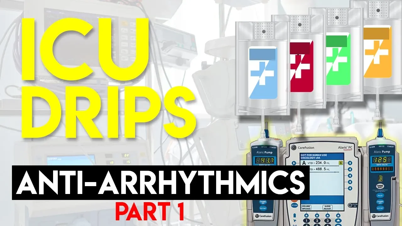
Antiarrhythmics (Part 1) - ICU Drips
117.5K
An action potential is the movement of ions across cell membranes that changes membrane potential. Normally, cardiac cells have negative interior and positive exterior due to ion concentration gradients (higher sodium/calcium outside, higher potassium inside). Action potentials involve brief depolarization followed by repolarization. Two distinct cardiac action potentials exist: the pacemaker action potential in SA/AV/Purkinje cells, and the myocyte action potential in contractile cells. Pacemaker cells exhibit automaticity through funny currents that slowly leak sodium into cells when voltage is below -40 mV, spontaneously increasing voltage toward threshold. This automaticity can be modified by autonomic nerves, hormones, drugs, ischemia, and hypoxia.
Click to watch →

POTENCIAL DE MEMBRANA Y POTENCIAL DE ACCIÓN
406.4K
During an action potential, sodium channels open and sodium ions enter the cell, making the intracellular environment more positive. This causes depolarization. During the spike phase and repolarization, potassium channels open and potassium ions exit the cell toward the extracellular environment, causing the membrane potential to decrease. This ion movement is fundamental to the generation of the action potential.
Click to watch →
Deep Dive
The difference between skeletal and cardiac muscle tissue, particularly the presence of intercalated discs and gap junctions that allow rapid signal transmission.

Locomotion & Movement | Most Expected Question | Class 12 | NEET Biology
449
Intercalated discs are the exclusive feature of cardiac muscle tissue. They contain gap junctions that allow rapid electrical signal transmission between adjacent cardiac muscle cells, enabling coordinated heart contractions. Both skeletal and cardiac muscles have striated appearance and excitable membranes, but only cardiac muscle has intercalated discs.
Click to watch →
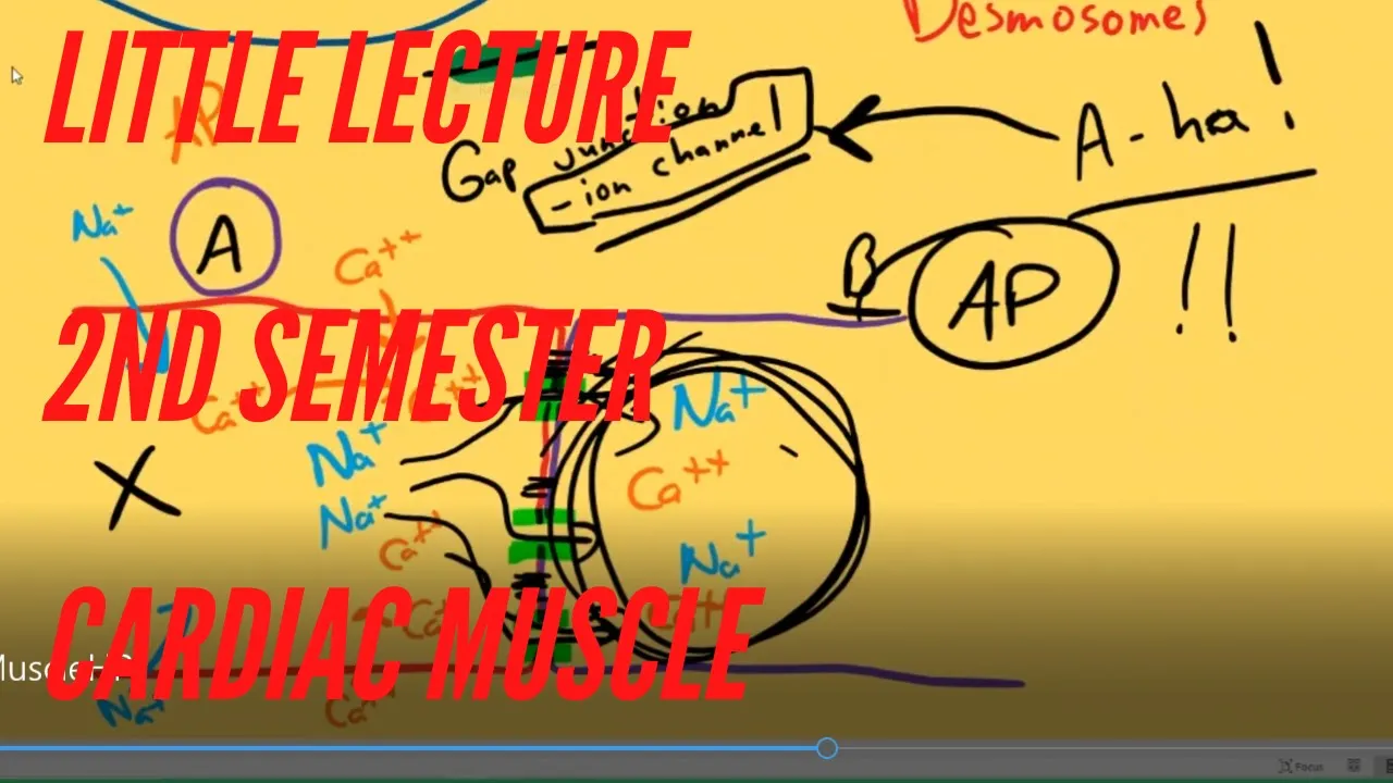
Little Lecture 2nd Semester Cardiac Muscle
984
Intercalated discs are the most significant structural difference between cardiac and skeletal muscle. They consist of gap junctions and desmosomes that connect neighboring cardiac muscle cells. Gap junctions are formed when proteins in the cell membranes of two neighboring cells fuse together to form a tunnel or channel. This channel allows ions to move directly between the cytoplasm of adjacent cells, facilitating the transmission of electrical signals throughout the heart muscle.
Click to watch →
TEST-03 I ANIMAL TISSUE l RPSC 1ST GRADE BIOLOGY MCQ #test_series_for_rpsc_first_grade_biology
5.2K
Cardiac muscle differs from skeletal muscle in several key ways. Cardiac muscle is involuntary (not under conscious control) while skeletal muscle is voluntary. Cardiac muscle cells are branched and connected by intercalated discs, which contain gap junctions that allow ions to flow between cells. These gap junctions enable coordinated contraction of the heart muscle. Skeletal muscle cells are long, cylindrical, and multinucleated without intercalated discs.
Click to watch →

Muscular revision pharmacy
106
Intercalated discs are specialized junctions between cardiac muscle fibers that contain gap junctions. These junctions allow rapid transmission of electrical impulses between cells, enabling the heart to contract as a synchronized unit. Intercalated discs are absent in skeletal muscle.
Click to watch →

9. sm. muscle, cardiac, disorders
340
Cardiac muscle tissue is the principal tissue in the heart. Like skeletal muscle, it contains actin and myosin filaments arranged in bands and zones, giving it a striated appearance. However, cardiac muscle is unique in having intercalated discs containing desmosomes (spot welds holding fibers together) and gap junctions (allowing action potentials to spread between cells). Unlike skeletal muscle, which contracts only when stimulated by acetylcholine from motor neurons, cardiac muscle contracts through its own autorhythmic cells that depolarize and cause contraction approximately 70 times per minute from heart formation until death.
Click to watch →
Deep Dive
The basic mechanical cardiac cycle, understanding the phases of systole (contraction) and diastole (relaxation).

Cardiac Cycle
82.2K
The cardiac cycle alternates between systole (contraction) and diastole (relaxation). Systole begins after ventricular filling during diastole and involves two phases: isovolumic contraction (pressure rises above atrial pressure, closing AV valves with first heart sound) and ejection (pressure exceeds aortic pressure, opening semilunar valves for blood ejection). Diastole follows with four phases: isovolumic relaxation (pressure drops below aortic pressure, closing semilunar valves with second heart sound), rapid inflow (AV valves open, passive filling to 70-80%), diastasis (minimal volume change), and atrial systole (atria contract adding final 20-30%). Diastole constitutes approximately two-thirds of the cardiac cycle.
Click to watch →

Basic Electrophysiology, part 2 - Mechanical Anatomy of the Heart, part 2
51.7K
The cardiac cycle consists of two main phases: systole (contraction) and diastole (relaxation). Systole refers to the contraction of the ventricles, which ejects blood into the aorta and pulmonary artery. Diastole is the relaxation phase where heart muscle fibers lengthen, allowing filling of the atria and ventricles, and coronary perfusion occurs. When discussing cardiac cycle phases without qualification, they typically refer to ventricular systole and diastole.
Click to watch →

cardiac cycle
82.5K
The cardiac cycle consists of two basic phases: systole and diastole. Systole begins with the actual contraction of the ventricular muscle and includes the period of ejection when blood is ejected into the outflow tracts. Near the end of systole, some muscles undergo relaxation and begin to lose their ability to generate force. Diastole is the longer of the two phases at normal heart rates and is initiated with relaxation. Once the ventricle relaxes sufficiently, it begins to fill with blood from the atrial chambers.
Click to watch →
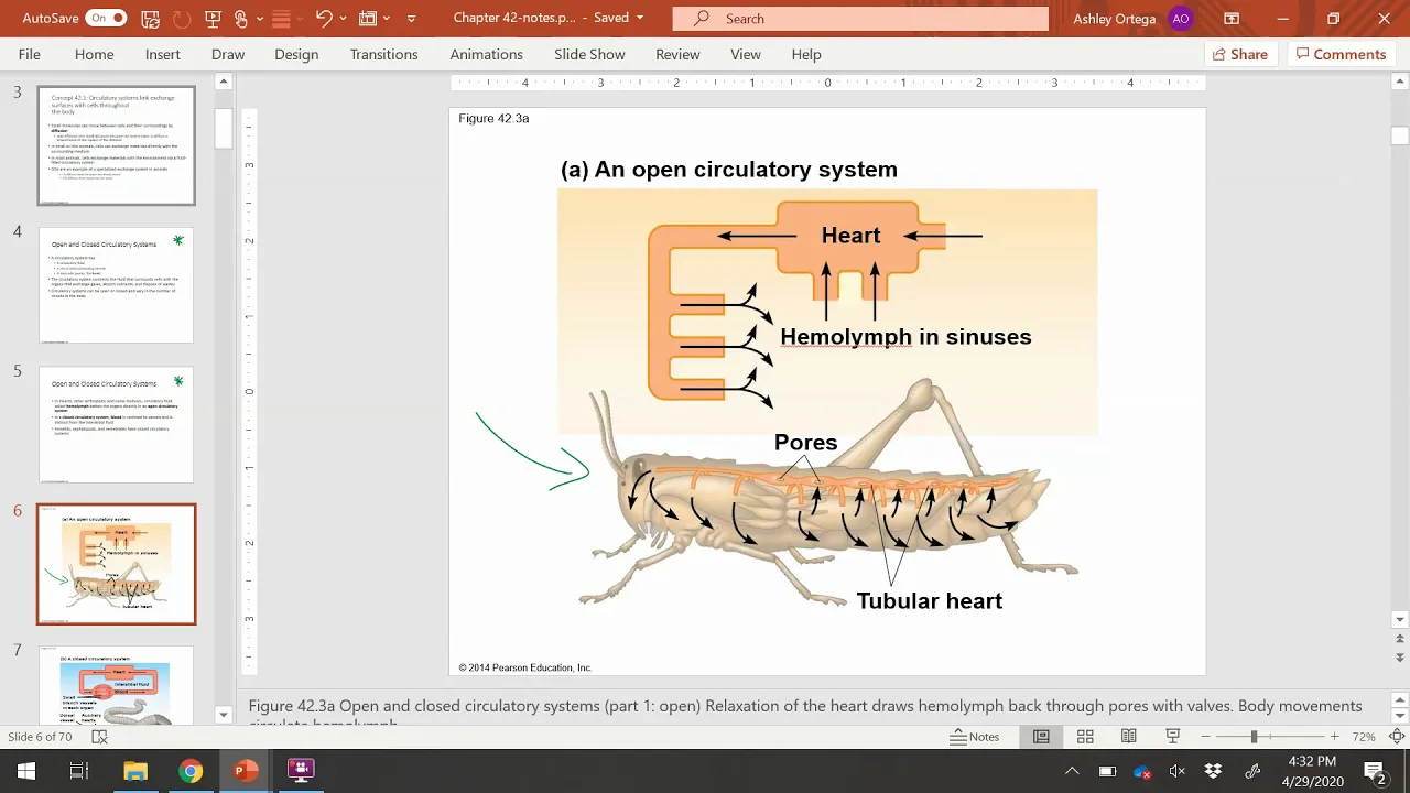
BIOL 1407 Chapter 42 Part 1
448
The cardiac cycle consists of alternating contraction (systole) and relaxation (diastole) phases. Systole (from Greek 'to contract') is the pumping phase when ventricles contract to push blood out. Diastole (from Greek 'to pull apart') is the filling phase when chambers relax and fill with blood. These phases create the heartbeat sounds.
Click to watch →

dr. Andi Adil, Sp.An-KAKV - KARDIOTORASIK I
766
The cardiac cycle consists of systole (contraction) and diastole (relaxation). Systole includes isovolumetric contraction (ventricular pressure rises without volume change), ejection phase (blood pumped into aorta), and isovolumetric relaxation. Diastole includes isovolumetric relaxation, rapid filling, diastasis, and atrial systole which contributes approximately 30% of ventricular filling.
Click to watch →
Prerequisite Knowledge
- Concept 01Basic gross anatomy of the heart, specifically the four chambers (atria and ventricles) and the major valves.
- Concept 02The concept of membrane potentials and action potentials, including the movement of sodium, potassium, and calcium ions across cell membranes.
- Concept 03The difference between skeletal and cardiac muscle tissue, particularly the presence of intercalated discs and gap junctions that allow rapid signal transmission.
- Concept 04The basic mechanical cardiac cycle, understanding the phases of systole (contraction) and diastole (relaxation).
Subsequent Learning
- Step 01Interpretation of Electrocardiograms (ECGs/EKGs) and how specific electrical waves (P, QRS, T) correlate with the conduction system's pathway.
- Step 02Pathophysiology of cardiac arrhythmias, such as heart blocks, atrial fibrillation, and ventricular tachycardia.
- Step 03The mechanism of action of antiarrhythmic medications (such as beta-blockers and calcium channel blockers) on the cardiac action potential.
- Step 04Clinical applications and technology behind artificial pacemakers and implantable cardioverter-defibrillators (ICDs) to manage conduction defects.
系统定位
核心组件
信号启动
心室传导
心电监测
临床意义
系统定位
0:09Playing Section
- 1
心肌特化细胞网络位于心脏壁内。
- 2
该网络负责发送信号引发心脏收缩。
- 3
此结构被命名为心脏传导系统。
The Syncytial Model and Mechano-Electrical Feedback
While traditional animations depict the cardiac conduction system as a series of discrete, insulated electrical 'wires' (the cable theory), modern cardiac physiology presents a more integrated view. Critics of the simple 'wiring' analogy point out that the heart is a functional syncytium, where electrical current propagates continuously through gap junctions across all cardiac muscle cells, not just specialized pathways. Furthermore, the 'Mechano-Electrical Feedback' (MEF) theory argues that heart rhythm is not purely driven by a top-down electrical hierarchy. Instead, mechanical forces, such as the stretching of chambers during filling, directly alter electrical properties via stretch-activated ion channels. Additionally, research shows the SA node is not a single, isolated 'spark plug' but a distributed, highly integrated pacemaker network. Understanding these complex, bi-directional, and diffuse interactions is crucial, as the simplistic 'wiring' model fails to fully explain complex arrhythmias and the heart's self-regulating adaptive mechanisms.
Deep Dive
Interpretation of Electrocardiograms (ECGs/EKGs) and how specific electrical waves (P, QRS, T) correlate with the conduction system's pathway.

Cardiovascular System - Cardiac arrhythmias | NORCET & Other Nursing Exams | Raju Sir
44.1K
An electrocardiogram (ECG) records the heart's electrical activity, not brain activity. The cardiac electrical conduction pathway follows: SA node generates impulses, which travel through atria causing atrial depolarization (P wave), then through AV node, bundle of His, and Purkinje fibers to ventricles causing ventricular depolarization (QRS complex). The T wave represents ventricular repolarization. The P wave indicates atrial contraction, QRS complex indicates ventricular contraction, and T wave indicates ventricular relaxation. Understanding these components is essential for cardiac diagnosis.
Click to watch →
L6: ECG Disorders of Circulatory System - Body Fluids and Circulation | 4 Din me Chapter Chitt
99.6K
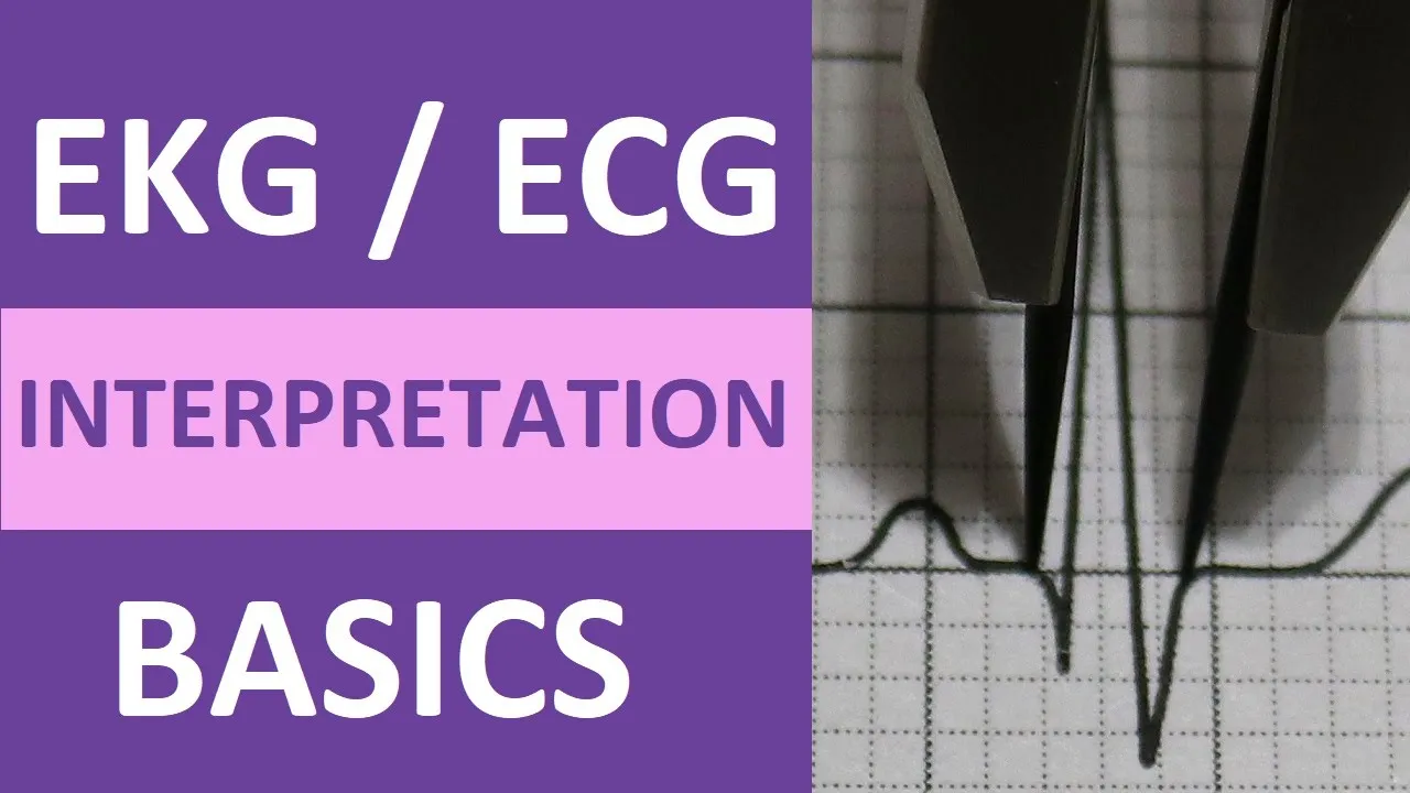
The P wave represents atrial depolarization. The QRS complex represents ventricular depolarization and is larger due to greater ventricular muscle mass. The T wave represents ventricular repolarization. Ventricular depolarization masks atrial repolarization signals. Understanding these waves is essential for cardiac diagnosis.
Click to watch →
EKG Interpretation Basics: QRS, P Wave, T Wave, PR Interval
1.5M
An electrocardiogram (EKG/ECG) assesses the heart's electrical conduction system, which coordinates heart contractions through depolarization (contraction) and repolarization (relaxation); the PQRST complex represents this process where the P wave indicates atrial depolarization, the QRS complex shows ventricular depolarization, and the T wave marks ventricular repolarization, with specific measurements (P wave <0.12s, PR interval 0.12-0.20s, QRS <0.12s, QT interval 0.35-0.44s) helping identify normal sinus rhythm or dysrhythmias.
Click to watch →
Cardiovascular System | ECG Waves Simplified | Plan C+ Gold Edition | Dr Deepak Marwah | NNL ONE
74.6K
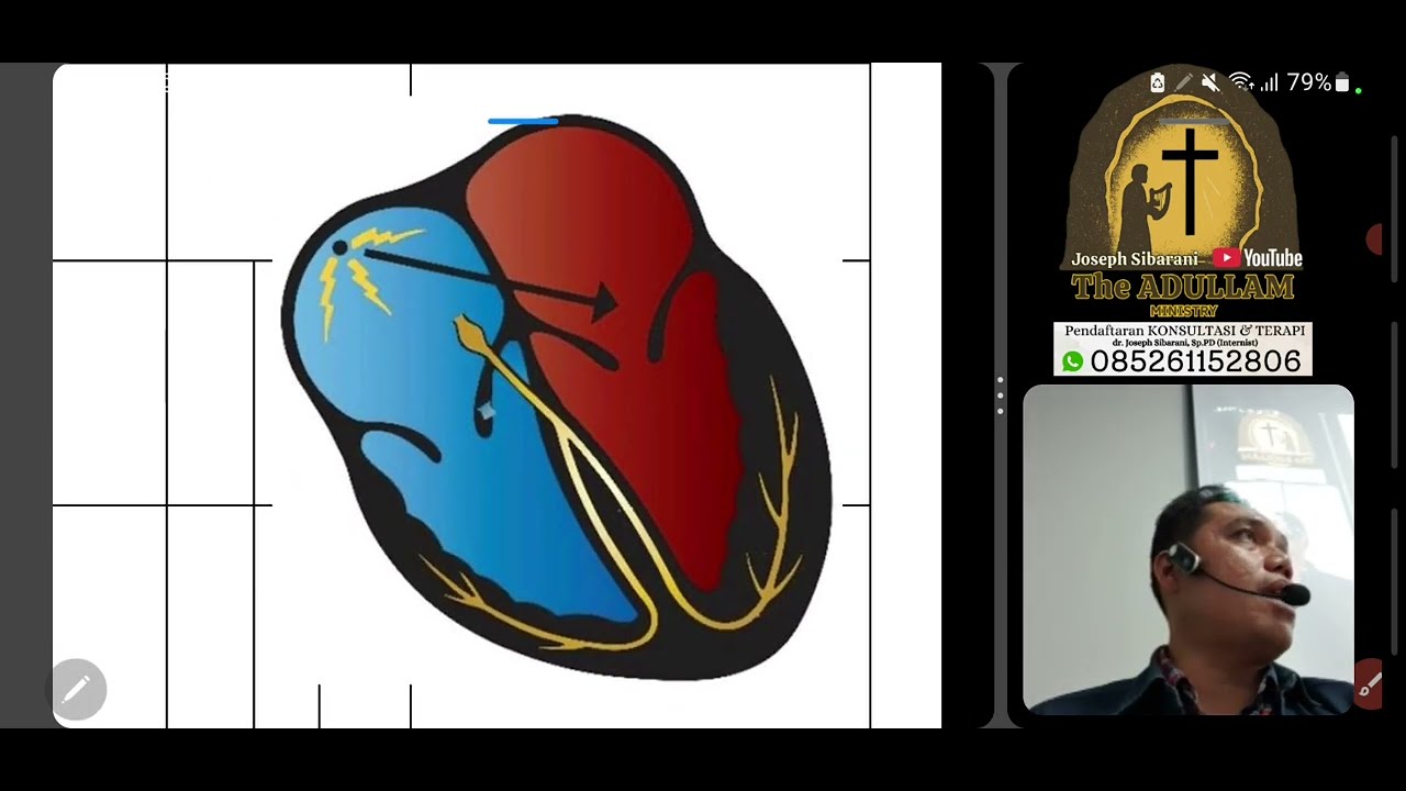
The heart's electrical conduction system includes the SA node (dominant pacemaker), AV node (gatekeeper with 0.1s delay), Bundle of His, and Purkinje fibers (fastest at 4 m/s). The ECG waves represent this system: P wave (atrial depolarization), PR interval (SA to AV conduction, 120-200ms), Q wave (septal activation), QRS complex (ventricular depolarization, 80-100ms), and T wave (ventricular repolarization). The PR interval begins at P wave start and ends at Q wave start, not R wave peak.
Click to watch →
Bagaimana Membaca EKG Patologis
460
The cardiac electrical conduction system follows a specific pathway: SA node (primary pacemaker, 60-100 bpm) → atria → AV node (creates delay) → Bundle of His → Purkinje fibers → ventricles. Each waveform represents a specific phase: P wave = atrial depolarization, QRS = ventricular depolarization, T wave = ventricular repolarization. The PR interval (3-5mm) reflects AV node conduction time. The QRS complex consists of Q (septal depolarization), R (left ventricular), and S (right ventricular) waves. Normal QRS amplitude in lead 2 is ≥5mm, in V3-V4 is ≥9mm. Duration should be 2-3mm. Prolonged QRS indicates ventricular enlargement or conduction abnormalities.
Click to watch →
Deep Dive
Pathophysiology of cardiac arrhythmias, such as heart blocks, atrial fibrillation, and ventricular tachycardia.

REPASO ENARM 2025 / CARDIOLOGÍA PT 2 - Preguntas generales
156
This comprehensive section covers cardiac arrhythmias including atrial fibrillation, preexcitation syndromes, and ventricular arrhythmias. Atrial fibrillation risk factors include aging, hypertension, diabetes, thyrotoxicosis, heart failure, coronary artery disease, valvular disease, COPD, sleep apnea, cardiac surgery, alcohol, and increased vagal tone. Pathophysiology involves premature depolarization from pulmonary veins leading to structural and electrical remodeling. Consequences include loss of atrial contribution to ventricular filling, blood stasis, and thrombus formation. Classification includes paroxysmal, persistent, and permanent types with symptom severity grading. Preexcitation syndromes involve accessory pathways bypassing normal AV conduction, with Wolff-Parkinson-White syndrome featuring delta wave on ECG. Brugada syndrome is hereditary with sodium channel mutations and characteristic ST elevation. Ventricular arrhythmias arise from automaticity abnormalities or reentry currents, ranging from premature beats to ventricular fibrillation.
Click to watch →
Physiology of cardiac arrhythmia (3-2015) by Dr Khaled A Abulfadle
3.4K
Cardiac arrhythmia refers to any abnormality in heart rate, which can be classified into sinus arrhythmias (originating from the SA node, including tachycardia, bradycardia, and sinus arrhythmia), atrial arrhythmias (including atrial extrasystoles, atrial tachycardia, and atrial fibrillation), and ventricular arrhythmias (including ventricular extrasystoles, ventricular tachycardia, and ventricular fibrillation); the underlying mechanisms involve abnormalities in the cardiac conduction system, including ectopic foci, re-entry circuits, and conduction blocks that disrupt the normal electrical impulse propagation through the heart.
Click to watch →
Fisiopatología de las Arritmias | Simposio de Taquiarritmias
98.5K
Las arritmias cardíacas se generan principalmente por alteraciones en el automatismo (trastornos del automatismo normal y anormal, y actividad desencadenada) o por alteraciones en la conducción (bloqueos y reentrada). El automatismo normal alterado ocurre cuando el nodo sinusal no funciona correctamente, causando taquicardia sinusal o bradicardia. El automatismo anormal ocurre cuando células de respuesta rápida adquieren propiedades de automatismo, generando focos ectópicos que producen extrasístoles o taquicardias. La actividad desencadenada se divide en potenciales tardíos (debidos a aumento de calcio intracelular, causados por frecuencia cardíaca elevada, isquemia, digital y catecolaminas, asociados a taquicardia ventricular polimórfica y taquicardia bidireccional) y potenciales precoces (asociados a prolongación del QT y taquicardia ventricular polimórfica tipo torsades de pointes). Los bloqueos son alteraciones en la conducción del impulso, mientras que la reentrada requiere un bloqueo unidireccional, un trayecto de conducción lento, una masa crítica y un desencadenante, y se clasifica en anatómica (calcificaciones, isquemias, vías accesorias) o funcional (alteraciones en la conducción del tejido).
Click to watch →

Chp#11 Robbins Patho | Heart Pathology | Arrythmias & Sudden Cardiac Death | Dr Asif Lectures
1.9K
When atrial myocytes become irritable and depolarize prematurely, they can serve as abnormal depolarization hubs, leading to atrial fibrillation characterized by random irregular heart rates. Heart block occurs when signal transmission between atria and ventricles is impaired. First-degree block shows prolonged PR interval on ECG. Second-degree block involves intermittent signal transmission. Third-degree (complete) block represents complete failure of signal transmission from atria to ventricles.
Click to watch →

Physiology Guyton Ch#13 | ECG Interpretation of Cardiac Arrythmias | Cardiac Physiology
25.8K
Cardiac arrhythmias arise from four primary mechanisms: abnormal pacemaker function, pacemaker shift, conduction system block, or ectopic impulse generation. The SA node normally generates 60-100 bpm impulses, but physiological variations like fever (10 bpm/°F increase), dehydration, anemia, and athletic training alter heart rate. Pathological causes include ischemia, inflammation, infections, and medications. Heart block occurs because the AV bundle is the sole pathway connecting atria and ventricles; conditions causing block include ischemia, compression, inflammation, vagal stimulation, degeneration, or drugs. First-degree block shows prolonged PR interval (>0.2 sec) with preserved conduction. Second-degree block involves intermittent non-conducted beats (Mobitz Type I shows progressive PR prolongation; Type II shows fixed ratios). Third-degree (complete) block creates complete dissociation with independent escape pacemakers. Stokes-Adams syndrome involves intermittent complete block causing syncope due to overdrive suppression. Premature contractions occur when abnormal foci generate impulses before the SA node's scheduled beat, showing characteristic ECG features depending on origin. Long QT syndrome involves prolonged QT interval from delayed repolarization, increasing susceptibility to torsades de pointes and sudden death.
Click to watch →
Deep Dive
The mechanism of action of antiarrhythmic medications (such as beta-blockers and calcium channel blockers) on the cardiac action potential.
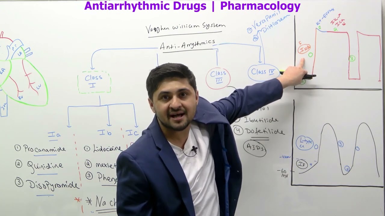
Intro | Classification | Antiarrhythmic Drugs | Part 1 | Pharmacology
141
Antiarrhythmic drugs exert their effects by targeting specific phases of the cardiac action potential. Class 1 drugs (1a, 1b, 1c) block fast sodium channels during Phase 0 in ventricular myocytes, slowing depolarization. Class 3 drugs block potassium channels during Phase 3, prolonging action potential duration by preventing potassium efflux. Class 2 drugs (beta blockers) affect funny current and calcium channels in SA/AV nodes, reducing automaticity. Class 4 drugs (calcium channel blockers) block L-type calcium channels during the plateau phase (Phase 2), reducing calcium influx. The effective refractory period—the time when cells cannot regenerate action potentials—is critical for drug efficacy and safety, as drugs can alter this period to prevent reentry circuits that cause arrhythmias.
Click to watch →

Arrhythmia Basics ( part 1 )
455
The heart contains different cardiac cells with distinct action potentials. Atrial muscle cells require stimulation to depolarize, with sodium influx followed by potassium efflux and calcium entry during the plateau phase. The SA node has automaticity due to calcium influx during phase 4. The AV node has a longer plateau phase that prolongs action potential duration. Beta-blockers reduce sodium channel activation by blocking beta receptors, prolonging phase 4 slope. Calcium channel blockers directly block calcium channels, reducing calcium influx. Adenosine increases potassium efflux, hyperpolarizing cells and delaying the next action potential, particularly effective for AV nodal reentrant tachycardia.
Click to watch →

Antiarrhythmic Drugs
207.3K
Antiarrhythmic drugs suppress abnormal cardiac rhythms by modifying action potentials through various mechanisms. Class 1 agents block sodium channels: 1A provides moderate blockade increasing action potential duration (quinidine, procainamide, disopyramide); 1B provides mild blockade decreasing duration (lidocaine IV, mexiletine oral); 1C provides marked blockade without duration change (flecainide, propafenone). These agents decrease phase 4 depolarization and increase threshold potential. Class 2 agents are beta blockers that counteract catecholamines, reducing heart rate, contractility, and automaticity while prolonging AV node repolarization. Class 3 agents block potassium channels (specifically DRK channels), prolonging phase 3 repolarization. Class 4 agents block L-type calcium channels, primarily affecting SA and AV nodes to decrease heart rate and terminate re-entrant rhythms.
Click to watch →
Antiarrhythmics (Part 1) - ICU Drips
117.5K
Antiarrhythmic medications treat abnormal heart rhythms by targeting the electrical conduction system of the heart, which consists of the SA node (primary pacemaker), Bachmann's bundle, AV node (which delays signals), bundle of His, bundle branches, and Purkinje fibers that coordinate synchronized contraction of cardiac myocytes. The heart relies on two distinct action potentials: the pacemaker action potential (found in SA/AV/Purkinje cells) features a continuous depolarization (phase 4) due to funny currents that slowly allow sodium to leak in, reaching threshold at -40mV and causing calcium influx; while the myocyte action potential (in contractile cells) has a resting phase at -90mV, followed by rapid depolarization (phase 0), early repolarization (phase 1), plateau phase (phase 2) where calcium influx triggers contraction via sarcoplasmic reticulum release, and final repolarization (phase 3). These medications are classified into five classes (Class I further divided into Ia, Ib, and Ic) based on their mechanisms of action, with each class affecting different phases of the action potential to restore normal cardiac rhythm.
Click to watch →
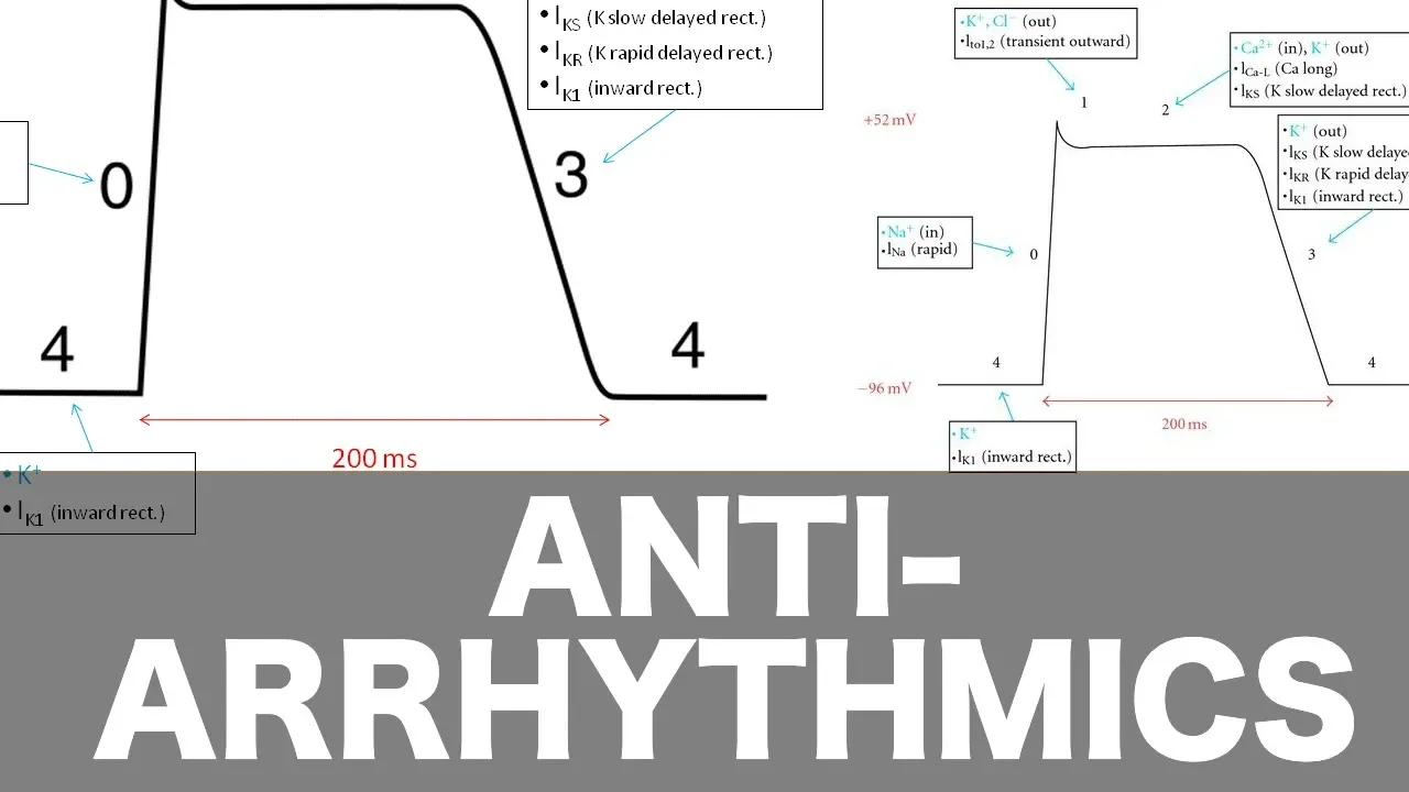
Antiarrhythmic Pharmacology
400K
Class 4 antiarrhythmics (calcium channel blockers) decrease the slope of phases 0, 3, and 4 of the cardiac action potential. They also prolong repolarization via the AV node. This is remembered using the fact that calcium has seven letters, and 0+3+4 equals seven.
Click to watch →
Deep Dive
Clinical applications and technology behind artificial pacemakers and implantable cardioverter-defibrillators (ICDs) to manage conduction defects.
CHAPTER 243 - Clinical Cardiac Electrophysiology: Rhythm Disorders and Treatments
145
Implantable devices are cornerstone therapies for bradyarrhythmias and life-threatening tachyarrhythmias. Pacemakers (since 1958) sense intrinsic cardiac activity and deliver pacing stimuli when rates fall below programmed thresholds. They're implanted percutaneously with leads in the right atrium and/or ventricle, or as leadless devices (capsule implanted directly in the right ventricle). Indications include symptomatic sinus node dysfunction and AV conduction disease. Implantable cardioverter defibrillators (ICDs) detect and treat life-threatening ventricular arrhythmias (VT/VF) through anti-tachycardia pacing (ATP) or high-energy shocks. Indications include primary prevention (high-risk patients without prior events) and secondary prevention (survivors of cardiac arrest). Subcutaneous ICDs avoid intravenous leads, reducing infection risk while providing defibrillation capability.
Click to watch →
![7. Arrhythmia Management: Device Therapy [Hong Kong University]](https://i.ytimg.com/vi/LtazIYNzBnY/maxresdefault.jpg)
7. Arrhythmia Management: Device Therapy [Hong Kong University]
154
Implantable devices include pacemakers (stimulating heart rhythm) and ICDs (delivering shocks or ATP to terminate fast rhythms). ICD technology evolved from abdominal epicardial patches (1985) to pectoral devices (1995) to modern transvenous systems. Modern ICDs offer programmable therapy, triple-chamber capability, and extended longevity. Clinical trials established ICD importance in secondary prevention for syncope, cardiac arrests, and VT. The SCD-HeFT trial demonstrated primary prevention effectiveness. ICD leads placed via subclavian vein or right atrium provide sensing, pacing, and defibrillation capabilities. This evolution reflects increasing sophistication in life-saving rhythm management.
Click to watch →

When Your Heart Needs Help: Interventions & Repair. Part 1
134
Pacemakers are electronic devices with pulse generators and leads placed in heart chambers. They remain inactive when the heart beats adequately but deliver electrical stimuli when heart rate drops below programmed thresholds. Implantable cardioverter defibrillators (ICDs) evolved from large abdominal devices requiring sternotomy to small chest implants placed through collarbone veins. ICDs can detect and terminate dangerous rhythms like ventricular fibrillation through electrical shocks. Survival from cardiac arrest depends critically on time to defibrillation—survival drops dramatically after 10-15 minutes without intervention. With rapid response systems, survival rates can reach 40%, compared to only 3-16% overall.
Click to watch →

Ask the Mito Doc - January 2026: Genetics and Clinical Insights to Jumpstart the Year
190
Pacemakers are implanted computers with batteries that monitor heart rate and deliver regular beats when electrical conduction fails. Modern leadless pacemakers eliminate wires, reducing infection risk. Ventricular tachycardia appears as broad, fast, disorganized rhythm where the heart pumps too fast to fill, potentially causing death. Defibrillation interrupts this rhythm. Implantable cardioverter-defibrillators (ICDs) are larger devices that automatically detect and defibrillate dangerous rhythms. ICD implantation criteria include prior cardiac arrest, family history of sudden death, syncope episodes, heart muscle thickening exceeding 3 cm, apical aneurysm, or ejection fraction below 50%. ICDs carry risks including surgical complications, lead failure, infection, and inappropriate shocks causing anxiety.
Click to watch →

어지럼증 해결하는 심장기계. Pacemaker 와 ICD
4.5K
Pacemakers and ICDs are implantable cardiac devices that help maintain heart rhythm; pacemakers regulate slow heart rates (bradycardia, typically below 50 bpm) by delivering gentle electrical impulses to ensure regular beating, while ICDs monitor heart rhythm and deliver stronger electrical shocks to restore normal rhythm when life-threatening arrhythmias occur, preventing sudden cardiac death.
Click to watch →
系统定位
核心组件
信号启动
心室传导
心电监测
临床意义
系统定位
0:09- 1
心肌特化细胞网络位于心脏壁内。
- 2
该网络负责发送信号引发心脏收缩。
- 3
此结构被命名为心脏传导系统。
The Syncytial Model and Mechano-Electrical Feedback
While traditional animations depict the cardiac conduction system as a series of discrete, insulated electrical 'wires' (the cable theory), modern cardiac physiology presents a more integrated view. Critics of the simple 'wiring' analogy point out that the heart is a functional syncytium, where electrical current propagates continuously through gap junctions across all cardiac muscle cells, not just specialized pathways. Furthermore, the 'Mechano-Electrical Feedback' (MEF) theory argues that heart rhythm is not purely driven by a top-down electrical hierarchy. Instead, mechanical forces, such as the stretching of chambers during filling, directly alter electrical properties via stretch-activated ion channels. Additionally, research shows the SA node is not a single, isolated 'spark plug' but a distributed, highly integrated pacemaker network. Understanding these complex, bi-directional, and diffuse interactions is crucial, as the simplistic 'wiring' model fails to fully explain complex arrhythmias and the heart's self-regulating adaptive mechanisms.
Basic gross anatomy of the heart, specifically the four chambers (atria and ventricles) and the major valves.
The concept of membrane potentials and action potentials, including the movement of sodium, potassium, and calcium ions across cell membranes.
The difference between skeletal and cardiac muscle tissue, particularly the presence of intercalated discs and gap junctions that allow rapid signal transmission.
The basic mechanical cardiac cycle, understanding the phases of systole (contraction) and diastole (relaxation).
Interpretation of Electrocardiograms (ECGs/EKGs) and how specific electrical waves (P, QRS, T) correlate with the conduction system's pathway.
Pathophysiology of cardiac arrhythmias, such as heart blocks, atrial fibrillation, and ventricular tachycardia.
The mechanism of action of antiarrhythmic medications (such as beta-blockers and calcium channel blockers) on the cardiac action potential.
Clinical applications and technology behind artificial pacemakers and implantable cardioverter-defibrillators (ICDs) to manage conduction defects.
112.7K views446likes1:24@AniMedical-AnimationsOriginal Release: 2016-02-14
[Music] a network of specialized muscle cells is found in the heart's walls these muscle cells send signals to the rest of the heart muscle causing a contraction this group of muscle cells is called the cardiac conduction system the main parts of the system are the SA node AV node node bundle of His bundle branches and pingi fibers let's follow a signal through the contraction process the SA node starts the sequence by causing the atrial muscles to contract that's why doctors sometimes call it the anatomical pacemaker next the signal travels to the AV node through the bundle of hisis down the bundle branches and through the penji fibers causing the ventricles to contract this signal creates an electrical current that can be seen on a graph called an electrocardiogram or EKG doctors use an EKG to see how well the cardiac conduction system works any changes on the EKG can mean serious problems
Up Next
14:15
How to Determine Heart Rate on ECG/EKG | Rate Calculation Basics
@ECGDoc
79.6K views•2011-11-13

48:47
Graphic Medicine: Comics for Collaborative Healthcare Communication
@nationalpatientadvocate
189 views•2023-12-04

10:28
Neuroanatomy: Central and Peripheral Nervous System Divisions Explained
@AKLECTURES
136.2K views•2014-09-20

3:05
Stages of Labor and Vaginal Birth | Childbirth Animation
@nucleusmedicalmedia
52.1M views•2017-08-18
Related Study Plans & Knowledge Roadmaps
Structured learning paths in Medicine
medicine
Abnormal LFTs: Diagnostic Workup & Pathology
01Liver Anatomy and Basic Physiology
02Introduction to Liver Function Tests (LFTs)
03Hepatocellular vs. Cholestatic Patterns
+ 3 more learning modules
12h6 modules
Explore Roadmap
medicine
Analyzing Blood Smears: RBC, WBC & Platelets
01Introduction to Blood Components & Smear Preparation
02Normal Morphology of Blood Cells
03Erythrocyte Pathologies & Morphological Variants
+ 2 more learning modules
12h5 modules
Explore Roadmap
medicine
Mechanical Ventilation: Waveforms & Alarms
01Anatomy and Physiology of Breathing
02Introduction to Mechanical Ventilation & Core Modes
03Ventilator Settings and Waveform Interpretation
+ 2 more learning modules
12h5 modules
Explore Roadmap
medicine
Neuro Exam: Localization & Stroke Syndromes
01Foundations of Neuroanatomy and Vascular Supply
02The Neurological Examination: Step-by-Step
03Principles of Neurological Localization
+ 3 more learning modules
18h6 modules
Explore Roadmap