The primary somatosensory cortex, located in the postcentral gyrus behind the central sulcus, processes somatic sensations including touch, proprioception, pain, and temperature; it consists of four interconnected areas (3a, 3b, 1, and 2) where area 3 receives direct thalamic input and areas 1 and 2 perform higher-level processing, with all areas exhibiting somatotopic organization where more sensitive body regions occupy larger cortical space.
Neuroscience Basics: Primary Somatosensory Cortex Areas 3a, 3b, 1, 2
Added:Deep Dive
Basic neuroanatomy of the cerebral cortex, specifically the parietal lobe and the postcentral gyrus.
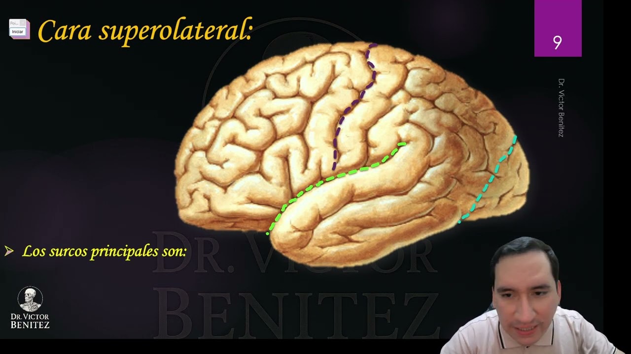
Neuroanatomía - Cerebro
954
The frontal lobe contains four gyri: precentral gyrus (primary motor area), superior frontal gyrus, middle frontal gyrus, and inferior frontal gyrus (containing Broca's area). The parietal lobe contains three gyri: postcentral gyrus (primary sensory area), superior parietal gyrus, and inferior parietal gyrus. The intraparietal sulcus divides the inferior parietal gyrus into supramarginal and angular gyri. The precentral and postcentral gyri ascend to the medial face, forming the paracentral lobule.
Click to watch →
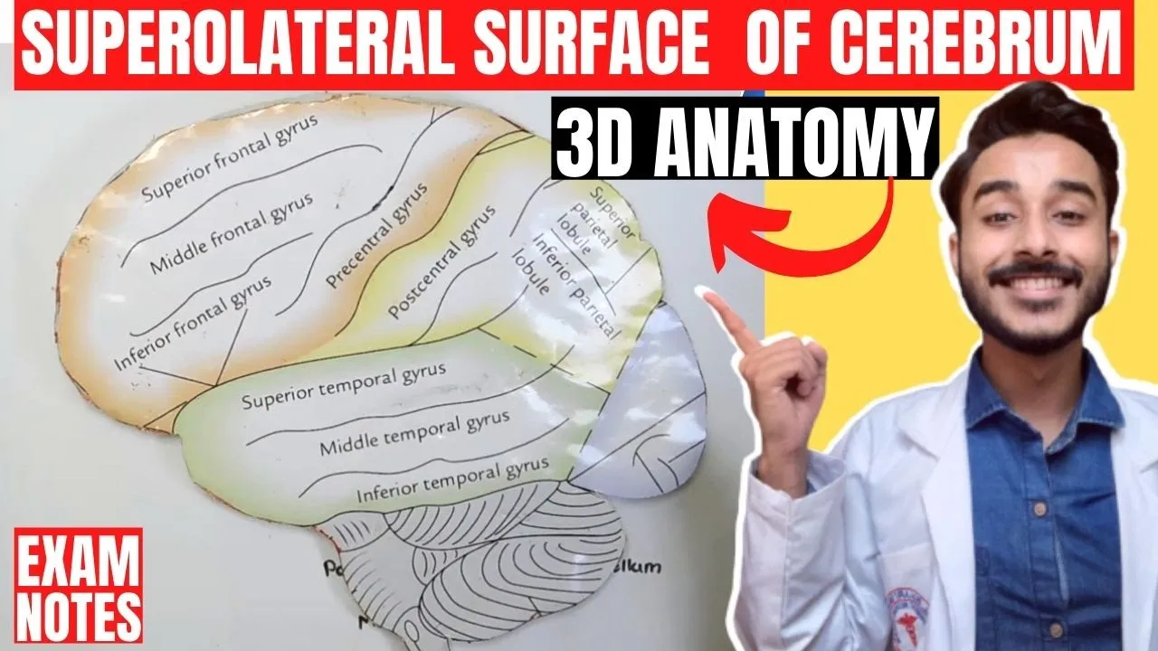
superolateral surface of cerebrum anatomy 3d | gyri and sulcus of cerebrum hemispheres anatomy
402.2K
This section covers the gyral organization of the temporal and parietal lobes. The temporal lobe contains two main gyri: superior temporal gyrus (above the lateral sulcus) and inferior temporal gyrus (below the lateral sulcus). The parietal lobe contains the postcentral gyrus (behind the central sulcus, named 'post-' meaning after/beyond the central sulcus) and the intraparietal sulcus (a horizontal sulcus within the parietal lobe). These structures represent distinct functional areas within each lobe.
Click to watch →
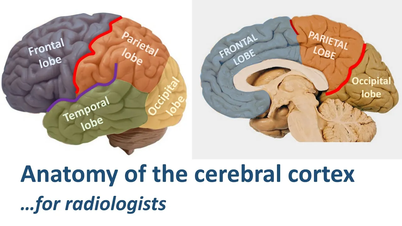
Radiological anatomy of the cerebral cortex... made easy.
71.3K
The parietal lobe contains the post-central sulcus posterior to the post-central gyrus. Posterior to this sulcus lies the superior parietal lobule, which is divided from the inferior parietal lobule by the intraparietal sulcus. The inferior parietal lobule consists of two gyri: the supramarginal gyrus (horseshoe-shaped, sitting on top of the posterior Sylvian fissure extension) and the angular gyrus (sitting on top of the superior temporal sulcus extension).
Click to watch →
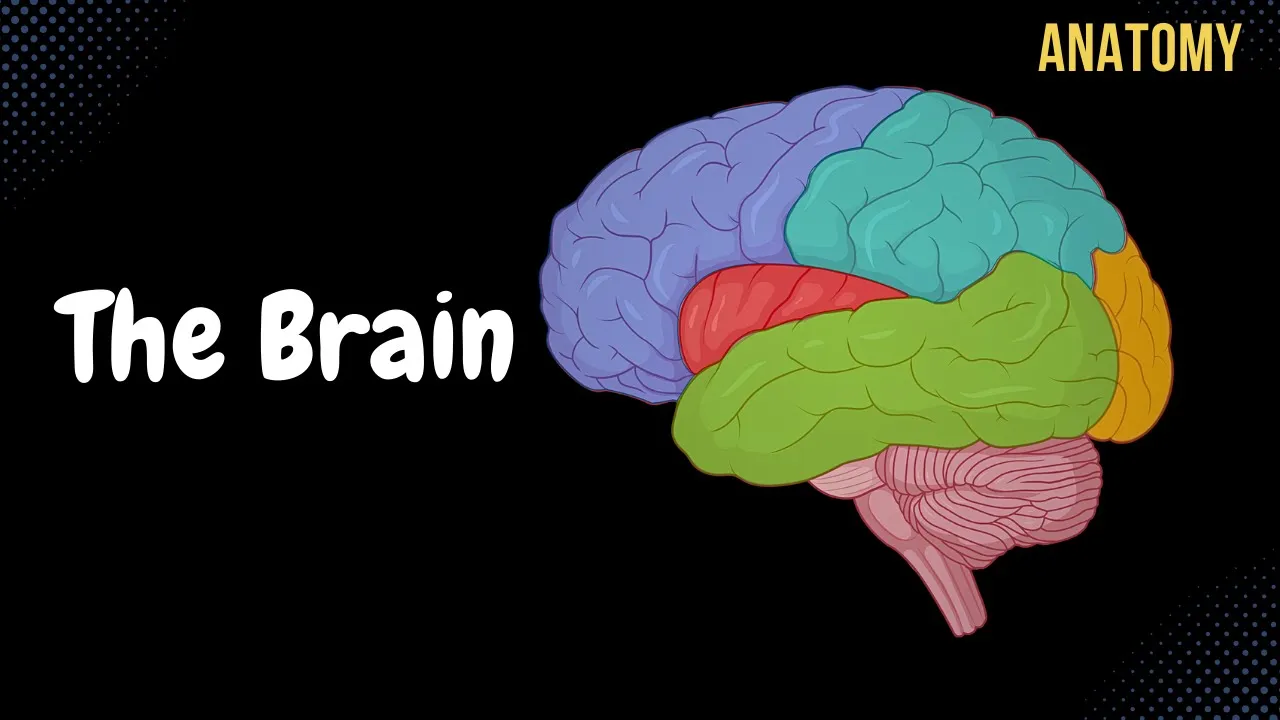
Cerebral Cortex (Function, Covering, Lobes, Sulcus, Gyrus, Fissures) | Anatomy
318.4K
The cerebral cortex is the outer layer of the telencephalon (cerebrum), consisting of six distinct layers (molecular, external granular, external pyramidal, internal granular, internal pyramidal, and multiform) that vary in thickness depending on function, with sensory areas being thinner and motor areas thicker due to the density of pyramidal cells; it is divided into five major lobes (frontal, parietal, temporal, occipital, and insular) separated by key sulci including the lateral sulcus, central sulcus, and parietooccipital sulcus, with each lobe containing specific functional regions such as the precentral gyrus (primary motor cortex) and postcentral gyrus (primary somatosensory cortex), and is protected by dural septae including the falx cerebri in the longitudinal fissure and tentorium cerebelli in the transverse fissure.
Click to watch →

Neurology | Cerebrum: Parietal Lobe Anatomy & Function
256.4K
The parietal lobe of the cerebrum contains three key functional areas: the primary somatosensory cortex (postcentral gyrus, Brodmann area 312) responsible for conscious awareness of somatic sensations like touch, pain, temperature, and proprioception; the somatosensory association cortex that analyzes and provides meaning to sensations, enabling object recognition and spatial awareness; and the posterior association area (multi-modal) that integrates visual, auditory, and somatic sensations for spatial coordination. Sensory information travels through the dorsal column medial lemniscus pathway (fine touch, vibration, proprioception) and spinothalamic tract (pain, temperature) to reach the contralateral primary somatosensory cortex. The somatotopic arrangement follows a sensory homunculus pattern, with lower limb representations in the medial cortex supplied by the anterior cerebral artery and upper limb/head/neck representations in the lateral cortex supplied by the middle cerebral artery. Clinical testing includes stereognosis (object identification by touch), graphagnosia (symbol recognition), and statognosis (body position awareness).
Click to watch →
Deep Dive
The concept of Brodmann areas as a system for dividing the cerebral cortex based on cytoarchitecture.
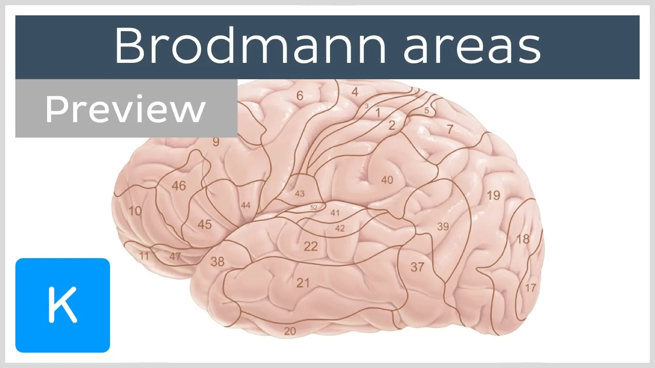
Brodmann areas of the cerebral cortex (preview) - Human neuroanatomy | Kenhub
61.8K
The Brodmann areas are 52 regions of the cerebral cortex classified by German anatomist Corbinian Brodmann based on cytoarchitecture (cellular layer organization), with each area having distinct histological structures and corresponding to specific cortical functions such as motor control, sensory processing, and cognitive tasks.
Click to watch →
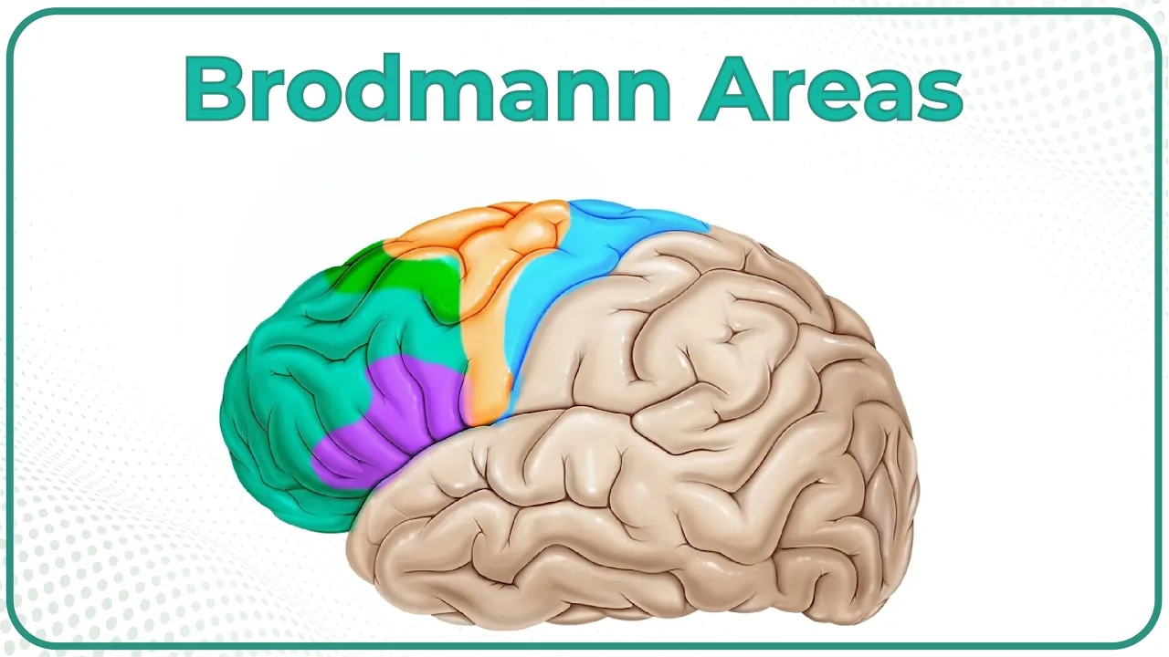
Brodmann areas of the cerebral cortex / Lobes of the brain: Structure and function / Neuroanatomy
64.6K
The cerebral cortex is divided into 52 cytoarchitectural Brodmann areas, with each lobe containing specific functional regions: the frontal lobe includes the primary motor area (area 4, controlling voluntary movement via the motor homunculus), premotor area (area 6), frontal eye field (areas 6 and 8), Broca's area (areas 44-45 for speech production), and prefrontal cortex; the parietal lobe contains the primary sensory area (areas 3-2, receiving somatosensory input via the sensory homunculus) and Wernicke's area (areas 22, 39-40 for speech comprehension); the occipital lobe houses the primary visual area (area 17, receiving input from the lateral geniculate body); and the temporal lobe contains the primary auditory area (areas 41-42, receiving input from the medial geniculate body). Lesions in these areas produce characteristic deficits: motor area lesions cause contralateral flaccid paralysis, Broca's area lesions cause expressive aphasia, Wernicke's area lesions cause receptive aphasia, and prefrontal lesions disrupt social behavior.
Click to watch →
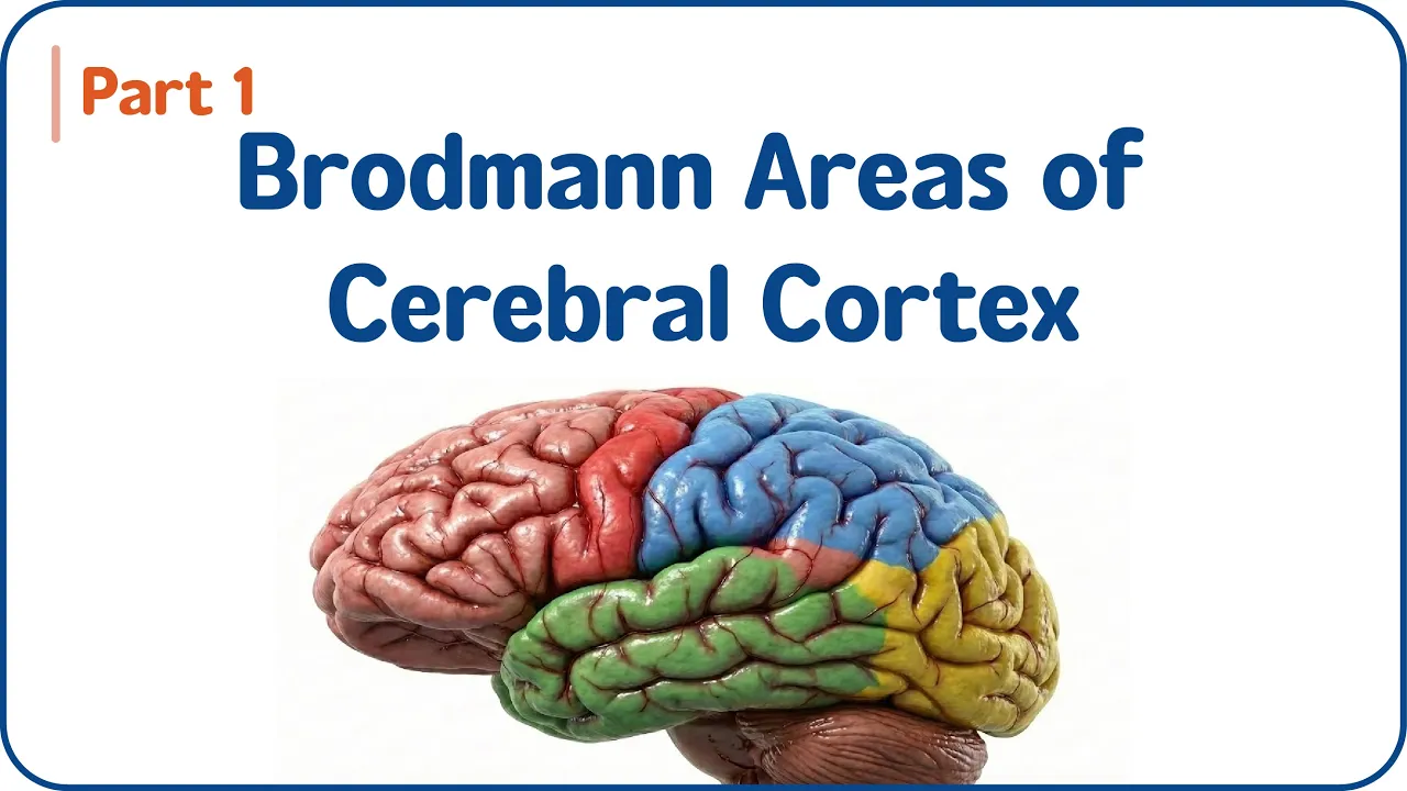
Brodmann Areas Of Cerebral Cortex (Part - 1) 🧠Motor, Sensory & Association Areas | USMLE Step 1
361
The cerebral cortex is divided into 52 Brodmann areas based on cytoarchitectonic features, with the neocortex (90%) having 9 layers and the allocortex (10%, including olfactory cortex) having 3-5 layers; the frontal lobe contains key motor areas including Brodmann Area 4 (Primary Motor Cortex in precentral gyrus controlling contralateral body movements somatotopically), Area 6 (Premotor Cortex for movement planning), and Area 8 (Eye Movement Control), while the parietal lobe contains sensory areas (Areas 3, 1, 2 for somatosensory input), the occipital lobe contains visual processing areas (Area 17 for primary vision, Areas 18, 19 for visual association), and the temporal lobe contains auditory areas (Areas 41, 42 for primary hearing, Area 22 for speech comprehension).
Click to watch →

Web 1 Curso de Neuropsicología- campos - Neuroanatomía- Áreas de Brodmann
1.4K
Brodmann areas are 47 numbered regions of the cerebral cortex classified by cytoarchitecture. They are distributed across primary, secondary, and tertiary areas based on function. Primary areas (red) receive sensory information, secondary areas (green) process and distribute information, and tertiary areas (orange) integrate information and generate responses. Functional classification includes: motor areas (pink) for voluntary movement control, emotional areas (orange) for emotion regulation, olfactory areas (dark orange) for smell processing, somatosensory areas (purple) for body sensation, attention areas (dark green) for attention regulation, visual areas (light green) for visual processing, and auditory areas (blue) for sound processing. Of the 47 areas, only 47 have been fully researched, with areas 48-52 still under investigation.
Click to watch →
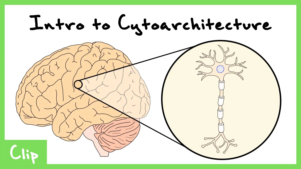
Introduction To Cytoarchitecture (Brodmann Areas/Map) | Clip
2.1K
Cytoarchitecture refers to the study of cellular properties in the brain's gray matter, where neurons are organized in layered structures; Brodmann mapped the cortex into 52 regions based on these cytoarchitectural differences, revealing that the neocortex has six layers (with layer 4 receiving sensory input and layer 5 mediating motor output) while allocortex has fewer layers, and that cortical regions can be classified as granular (with prominent layer 4, like visual cortex) or agranular (with minimal layer 4, like motor cortex) based on their functional specialization.
Click to watch →
Deep Dive
Fundamental somatosensory pathways, such as the dorsal column-medial lemniscal system and the spinothalamic tract, which transmit sensory information from the body to the brain.

Three Critical Vertical Pathways: Neuroanatomy Video Lab - Brain Dissections
54.4K
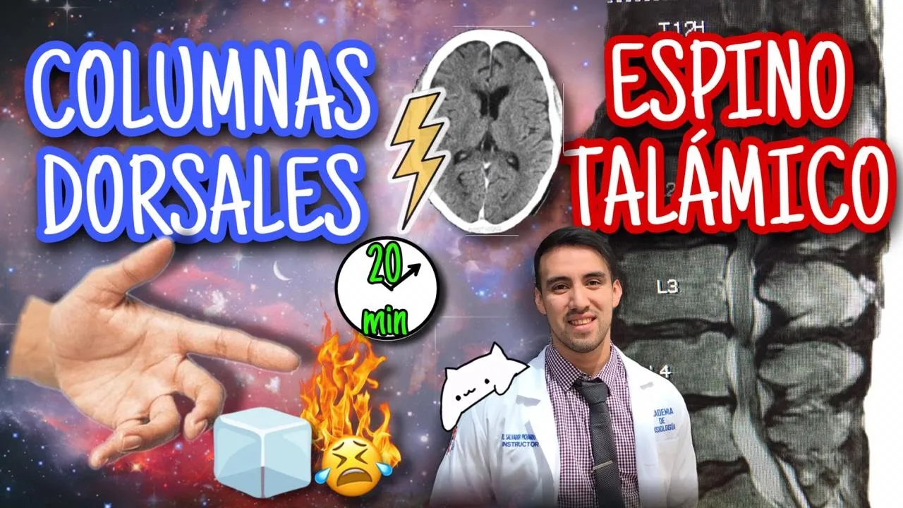
Two major sensory pathways transmit somatic information from the body to the brain. The spinothalamic tract carries pain, temperature, and crude touch, while the dorsal column-medial lemniscus pathway transmits proprioception, vibration, joint position sense, and fine touch. Both follow a three-neuron structure: first neuron in dorsal root ganglion, second neuron relaying in thalamus, third neuron projecting to postcentral gyrus. The key difference is decussation timing—the spinothalamic crosses in spinal cord, while the dorsal column crosses in caudal medulla. Both terminate in the ventral posterior lateral thalamic nucleus before reaching somatosensory cortex.
Click to watch →
¡VÍAS SENSITIVAS! RESUMEN PARA ESTUDIANTES
75.8K
The somatosensory system consists of two main ascending pathways: the dorsal column-medial lemniscus pathway (lemniscus medial) responsible for fine touch, vibration, pressure, and proprioception, and the spinothalamic pathway (anterolateral) responsible for pain and temperature sensation. Both pathways use a three-neuron relay system where the first neuron's cell body is in the dorsal root ganglion, the second neuron's cell body is in the spinal cord or brainstem, and the third neuron's cell body is in the thalamus (ventral posterolateral nucleus). A key rule is that the second neuron always decussates (crosses to the opposite side) after synapsing. The dorsal column pathway carries information from the lower body through the fasciculus gracilis and from the upper body through the fasciculus cuneatus, while the spinothalamic pathway carries pain and temperature information through the anterior and lateral funiculi of the spinal cord.
Click to watch →

Mesencephalon Yüksel AYDAR
32K
The lemniscal pathways include the dorsal column-medial lemniscus system and the spinothalamic tract. The dorsal column-medial lemniscus pathway carries information about proprioception, vibration, and fine touch from the body to the brain. The pathway begins in the dorsal columns of the spinal cord and ascends to the medulla, where it synapses in the nucleus gracilis and nucleus cuneatus. The second-order neurons then project via the medial lemniscus to the thalamus. The medial lemniscus is located in the mesencephalon and carries sensory information from the body to the thalamus. The spinothalamic tract carries pain and temperature information from the body to the brain. The pathway begins in the dorsal horn of the spinal cord and ascends to the thalamus. The pathway synapses in the substantia gelatinosa of the spinal cord, and the second-order neurons project via the spinothalamic tract to the thalamus. The spinothalamic tract is located in the mesencephalon and carries sensory information from the body to the thalamus.
Click to watch →

4/ Physiologie de la Somesthésie (Bonne Qualité)| Physiologie 2éme MED
8.1K
The dorsal column-medial lemniscus pathway transmits fine touch, vibration, and proprioceptive information from the body to the brain. First-order neurons enter the spinal cord and ascend in the dorsal columns without synapsing. Second-order neurons synapse in the dorsal column nuclei of the medulla and cross to the opposite side. Third-order neurons project from the thalamus to the somatosensory cortex. This pathway provides precise spatial localization of tactile stimuli. The spinothalamic tract transmits pain and temperature information from the body to the brain. First-order neurons enter the spinal cord and synapse in the dorsal horn. Second-order neurons cross to the opposite side and ascend in the spinothalamic tract. Third-order neurons project from the thalamus to the somatosensory cortex. This pathway allows the brain to perceive pain and temperature sensations from different body regions, enabling protective responses to harmful stimuli. The decussation (crossing to the opposite side) means that sensory information from one side of the body is processed in the opposite cerebral hemisphere.
Click to watch →
Neuroanatomie 11 : Principes d'organisation des voies sensitives
52.2K
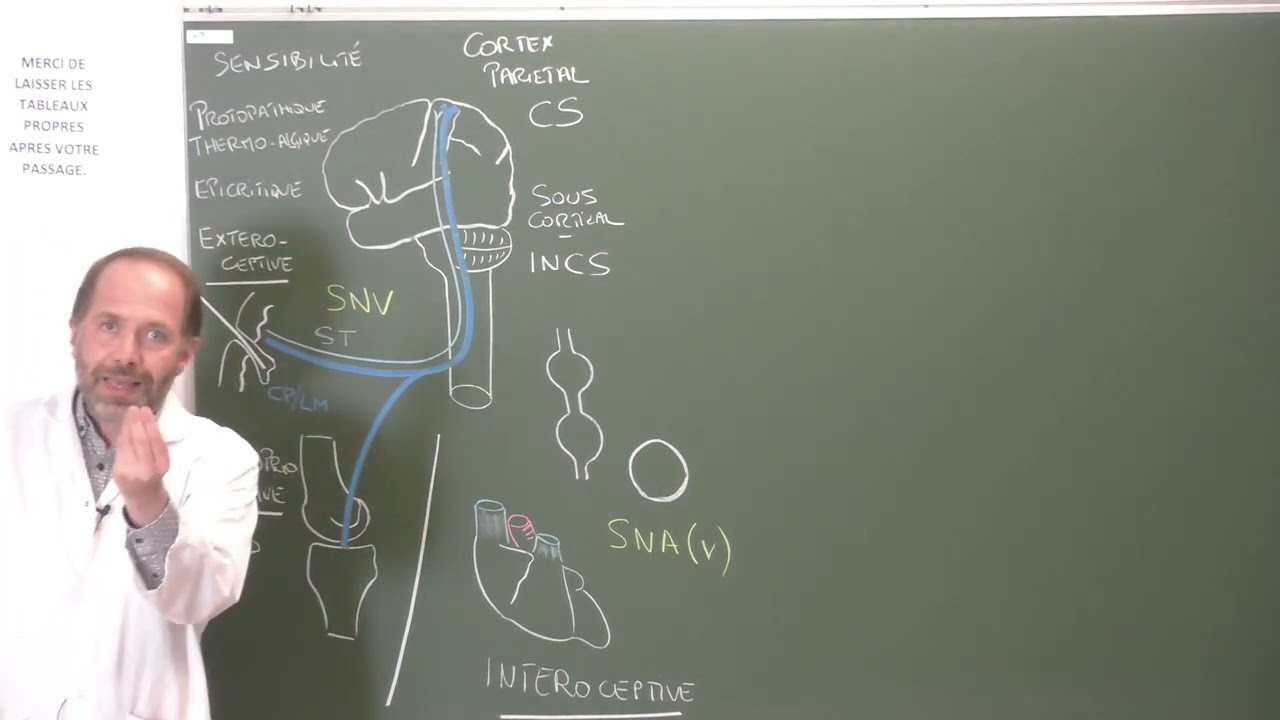
Two major conscious sensory pathways transmit information to the cerebral cortex. The spinothalamic tract carries crude touch, pressure, and pain/temperature sensations using a three-neuron chain with midline crossing at each spinal segment. The dorsal column-medial lemniscus pathway transmits fine discriminative touch and conscious proprioception without crossing until the medulla. Both pathways synapse in the ventral posterior lateral thalamic nucleus before projecting to the primary somatosensory cortex. These systems enable conscious perception of body position and environmental contact.
Click to watch →
Deep Dive
Types of peripheral sensory receptors, including mechanoreceptors (touch) and proprioceptors (body position).

Tp34
119
Sensory receptors are classified into four main types: mechanoreceptors (detect pressure, vibration, and texture), thermoreceptors (detect temperature changes), nociceptors (detect pain), and proprioceptors (detect body position and movement). Mechanoreceptors include Merkel discs (slowly adapting, detect pressure and texture), Meissner's corpuscles (rapidly adapting, detect light touch and texture), Ruffini endings (detect skin stretch), and Pacinian corpuscles (detect deep pressure and vibration). Meissner's corpuscles are most sensitive and are located in the dermal-epidermal junction, particularly in hairless skin, lips, tongue, and fingertips. Merkel discs are also slowly adapting and are important for detecting pressure and duration of stimulation.
Click to watch →

Sensory receptors and proprioception
15.8K
Proprioception is the sense of position, posture, and motion, transmitted from peripheral mechanoreceptors via afferent nerves to the CNS for processing and motor output. Four types of articular mechanoreceptors exist: Type 1 (Ruffini corpuscles) in superficial capsule layers respond to stretch at rotation limits with low threshold and slow adaptation; Type 2 (Pacinian corpuscles) in deeper capsule layers and fat pads respond to compression with dynamic, low-threshold responses; Type 3 (Golgi tendon organs) in tendons and ligaments respond only to active tension with high threshold and slow adaptation; Type 4 (free nerve endings) respond to pain and inflammation as nociceptors. Skin contains Meissner's corpuscles (touch/pressure, rapid adaptation), Pacinian corpuscles (deep pressure/vibration, slow adaptation), Merkel's disks (touch/pressure, slow adaptation), and free nerve endings (pain/temperature/pressure, high threshold). Muscle spindles contain intrafusal and extrafusal fibers responding to stretch amount and rate. The stretch reflex involves muscle spindle activation causing contraction, while Golgi tendon organs provide inhibitory responses causing relaxation.
Click to watch →
LECTURE: The Peripheral Nervous System
40.1K
Sensory receptors are classified by the type of stimulus they detect: (1) Thermal receptors detect hot and cold; (2) Mechanoreceptors detect touch and physical stimuli (including Merkel cells, Pacinian corpuscles, and Meissner's corpuscles); (3) Proprioceptors detect body position and movement from muscles and joints; (4) Photoreceptors detect light for vision; (5) Chemoreceptors detect blood chemistry including oxygen, CO2, calcium, magnesium, phosphorus, sodium, and hormone levels; (6) Baroreceptors detect blood pressure, particularly in the kidneys.
Click to watch →

Nervous Coordination | Receptors in skin | Receptors and it's types |Hindi Urdu|Pro.Suresh Kumar
1.3K
Mechanoreceptors detect mechanical energy including pressure, touch, and movement. Types include: baroreceptors (blood pressure), tactile receptors (Merkel discs, Meissner's corpuscles, Pacinian corpuscles), stretch receptors (proprioceptors for body position and movement), phonoreceptors (sound detection in ears), and lateral line receptors (water currents in aquatic animals). Proprioceptors allow us to know body position without looking and detect movement direction and speed.
Click to watch →

Somatosensory Receptors by Dr. Soumen Manna | Physiology Mind | StupireMed
7.4K
Mechanoreceptors can be classified into four main types: tactile receptors (for touch and pressure), nociceptors (for pain), proprioceptors (for position sense), and hair cells of the organ of Corti (special sense for hearing).
Click to watch →
Prerequisite Knowledge
- Concept 01Basic neuroanatomy of the cerebral cortex, specifically the parietal lobe and the postcentral gyrus.
- Concept 02The concept of Brodmann areas as a system for dividing the cerebral cortex based on cytoarchitecture.
- Concept 03Fundamental somatosensory pathways, such as the dorsal column-medial lemniscal system and the spinothalamic tract, which transmit sensory information from the body to the brain.
- Concept 04Types of peripheral sensory receptors, including mechanoreceptors (touch) and proprioceptors (body position).
Subsequent Learning
- Step 01The structure and function of the Secondary Somatosensory Cortex (S2) and its role in higher-order sensory integration and tactile memory.
- Step 02Cortical plasticity and reorganization of somatotopic maps, including clinical phenomena like phantom limb pain.
- Step 03Sensorimotor integration, detailing how the primary somatosensory cortex communicates with the primary motor cortex (M1) to coordinate movement.
- Step 04Clinical deficits resulting from localized lesions in S1, such as astereognosis (inability to identify objects by touch) and agraphesthesia.
Location
Functions
Subdivisions
Area Roles
Connections
Somatotopy
Sensitivity
Location
0:05Playing Section
- 1
Primary somatosensory cortex sits in postcentral gyrus.
- 2
Located just behind the central sulcus fissure.
Distributed Network and Dynamic Plasticity Models of Somatosensation
The traditional view of the primary somatosensory cortex (S1)—consisting of Brodmann areas 3a, 3b, 1, and 2—emphasizes a rigid, feedforward hierarchy where sensory information is processed serially alongside a static somatotopic map (the homunculus). However, contemporary neuroscience presents an alternative model emphasizing distributed parallel processing and dynamic plasticity. Opposing theories argue that areas 1 and 2 receive direct thalamic inputs and process features in parallel with area 3, rather than strictly downstream. Furthermore, instead of a static map, the somatosensory cortex exhibits continuous, experience-dependent plasticity, meaning receptive fields constantly reorganize based on behavior, learning, and injury. Additionally, 'active sensing' paradigms suggest S1 is functionally integrated with motor execution networks in a recurrent feedback loop, challenging the classical view of S1 as a passive, isolated sensory processor.
Deep Dive
The structure and function of the Secondary Somatosensory Cortex (S2) and its role in higher-order sensory integration and tactile memory.
SENSIBILIDAD SOMÁTICA definitivo
2.5K
The thalamus is a double encephalic structure where all sensory references (except olfactory) relay before reaching the cerebral cortex. It has a predominant role in somatosensory functions - destruction of somatosensory cortex loses tactile sensitivities but some coarse tactile sensitivity recovers, suggesting thalamic capacity to distinguish tactile sensations. The somatosensory cortex is in the postcentral gyrus behind the central sulcus, divided into Brodmann areas: 3, 1, 2 (primary somatosensory area S1) and 40 (secondary somatosensory area S2). Areas 5 and 7 are association areas. S1 has highly accurate body part localization with each sector corresponding to different body locations well discriminated. S2 is more extensive with less precise localization (face anterior, arms central, legs posterior). S1 receives mainly from dorsal column system, has developed granular cortex (layer 4), developed thalamocortical connection, large somatotopy, and ability to detect shape and size. S2 has bilateral information, lower somatotopy, higher latency, and sensory convergence with visual and auditory areas. Representation in S1 shows zones with smaller peripheral fields (greater two-point discrimination) have greater representation. Hands, mouth, lips, and tongue have greatest sensory representation. S2 may be a higher-order area related to tactile learning and memory through interhemispheric transfer and sensory-motor integration. Projections from S1 are necessary for S2 function.
Click to watch →

Neuro series: Cerebral hemisphere 3 || Somatosensory processing at cortical level
365
The secondary somatosensory cortex (S2) is located in the parietal operculum, adjacent to S1. Unlike S1, which receives primarily contralateral input, S2 receives bilateral sensory information from both hemispheres, allowing more complex object representations. S2 receives most input from S1 with some direct spinal cord input. Parietal association areas (Brodmann areas 5 and 7) receive input from S1, S2, and visual areas. Area 5 processes tactile information related to object manipulation and spatial relationships. Area 7 integrates visual and somatosensory information for spatial awareness and action planning. The somatosensory system has two major processing streams: the dorsal stream (parietal pathway) involved in spatial awareness and action guidance, and the ventral stream (temporal pathway) involved in object recognition. Associative agnosia involves inability to integrate sensory perception with stored knowledge to identify objects, despite intact basic tactile discrimination. Tactile agnosia involves inability to perceive tactile properties.
Click to watch →

SISTEMA SENSORIAL SOMÁTICO (SOMATOSSENSORIAL): TATO E TEMPERATURA | MK Fisiologia
100.6K
Information from the primary somatosensory cortex projects to secondary sensory areas (S2) in the parietal lobe. These areas integrate tactile, proprioceptive, and temperature information with visual and auditory inputs. This integration enables object identification, memory formation, and emotional responses to objects.
Click to watch →
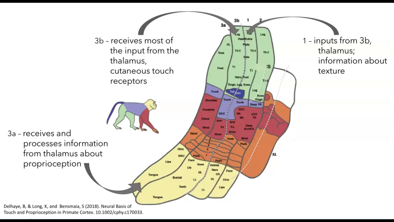
Somatosensory cortex organization and function
16.9K
The somatosensory cortex, located in the postcentral gyrus of the parietal lobe, contains four distinct homunculi (areas 3a, 3b, 1, and 2) that process somatosensory information through hierarchical processing: area 3b receives direct thalamic input for basic touch and proprioception, areas 1 and 2 extract higher-order features like texture and object shape, and information projects to secondary somatosensory cortex (S2) and posterior parietal cortex for multimodal integration and movement planning; this cortical organization demonstrates remarkable plasticity, as shown by experiments where amputated digits cause adjacent cortical regions to expand into the vacated space.
Click to watch →
LA PHYSIOLOGIE DE LA SOMESTHÉSIE
1.4K
The somatosensory cortex is organized somatotopically, with different body parts represented in specific regions. The primary somatosensory cortex (S1) receives thalamic input and is organized into a homunculus where body parts with higher sensory importance occupy larger areas. The secondary somatosensory cortex (S2) integrates sensory information with memory and spatial awareness. Lesions to these areas produce specific syndromes including loss of sensation, ataxia, and agnosia.
Click to watch →
Deep Dive
Cortical plasticity and reorganization of somatotopic maps, including clinical phenomena like phantom limb pain.
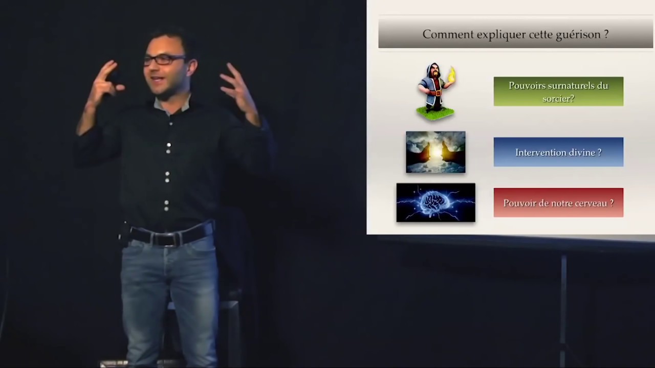
Neurosciences - Thierry Gallopin- Hypnologie 2015
8.8K
The brain's somatosensory cortex contains a map of the body where different body parts occupy different areas based on their sensitivity (hands and lips are highly represented, while the trunk is less represented). When a limb is amputated, the brain area that previously coded for that limb becomes unused and can be 'colonized' by adjacent brain areas. This causes phantom limb pain, where patients feel sensations in their missing limb at locations where other body parts are represented. This demonstrates the brain's remarkable plasticity and its ability to reorganize in response to changes in the body.
Click to watch →
Neuro series: Cerebral hemisphere 3 || Somatosensory processing at cortical level
365
The pain pathway involves the spinothalamic tract carrying pain and temperature information to the thalamus (VPL), then to S1 for localization. S1 neurons have small receptive fields allowing precise pain localization. Pain processing involves two components: sensory-discriminative (S1, insula) for localization and identification, and emotional-affective (anterior cingulate, insula) for unpleasant feelings. Phantom limb sensation occurs because the somatosensory cortex maintains representation of amputated limbs. When amputated, adjacent body parts expand into this area, causing sensations in the phantom limb. This demonstrates cortical plasticity, where the brain's body representation can reorganize based on experience and input. The somatosensory cortex exhibits plasticity, meaning it can reorganize its representation of the body in response to changes in sensory input or use. This plasticity allows for phantom sensations and explains how the brain adapts to changes in the body.
Click to watch →

Embodied Souls — Lessons from Neurology V.S. Ramachandran
61.6K
After amputation, patients often continue to feel sensations in the missing limb (phantom limb). Research shows that sensory signals from adjacent body areas (such as the face) can invade the territory vacated by the amputated limb in the somatosensory cortex. This cross-wiring occurs because the brain region deprived of input becomes 'hungry' for new sensory information. When touched, these invading signals are misinterpreted as coming from the phantom limb, demonstrating how the brain reorganizes itself following injury.
Click to watch →
Nerve Physiology : Structure and Types of Neurons, Blood brain barrier, Resting membrane potential
37.6K
Sensations are not felt at the receptor level but in the somatosensory cortex. This explains phantom limb pain after amputation—patients feel sensations in limbs that no longer exist because the second and third-order neurons remain intact and can still activate the somatosensory cortex. Direct electrical stimulation of the somatosensory cortex can produce the sensation of being touched, while stimulation of the motor cortex produces actual muscle movements.
Click to watch →
V.S. Ramachandran - What are Breakthroughs in Biology?
15.7K
The brain's plasticity allows sensory maps to reorganize after amputation, causing adjacent body areas to invade the vacated territory and produce phantom sensations; mirror neurons enable us to simulate others' sensations, which explains why phantom limb pain can be alleviated through mirror therapy and why some individuals experience congenital intersensory referral, where they feel others' sensations as their own.
Click to watch →
Deep Dive
Sensorimotor integration, detailing how the primary somatosensory cortex communicates with the primary motor cortex (M1) to coordinate movement.

8.2 Sensorimotor Cprtex and Pathways
1.7K
The sensory motor association cortex includes the posterior parietal association cortex (integrating visual, auditory, and somatosensory information about body location, external objects, and spatial relationships) and the dorsolateral prefrontal association cortex (evaluating stimuli and initiating voluntary reactions). Damage to the posterior parietal cortex produces apraxia (inability to perform specific movements despite intact motor ability) and contralateral neglect (inability to respond to stimuli on the opposite body side). The primary motor cortex, located at the precentral gyrus, serves as the major convergence point for sensory motor signals. Penfield's 1937 studies revealed a somatotopic map where specific cortical sites activate contralateral body parts, with more cortex devoted to body parts capable of complex movements (hands, mouth, face).
Click to watch →
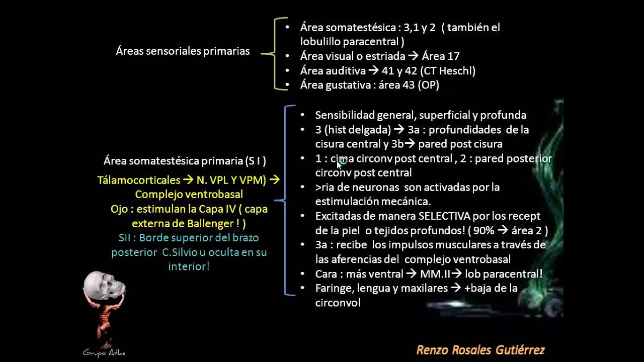
Neuroanatomia - Hemisferios cerebrales - Parte 3
228
The primary somatosensory cortex (areas 3, 1, 2 of Brodmann) processes tactile and proprioceptive information, with area 3b receiving superficial skin mechanoreceptor input and area 3a receiving deep muscle and joint receptor input, while the primary motor cortex (area 4) controls voluntary movement through corticofugal fibers including corticospinal, corticobulbar, and corticoreticular pathways; these areas are organized according to the homunculus of Penfield, with the face represented ventrally and lower limbs in the paracentral lobule, and they work in integration with sensory association areas (areas 5 and S) and limbic structures to coordinate voluntary movements.
Click to watch →

Brain 5- Primary cortexes
35.6K
The brain integrates sensory input with motor output through a coordinated pathway. Sensory information from general senses (touch, pressure, temperature) travels via afferent neurons to the spinal cord and then to the brain. If deemed worthy of consciousness, this information reaches the primary somatosensory cortex in the postcentral gyrus. After processing and interpretation, the brain generates a response plan. The primary motor cortex in the precentral gyrus then coordinates the activation of specific muscles to execute the response, demonstrating how sensory perception directly influences motor behavior.
Click to watch →
Neurology | Cerebrum: Parietal Lobe Anatomy & Function
256.4K
Two major pathways carry somatic sensations to the primary somatosensory cortex (Brodmann area 312): the dorsal column medial lemniscus pathway carries fine touch, discriminative touch, proprioception, and vibration sensations; the spinothalamic tract carries pain, temperature, crude touch, and pressure sensations. Both pathways cross in the medulla and project to the contralateral side of the cortex. Interestingly, the primary somatosensory cortex contributes approximately 40% of the corticospinal and cortical bulbar motor tracts, demonstrating bidirectional sensory-motor connectivity.
Click to watch →

Somatosensory system Part 3 slides 9 12
143
The primary somatosensory cortex (S1), located in the anterior parietal cortex and containing Brodmann areas 3a, 3b, 1, and 2, processes tactile information through somatotopic maps where specific body regions are represented regardless of their physical size; Area 2 integrates tactile input with limb position to enable object recognition, while the cortex projects to motor control areas and the cerebellum, which serves as an error-correcting mechanism for predictive motor control by adjusting outgoing motor signals based on sensory feedback and movement history.
Click to watch →
Deep Dive
Clinical deficits resulting from localized lesions in S1, such as astereognosis (inability to identify objects by touch) and agraphesthesia.
Neuro series: Cerebral hemisphere 3 || Somatosensory processing at cortical level
365
S1 is divided into three main subdivisions with distinct functions: Area 3A receives proprioceptive information from muscle spindles and joint receptors, providing awareness of body position. Area 3B receives tactile information from the skin, including texture and surface properties. Areas 1 and 2 receive input from both 3A and 3B, integrating tactile and proprioceptive information to create comprehensive body position and tactile property representations. Area 3B2 represents a higher-order subdivision involved in texture discrimination and shape perception. Stereognosis is the ability to identify objects by touch alone, using spatial and shape information. Damage to S1 can cause stereognosis deficits, where patients cannot recognize objects by touch despite intact basic tactile sensation. This occurs because integration of tactile and proprioceptive information necessary for object recognition is disrupted. Different S1 subdivisions have distinct functions, so lesions to specific areas produce different sensory deficits.
Click to watch →

Trastornos de la percepción psicopatología
9.8K
Perception defects from localized brain lesions occur after strokes, craniocerebral trauma, neurosurgery, cerebral anoxia, brain tumors, or infections. When lesions affect projection areas, specific senses are abolished (cortical blindness or deafness). When lesions affect adjacent areas, agnosias result: visual agnosia (damage to Brodmann areas 18-19) prevents recognizing objects, colors, words, or faces; auditory agnosia (areas 41-42) prevents recognizing sounds; tactile agnosia (astereognosia) prevents recognizing objects by touch. Asymbolia (area 43) creates disconnection between tactile sensation and limbic system, causing patients to feel pain without appropriate reaction. Hemineglect causes failure to recognize half the body.
Click to watch →

Les Syndromes Topographiques _ Dr Kohil ❤️
4.4K
Parietal lobe syndromes cause sensory disorders (contralateral sensory loss, astereognosis, agraphesthesia) and spatial disorders (astereognosis, constructional apraxia, neglect). Parietal lobe lesions in the non-dominant hemisphere cause body image disorders: somatoparaphrenia (denial that part of the body belongs to oneself), anosognosia (denial of illness), and left-right disorientation. Temporal lobe syndromes cause auditory disorders (cortical deafness, auditory hallucinations), memory disorders (amnesia, recognition deficits), and language disorders (Wernicke's aphasia in the dominant hemisphere). The temporal lobe is responsible for auditory processing, memory, and emotional responses.
Click to watch →

3 Physiology of Posterior column sensations-SBDeshpande
561
S2 (above lateral sulcus) requires S1 input and handles tactile discrimination, connecting to limbic area for memory. Posterior parietal area (5 and 7) analyzes object shape, size, texture, motion, and weight, communicating with visual, auditory, limbic, and motor areas. This enables stereognosis (recognizing objects by touch). Limbic association area provides emotional/memory attributes. Lesions of S1 cause loss of fine touch, vibration, discrimination, and localization on opposite side. Stereognosis (tactile agnosia) results from association area damage. Ideomotor apraxia from superior parietal lesions causes loss of purposeful skilled movements.
Click to watch →
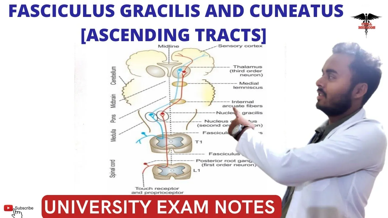
Fasciculus Gracilis and Fasciculus cuneatus Tract || Posterior ascending Tracts| CNS Physiology.
27.5K
Posterior column lesions cause ipsilateral sensory deficits because fibers do not cross in the spinal cord. Clinical features include loss of two-point discrimination, astereognosis (inability to identify objects by touch), loss of vibration sense, and impaired proprioception. These deficits result in sensory ataxia—uncoordinated, clumsy voluntary movements due to loss of position sense. Understanding these patterns is essential for neurological diagnosis.
Click to watch →
Location
Functions
Subdivisions
Area Roles
Connections
Somatotopy
Sensitivity
Location
0:05- 1
Primary somatosensory cortex sits in postcentral gyrus.
- 2
Located just behind the central sulcus fissure.
Distributed Network and Dynamic Plasticity Models of Somatosensation
The traditional view of the primary somatosensory cortex (S1)—consisting of Brodmann areas 3a, 3b, 1, and 2—emphasizes a rigid, feedforward hierarchy where sensory information is processed serially alongside a static somatotopic map (the homunculus). However, contemporary neuroscience presents an alternative model emphasizing distributed parallel processing and dynamic plasticity. Opposing theories argue that areas 1 and 2 receive direct thalamic inputs and process features in parallel with area 3, rather than strictly downstream. Furthermore, instead of a static map, the somatosensory cortex exhibits continuous, experience-dependent plasticity, meaning receptive fields constantly reorganize based on behavior, learning, and injury. Additionally, 'active sensing' paradigms suggest S1 is functionally integrated with motor execution networks in a recurrent feedback loop, challenging the classical view of S1 as a passive, isolated sensory processor.
Basic neuroanatomy of the cerebral cortex, specifically the parietal lobe and the postcentral gyrus.
The concept of Brodmann areas as a system for dividing the cerebral cortex based on cytoarchitecture.
Fundamental somatosensory pathways, such as the dorsal column-medial lemniscal system and the spinothalamic tract, which transmit sensory information from the body to the brain.
Types of peripheral sensory receptors, including mechanoreceptors (touch) and proprioceptors (body position).
The structure and function of the Secondary Somatosensory Cortex (S2) and its role in higher-order sensory integration and tactile memory.
Cortical plasticity and reorganization of somatotopic maps, including clinical phenomena like phantom limb pain.
Sensorimotor integration, detailing how the primary somatosensory cortex communicates with the primary motor cortex (M1) to coordinate movement.
Clinical deficits resulting from localized lesions in S1, such as astereognosis (inability to identify objects by touch) and agraphesthesia.
222.6K views2.3Klikes2:00@neurochallengedOriginal Release: 2016-11-29
Welcome to 2 minute neuroscience, where I simplistically explain neuroscience topics in 2 minutes or less.
In this installment I will discuss the primary somatosensory cortex.
The primary somatosensory cortex is located in a ridge of cortex called the postcentral gyrus.
It is situated just posterior to the central sulcus, a prominent fissure that runs down the side of the cerebral cortex.
The primary somatosensory cortex is responsible for processing somatic sensations, or sensations from the body that include touch, proprioception or the position of the body in space, nociception or pain, and temperature.
When receptors detect one of these sensations, the information is sent to the thalamus and then to the primary somatosensory cortex.
The primary somatosensory cortex is typically divided into 4 areas: area 3a, 3b, 1, and 2.
Area 3 receives the majority of somatosensory input directly from the thalamus, and the initial processing of information occurs here.
Area 3b is primarily concerned with basic processing of touch sensations, while area 3a responds to information from proprioceptors.
Area 3b is densely connected to areas 1 and 2, and when area 3b receives touch information, that information is then sent to areas 1 and 2 for more complex processing.
Area 2 is also involved with proprioception.
Each of the four areas of the primary somatosensory cortex is arranged such that a particular location in that area receives information from a particular part of the body.
This arrangement is referred to as somatotopic, and the full body is represented in this way in each of the four regions of the somatosensory cortex.
More sensitive areas of the body take up a disproportionate amount of space in this somatotopic arrangement.
Up Next
24:13
Pain Modulation: Gate Control Theory & Descending Analgesic System
@NinjaNerdOfficial
656K views•2018-01-09

21:55
Deciphering Neural Circuits Controlling Anorexia in Mice | Richard Palmiter
@AllenInstitute
2.3K views•2011-10-10
1:59
How Smell Works: Olfaction Explained in 2 Minutes | Neuroscience
@neurochallenged
303.7K views•2018-01-31

13:03
How Exercise Benefits Your Brain: Science Explained
@TED
11.4M views•2018-03-21
Related Study Plans & Knowledge Roadmaps
Structured learning paths in Neuroscience
neuroscience
Appetite Regulation: Brain Circuits & Hormones
01Foundations of Endocrinology and Brain Anatomy
02Introduction to Energy Homeostasis
03Peripheral Signals: Leptin, Ghrelin, and Insulin
+ 2 more learning modules
14h5 modules
Explore Roadmap
neuroscience
Neuroeconomics: Value, Risk & Brain Mechanisms
01Foundations of Neuroscience and Brain Anatomy
02Introduction to Neuroeconomics and Subjective Value
03Dopaminergic Prediction Errors & Reinforcement Learning
+ 3 more learning modules
18h6 modules
Explore Roadmap
neuroscience
Spatial Navigation: Cells, Grids & Brain Maps
01Foundations of Brain Anatomy and Memory
02Place Cells and Head-Direction Cells
03Grid Cells: The Brain's Coordinate System
+ 2 more learning modules
12h5 modules
Explore Roadmap
neuroscience
BCIs: Neural Decoding & Prosthetic Control
01Neuroscience Foundations: How the Brain Signals Intent
02Signal Acquisition: Recording the Brain's Activity
03Neural Decoding: Translating Brain Waves into Data
+ 2 more learning modules
25h5 modules
Explore Roadmap