

![[#2] POTENCIAL DE AÇÃO: Período Refratário Absoluto e Relativo | MK Fisiologia](https://i.ytimg.com/vi/7djL_elQP8I/maxresdefault.jpg)















Peptide neurotoxins from spiders and other organisms serve as powerful molecular probes for studying voltage-sensitive sodium channel function, with some toxins like protoxin 2 demonstrating unique mechanisms of action that involve hydrophobic interactions with lipid membranes rather than direct protein-protein binding to the channel, offering promising templates for developing new drugs to treat chronic pain and other channelopathies.
Peptide Neurotoxin Probes of Ion Channel Function
Added:Understanding the structure and biophysical properties of voltage-gated ion channels, particularly sodium channels.
Basic principles of membrane potential, action potential generation, and propagation in excitable cells.
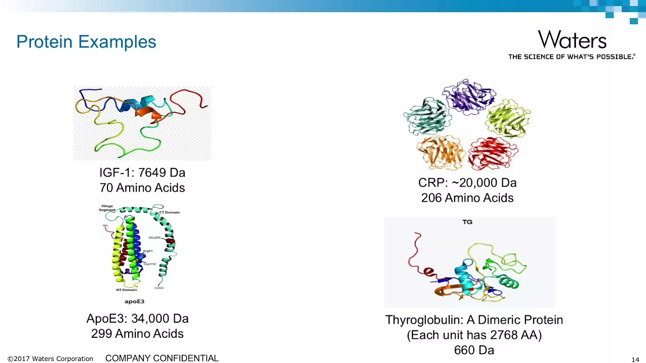
Fundamental concepts of biochemistry, specifically peptide structure, folding, and protein-protein interactions.
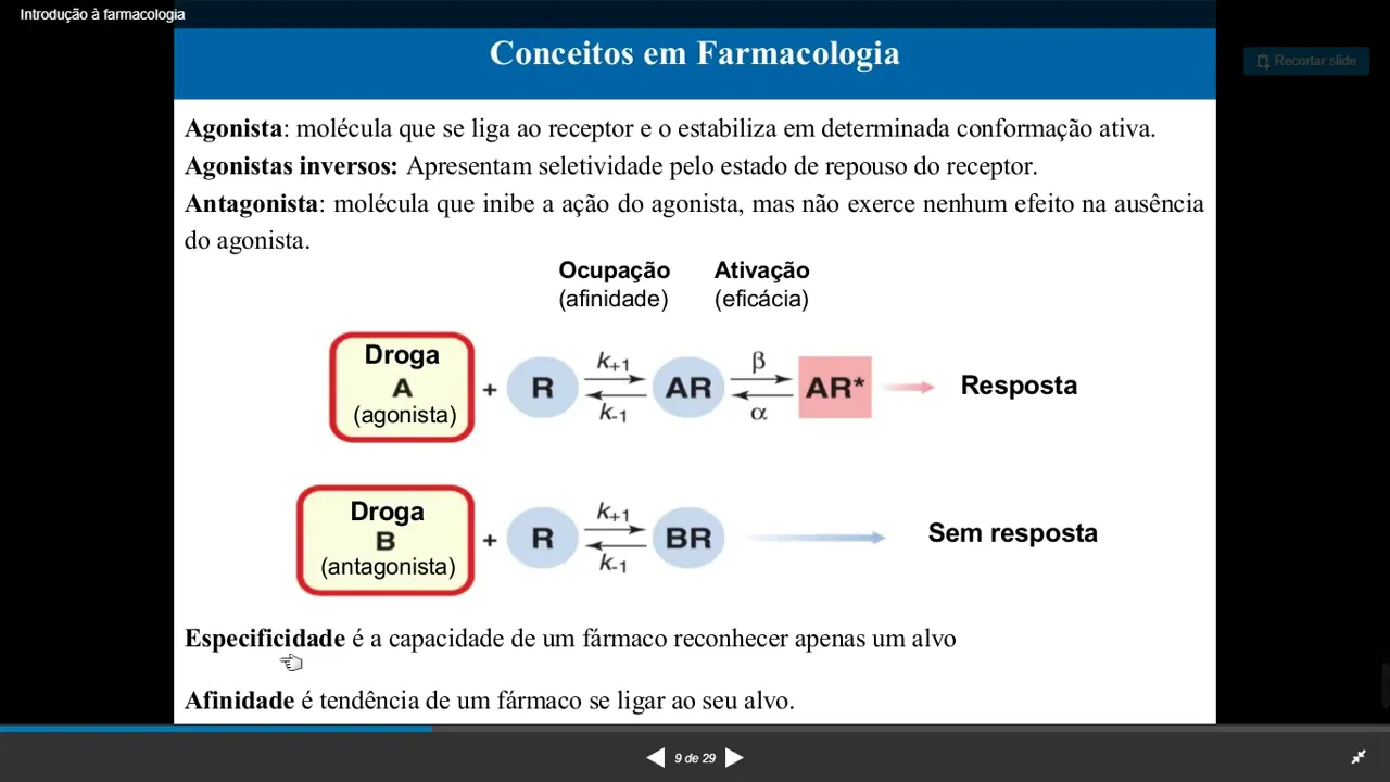
General pharmacological concepts such as receptor-ligand binding, affinity, selectivity, and the difference between agonists and antagonists.
Exploring advanced biophysical techniques used to study ion channels, such as patch-clamp electrophysiology and cryo-electron microscopy.
Investigating channelopathies—diseases resulting from ion channel mutations—and how neurotoxin probes help characterize these dysfunctions.
Translational drug design, focusing on how venom-derived peptides (e.g., conotoxins) are modified to create targeted therapeutics like non-opioid analgesics.
Analyzing the evolutionary biology and chemical ecology of venomous organisms and how their toxin arsenals co-evolved with prey ion channels.
324 views5likes33:24Original Release: 2010-09-12
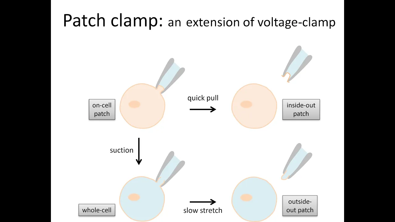
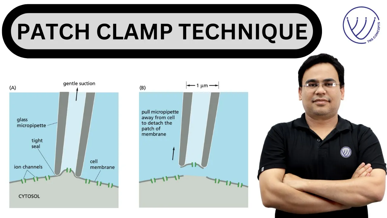
[Music] I would like to introduce our first Speaker Dr Ken uh Blumenthal who's professor and chair at the depart at the uh School of Medicine uh and biomedic Sciences Sunni Buffalo uh his talk this morning will be peptide uh neurotoxin probes of ION channel function uh Dr Blumenthal I'm going to talk to you today a little bit about uh voltage sensitive sodium channels and how they work uh and uh the title here really alludes to the to the fact that uh we all we've all heard the old the old expression that one man's uh meat is another man's poison uh and and and this is uh a case where a variety of different poisons and I'm going to tell you really only about one of them in detail uh has given us a lot of insight into how voltage sensitive sodium channels function and um um also has a good deal of potential for uh giving us templates for Designing drugs that modulate channel functions so um there are lots of different poisons that that uh and lots of different poisonous organisms that that U uh mod that secrete Things That modulate these channels uh pu from puffer fish to scorpions to anemones various kind of toades and some interesting spiders that I'm going to be talking to you about later you probably want to stay away from spiders that do things like that um so voltage sensitive sodium channels uh serve many important functions but most importantly they are responsible for the rising phase of the action potential and they function and there are there are isoforms of sodium channels that that initiate Action potentials in contraction in muscle and that initiate Action potentials in nerve uh as you might imagine there are disease there there are mutations that are associated with Channel malfunctions uh and these these have been identified now in at least four different classes of sodium channels muscle sodium channels uh have a variety of mutations that give rise to periodic paralyses uh cardiac sodium channels uh have mutations have been have been identified that that correlate to Long QT syndrome uh brigada syndrome and a variety of aymas uh CNS sodium channels scn one and two um mutations in these have been associated with um uh epilepsy and and various seizure disorders and more recently in the sodium Channel that's found in peripheral nerves it's expressed in peripheral nerves uh a couple of diseases have been identified that are associated with chronic pain with ability to sense chronic pain and the the the toxin that I'm going to tell you the most about today is actually one that targets this sodium channel uh and there there are a number of of pharmaceutical companies that are very interested in how this toxin uh modifies Channel function so here is the the the big big big picture of voltage sensitive uh sodium channels so voltage sensitive sodium channels are members of a large super family of voltage gated ion channels these channels pass selectively either sodium potassium or calcium in response to a change in membrane potential um they all have a basic structure which is probably pretty hard to see in this in this slide but if you look very closely you will see that for sodium and calcium channels which are shown up in this in this quadrant there are four domains four homologous domains and here's one domain of a voltage sensitive sodium or uh or calcium channel uh and what you can't see in this uh in this uh rendering is that each of these domains has six transmembrane regions in it so the whole protein has 24 transmembrane passages um and that there's a good deal of internal homology obviously doesn't show up in this between each domain so we know that these things or we we can we can deduce that these things are derived from a common Gene by two duplications and Gene fusions um one of these six transmembrane domains in each so one of these six transmembrane passages in each domain has a very high density of positive charges and is thought to be responsible for volt for voltage sensing um the whole protein is about 250,000 molecular weight depending on on which channel and which organism and which isoform we're talking about so it's a fairly complicated protein to work with um a a simplistic view of of how the channel functions and I'm going to I'm going put putting this up here so that we can talk about all the various sites that are involved and I'll tell you about the the sites that we're most interested in so the channel is a transmembrane protein most of its sequen is actually within the membrane um it has a a a large we think we don't we don't have a threedimensional structure of this channel although we do have threedimensional structures of some more simplistic homologues so we think it's w it has a wider pore at the outside than at the inside and in the pore there is a binding site for for a guanidinium compound called tetrodotoxin which is made by puffer fish and which blocks the channel so so tetrodotoxin is a blocker if you ingest uh improperly prepared uh Sushi uh that's made from puffer fish it will kill you um very quickly actually so tetr toxin is a channel blocker and it binds to what is called site one there is further down in the channel a selectivity filter so the selectivity filter is what determines whether this is a sodium Channel a calcium channel or a potassium channel so different ions are allowed through uh with different efficiencies sodium channels conduct sodium about 30 to1 preference over over potassium and don't conduct calcium hardly at all um there is also below the selectivity filter what's called an activation gate so the activation gate is that part of the channel which under goes a confirmational change the first confirmational change which causes the channel to open and if you think of the channel as sort of an inverted Tepe kind of a structure what happens during opening is is that the bottom is is pulled apart so that the pore opens all the way through and you can see sort of down here you could imagine that if these two part parts moved further apart this would open the pore so there are toxins that bind to this site called the acation gate at site 2 there's an inactivation particle so the channel has three states open closed and inactive and the inactive state is different from the closed state so that after the channel activates after these two pieces move further apart they they uh generate there is generated a docking site for what's called an inactivation particle and the inactivation particle jumps up into this into the inside of the pore here and closes it off and this is why Action potentials can only move in one Direction down either a nerve or a muscle so the channel becomes unable to fire for a transient period of time after it's been activated sodium can go through the channel as long as the inactivation particle is out of the way and the activation gate is opened there are also sites on the outside of the channel and I'm not showing you where they are right here although that's what a good deal of this talk is going to be about where peptides bind so there are a variety of different polypeptides made scorpions spiders anemones that bind to sites on the outside side of the channel designated as site three or site 4 site three is associated with Channel inactivation so when a toxin is bound to site 3 the inactivation particle is inhibited from moving into the into the inner part of the pore and site 4 is associated with activate with Channel Activation so cite four toxins typically are thought of as peptides that change activation in some way usually they make it easier to activate we're going to see that that's not all always the case today so the pharmacology of the sodium Channel very briefly is shown here and there are a variety of different sites and actually there seem to be additional sites coming up all the time some of this is just due to to inability to distinguish one from the other uh particularly with some of these these local anesthetics and things that buy into site six which are fairly and five which are fairly hydrophobic and it's it's sometimes not simple to distinguish in binding essays or in functional essays between these but certainly site one sites one through four are distinct from each other site one I already told you binds tetrodotoxin and it's in the pore so The Binding site is some is in the S5 S6 so the S the fifth and sixth transmembrane passages of each domain constitute the pore txin binds in that region bet troxin which comes from these these toads uh binds somewhere near the inside of the S6 and these two toxins here CNM scorpion toxins bind in domain 4 in the S3 S4 extracellular Linker uh beta scorpion toxins bind to the same region but in domain two these affect Channel inactivation these affect activation most of what I'm going to tell you is going to relate to things that affect Channel Activation so the story today is how do these gating modifiers so these toxins that I'm telling you about these polypeptide toxins are what we call gating modifiers they change the kinetics of activation and inactivation without affecting the intrinsic ability of the of the channel to conduct ions so how do they do this um and what we're going to see I'm going to tell you most about this one here um but in some cases they'll involve protein protein interactions but it looks like in some cases the interactions that are most important are not necessarily protein protein they may be protein lipid interactions so how do these gating modifiers work so let's take the two extreme examples here is domain a a rendering of domain four of the voltage sensitive sodium Channel and you can see the six transmembrane regions here the pore region is formed by S5 here this re-entrant Loop and S6 here so when a site3 toxin binds to the sodium Channel it inhibits the movement of this gating particle of of this gating uh domain there thereby preventing the inactivation particle from having from from finding its binding site and electrophysiologically what we see when this happens is that the normal uh uh current trace for an action potential which shows a rapidly developing inward current followed by a rapid decay of that current in the presence of a site3 toxin the current the channel activates so the current develops just fine but doesn't turn off so this is this is the the electrophysiological Hallmark of a delayed inactivation so that you can very readily measure the presence of toxin on the channel by simply measuring current at a time between 5 and 10 milliseconds after the initial stimulation normally the channel a normal channel will have no current at this time a toxin treated channel will have appreciable current so that's a site 3 Toxin and this is a little bit confusing in nomenclature the site3 toxin binds in domain four I will remind you okay site four toxins site four toxins unfortunately bind in domain 2 to make things a little bit more complicated uh but so here's a rendering of domain 2 and again you see the same remember these are homologous each of these domains is homologous so here we see the same S4 High highly positively charged region uh which is a voltage sensor so when the channel activates in the presence when the channel activates this uh Helix is thought to move outward in the presence of a site four toxin it gets trapped in this outward configuration and the so the electrophysiologic Hallmark which I'm showing you in a slightly different format here uh this format is called a current voltage relationship so a normal Channel an untreated channel will have a current voltage relationship that looks like this where maximum current uh is at about min-2 molts in the presence of a site 4 toxin which stabilizes this this configuration of the channel this curve is shifted to the left so that the channel activates with less voltage with with less depolarization required we can identify interactions in these in these uh toxin channel uh complexes using a formalism called mutant cycle analysis which was developed about 20 years ago by alen first to look at at enzyme substrate uh complex formation and basically mutant cycle analysis just says you can measure if you can measure the dissociation constants for the the wild type life and wild type receptor in this case Toxin and channel and if you can mutate one pair in this case let's say it's the the the mutant is in the toxin in this case it's in the channel you should be able to see a dis if you should be you should see A disruption in binding Affinity which you can measure by some some essay um and that disruption should be more or less the same if two residues one from the toxin one from the channel are interacting with each other and you shouldn't see any addied effect when you measure measure mutant versus mutant we can do this in a in a channel essay simply by measuring if we're looking at a site3 toxin for example we can measure current remember I said you could measure current at this in this sort of 8 Mill 7 to 8 millisecond time window and toxin binding is strictly proportional to the current that develops in this time window so you can watch the current increase with time after washing of toxin you can measure that you can get a K on from that and you can get a k off by just washing it out and measuring the wash out it's fairly simple assay um and when you do that in a in a in a couple in a in a in a case where you're seeing uh uh a where where you're demonstrating the existence of a of a of a pair of residues interacting with each other what you will see is shown here so here is a wild type site 3 toxin binding Affinity less than one nanomolar binding to a wild type cardiac sodium channel in the in the filled block if you mutate a particular residue in the S3 S4 Linker of domain 4 which is The Binding site for this toxin you decrease the Affinity by almost two orders of magnitude so the open blocks here are the mutated channel uh and the closed blocks are the wild type Channel if we mutate a particular amino acid residue lysine 37 in this case to an alanine we find that we lose a great deal of binding affinity for the wild type Channel but we lose no binding affinity for the mutant Channel and this is an indication that these two residues spartic acid 1612 of the channel and lysine 37 of the toxin format charge pair uh we can actually use a a the Gibbs equation to calculate a Delta Delta G and uh what we see in that case is that it's about the energ the energetics of this are about the energetics that you would expect for for a for an electrostatic interaction a kilo calorie and a half or so per mole um this is pretty old data we this was published almost 10 years ago actually it was published 10 years ago um but this is the same formalism that you would use in general to look at at protein protein interactions um this was all done with a site3 toxin and it's the same method that we chose to address The Binding of this new toxin that I'm going to tell you about for the rest of the talk so about five years ago we got interested in a family of spider toxins uh so spider toxins are spider toxins are very interesting they have an enormous pharmacologic diversity um and some of that's this is a very small snip of what the diversity the pharmacologic diversity of spider toxins is but there are spider toxins that interact with virtually every kind of ION channel that's been identified to date voltage sensitive mechanosensitive acid sensitive all of these things have not one but families of toxins that interact with them and some of these families are shown here that so they'll interact with potassium channels sodium channels calcium channels mechanos sensing asex and others these toxins are all related to each other and it's it's interesting that these guys have a basic structure so they're all co-evolved um and yet the only essentially the only thing that's conserved among all of these things is the disulfide framework that holds them together pretty much everything else in them is hypervariable so single spider Venoms have between 50 and 100 Rel toxins as assessed by mass spectrometry so single spider one animal um most of these are about between 30 and 40 amino acids in length they all have three disulfide bonds as far as we can tell they lack regular secondary structure if you do any of the the sort of typical secondary structure determinations you see pretty much they look like denatured proteins but they're not um and they have these three disulfide bonds that are always linked 1 to 4 2 to 5 and here's 1 to four uh two to 5 and 3 to six so they basically form an a a knot structure so ick stands for inhibitory cine not Motif the identified molecular targets I already told you all forms of voler sensitive sodium channels all forms that we've looked at of vard sensitive potassium channels most forms of calcium channels and several others some most of these things are gating modifiers some of them are chant are pore blockers so they do of all kinds of things and there are estimates are thousands of these toxins for uh for which targets have not been identified yet so what did they do we started and I should I should give credit at this point to Jamie Smith who's a graduate student in my lab who is now a postto at NIH who did most of the work that I'm going to show you about um we started to look at one of these compounds called protox 2 and we find that protox 2 as an initial characterization inhibits the activation of sodium channels a voltage sensitive sodium channels so here we're going to look at this is our protocol so for those of you who aren't familiar with electrophysiology um basically this is these assays are all done by wh cell patch clamp analysis and there are two different forms one of which holds the cell at- 130 molts and steps it to-30 molts for 10 second 10 milliseconds and measures the resulting current that flows and that's that generates what's called a train file and this is a train file here where no toxin is here and as you increase toxin current decreases okay the other way you can measure this is by doing steps in 5 or 10 molt increments from 130 fromus 130 up to plus 20 uh and when you do that you generate what's called an IV curve and most of what I'm going to show you is generate the data come from IV curve so here's an IV curve for a normal cardiac sodium Channel expressed in hexels and you can see maximum current is is at a about uh -30 to -40 molts in the presence of protox 2 that's the maximum current is greatly inhibited so it's about 80% inhibition and there's a shift in the maximum uh in the voltage required for maximum current to the right so this means what this means is that this toxin inhibits Channel opening even though it affects activation it does not stimulate Channel opening as the typical site 4 toxins do it inhibits Channel opening so how does it do that and what's more how does it how do we account for this so here's a here's some data from from uh Charlie con's lab uh in which he looked at the effect of this of this toxin protox 2 on several different channels and finds uh that it modifies uh activation of virtually any any sodium or calcium channel he looks at this is a little unusual because because these things are although they're homologous they're not that homologous they're only about 20% homologous um so how do you so we have to account for the fact that not only can this this peptide which has only 4 31 residues in it modify so many different Channel ISO forms but it does so with a very high degree of isopor selectivity so there's some specificity built into this you notice here that the nav 1.7 channel is modified probably at subnanomolar concentrations whereas nav 1.5 is modified at about.1 micromolar so there's 100-fold difference here and yet there's virtually no difference here even though the calcium channel and the sodium channel are much more distantly related than these two sodium channels so this is was a hint that we missed actually that this must involve something other than protein protein interactions um so we set out on what seemed to be a fairly simple quest which was to identify the binding site after all we had clones we knew how to do Cy directed met Genesis and the likelihood the initial thought was well this must be because it's mod ifying activation it must be either in domain two The Binding set must either be in domain two somewhere near site 4 or because it's inhibiting current it must be somewhere in or near the pore so we started out Jamie started out by wondering whether or not protox binds to site 4 and the experiment that she chose to do was a fairly simple one simply to to mutate residues in this Linker and the way in which she ended up doing this was simply by swapping the Linker in domain four for the Linker in domain 2 so she made constructs that had no domain 2 Linker and the construct that I'm going to show you the data for is this one here designated S4 in which the domain two Linker is completely replaced and then all she has to do in order to measure uh activity is to say does protox 2 still modify this Channel and that's shown here so here's the IV relationship current voltage relationship for a channel for a 44 4 channel no domain to S3 S4 Linker uh untreated and treated with protox and pretty much what you can see here is the prot it looks like the effect of protox is pretty much identical on the 44 channel to what it is on wild type Channel and more extensive data bear bear that out um and now you want to know as a control have we really succeeded in abolishing site 4 and in order to test that Jamie took a known site 4 toxin derived from cides from scorpion venom uh and demonstrated that this toxin which normally will do the same thing as protox now has no effect so site 4 has been abolished but protox activity has not been abolished therefore protox cannot bind in domain 2 S3 S4 okay we said that was one candidate the other candidate is the poor because of inhibition of current does it bind in the poor um so the answer to this unfortunately is no um and the way in this which this experiment is done is as follows again we're looking at IV curves so here's the IV curve of the normal Channel untreated with toxin we throw on protox at 500 nanomol we shift to the right we inhibit current just as we expected and we then ask what happens if we give the channel tetrodotoxin so tetrodotoxin is a Poe blocker it binds in the pore if protox binds in the pore it ought to inhibit tetrodotoxin binding and and we ought to see no inhibition when we Chase protox with tetrodotoxin but instead when she chases protox with tetrodotoxin the channel is completely wiped out Channel activity is completely wiped out therefore tetrodotoxin can access its binding site in the presence of protox therefore protox shouldn't be binding in the poor either so now we are in sort of the heads scratching stage because it's not in either of the two likely places so now how do you how do you where how do you proceed from here we're dealing with a peptide the peptide is fairly positively charged so the notion is it must be the binding site must be in some other extracellular Linker region so in order to test this Jamie made a a large I I would say a heroic number of mutants um in extracellular linkers and I'm not going to go through all of these except to say that in all of the S1 S2 and all of the S3 S4 linkers in the sodium Channel we have been she's was unable to detect a binding site for this toxin so notice that all of these different mutant constructs display inhibition by protox the degree of inhibition varies a little bit part of this is due to the to the difficulty in working with some of these mutant channels but the bottom line is that all of these things um behave as though they have a complete an intact binding site so at this point we said Well there must be something unusual about this how can we how can we can we come at this from the other side can we come with this from the toxin side and maybe get some insight from that so because Jamie has had by this time gotten very good at doing mutagenesis she then did an alanine scan of the entire toxin there's only 30 residues there you can't mutate the cysteines so it's it's really boils down to only making about two dozen mutants and what's shown here and you don't have to worry about what the numbers say what's shown here is the result in in terms of KD values for all of these mutants and the red ones are the ones that are important the red ones are the ones that have a major effect on binding affinity and if you look at what the red ones are methionine tryptophane methionine veine tryptophane Lucine tryptophane most of them are hydrophobic there are a few there are a few polar ones there's Arginine 13 and Arginine 22 but other than that virtually everything that has a major effect on binding is hydrophobic and this is a clue as to what's going on we think um what's more when you look at the and this probably doesn't doesn't show up very well but when you look at at where these things are on the on on the solution structure of the toxin they all map to one face so everybody that seems to be important maps to this one side of the Toxin and what's more these residues here glue 17 ser11 38 and some others that are not shown here which do not have an effect on binding Affinity all mapped to the other side so we now have sort of an amphipathic kind of molecule that's got a very clear hydrophobic side to it and a very and and that site is important for activity so the question then becomes does this toxin access its binding site by first going into the membrane or is its binding site really largely lipid derived and so jimie devised an essay uh a liposomal essay and it's a very simple essay you simply make uh pcps liposomes uh incubate toxin or mutant toxin with these things spin the liposomes out and assay toxin in the supernate by hplc so it's a it's a simple depletion assay and the results that you got are shown here so let's start at the bottom so here's a typical site 3 toxin before and after liposome treatment and what you can see is that the site 3 toxin is not depleted by treatment of liposome so before I can't remember whether the solid line is before the dotted line is before uh but they're the same protox 2 notice is completely depleted there is no protox 2 left in the supernatent after treatment with liposome so it's completely soaked up by the liposomes and what's odd what what became odd about this was the next set of experiments so we now said well these are pure peptides suppose we take a whole Venom that we know contains different kinds of toxins and ask is there some discrimination between toxin classes in terms of whether or not they'll bind lipid so on the left here is uh Venom of the the Scorpion lurus on the right is the Venom from the Scorpion cides which we received from lur of alp pasani in Mexico City and we do the depletion assay we characterize each of these Peaks by mass spectrometry compare it to the known molecular weights of of toxins that exist in this in this Sol in this in these Venoms and then asay selected fractions for their activity and what we find basically is there's a cy3 toxin known site3 toxin that runs right here it has this peak has exactly the same mass and if we assay the contents of this peak electrophysiologically we see a site 3 Type effect delayed inactivation inhibit inhibition of inactivation here we see one that is completely depleted the one that's completely depleted turns out to be css2 by molecular weight and css2 by electrophysiology and so what this is telling us is that cy4 toxins things that affect activation seem to bind lipids efficiently site3 toxins don't and what's more what makes this even more confusing is that the the site three and site four scorpion toxins are structurally related and you can see you can see that sort of here they all have this Helix and Sheet Motif you can't see all the sheet in this one because the way the molecule is oriented but there's a three stranded beta sheet with an alpha Helix stocked against it these things behave differently when it comes to ability to bind lipids whereas these things that have no structural similarity and these things that have no structural similarity behave the same when it comes to binding lipids which suggests that there may be a different some fairly subtle confirmational differences in the domains of the channel that are most associated with activation and inactivation and I'm going to just skip this one uh because what we did here was to make a number of of mutations in the transmembrane regions to see if we could find a site and the short answer is that we did not find a site so we are left with a model that looks like this where the where where this little diamond shaped thing is protox and what we think may be going on here is that protox binds to lipid initially and then either diffuses by laterally within the membrane to access a binding site which we have not been able to identify on the channel itself or by immobile izing annular lipids in the vicinity of the channel inhibits the ability of the channel to activate remember activation involves outward movement of this S4 Helix so that if you put enough energy in the form of depolarization into the system you can get it to activate but normally under under under normal voltage conditions it won't activate um so we're still working on trying to figure out whether this model that has no direct interaction between the two proteins is the valid model or whether uh there are sites on the on the on the channel That interact with the toxin as well and it looks like there are but we don't have good enough data yet just to to to to uh reach that conclusion so basically this is an interesting toxin because it modifies two different channels that are distantly related by homology it modifies them with very high affinity and with very high isopor selectivity it doesn't interact with site four we think it it will therefore allow us to identify new sites that are important for gating transitions its binding interactions are mostly hydrophobic and they could involve transmembrane and and probably do involve transmembrane regions of the channel and there's probably some composite interaction with phospholipids as well uh this toxin remember has a very high affinity for channels involved in sensing chronic pain and is therefore a reasonably good Target in terms of uh uh uh templating for drug design and with that I will close I thank I think I probably ran a little bit long uh so the people who did the work uh most of the work was done by Jamie Smith who graduated uh last summer and is now in Ken Schwartz's Lab at nincds uh she was assisted in some of this by suji Alfie who is still in the lab uh collaborators we have a long-term collaboration with Daddy Hank at the University of Chicago uh Ted cumins uh at IU and uh lurel pasan and herard Coro in Mexico City so thank you [Music]
Up Next

7:59
Membrane Receptors Explained: Signal Transduction & Ligand Binding
@khanacademymedicine
231.5K views•2014-05-04

21:55
Deciphering Neural Circuits Controlling Anorexia in Mice | Richard Palmiter
@AllenInstitute
2.3K views•2011-10-10

4:10
Vagus Nerve (CN X): Anatomy, Nuclei & Functions Explained
@Alilamedicalmedia
305.2K views•2022-10-31

13:03
How Exercise Benefits Your Brain: Science Explained
@TED
11.4M views•2018-03-21
Related Study Plans & Knowledge Roadmaps
Structured learning paths in Neuroscience
neuroscience
Appetite Regulation: Brain Circuits & Hormones
01Foundations of Endocrinology and Brain Anatomy
02Introduction to Energy Homeostasis
03Peripheral Signals: Leptin, Ghrelin, and Insulin
+ 2 more learning modules
14h5 modules
Explore Roadmap
neuroscience
Neuroeconomics: Value, Risk & Brain Mechanisms
01Foundations of Neuroscience and Brain Anatomy
02Introduction to Neuroeconomics and Subjective Value
03Dopaminergic Prediction Errors & Reinforcement Learning
+ 3 more learning modules
18h6 modules
Explore Roadmap
neuroscience
Spatial Navigation: Cells, Grids & Brain Maps
01Foundations of Brain Anatomy and Memory
02Place Cells and Head-Direction Cells
03Grid Cells: The Brain's Coordinate System
+ 2 more learning modules
12h5 modules
Explore Roadmap
neuroscience
BCIs: Neural Decoding & Prosthetic Control
01Neuroscience Foundations: How the Brain Signals Intent
02Signal Acquisition: Recording the Brain's Activity
03Neural Decoding: Translating Brain Waves into Data
+ 2 more learning modules
25h5 modules
Explore Roadmap