


















This webinar presents three cutting-edge studies on plant biotic interactions: (1) Plant extracellular RNA in the apoplast is primarily protected by RNA-binding proteins (AGO2 and GRP7) rather than extracellular vesicles, with m6A-modified circular and linear RNAs potentially functioning in host-pathogen communication; (2) Broad-spectrum fungal resistance in sorghum is conferred by the ARG1 NLR gene, which is embedded within a natural antisense transcript (CARG) and regulated by MITE transposable elements in its promoter; (3) Helper NLR proteins (ADR1 and NRG families) function downstream of TIR-NLR immune receptors through distinct EDS1/PAD4 or EDS1/SAG101 complexes, with truncated helper NLRs serving as negative regulators to prevent overactivation of immune responses.
Plant Biotic Interactions: Highlights from The Plant Cell Focus Issue
Added:Hello, welcome to the next in our series of scientific webinars. Today's webinar is hosted by the Plant Cell reviewing editor Cris Argueso and moderated by Plant Cell Assistant Features Editor, Thomas DeFalco. We have three speakers who will be sharing their work on the topic of plant biotic interactions. I will let Cris and Thomas introduce the speakers So, hi everyone, So it's my pleasure. It's One second first. You're fine. Today's webinar is a celebration of the May 22 2022 Focus Issue on plant biotic interactions, edited by Roger Innes, Dan Kliebenstein, Cris Argueso, Yangnan Gu, Libo Shan, Dorothea Tholl, and Mary Williams. This focus issue is now available online. After the webinar, we will be posting the recording to our YouTube channel. There you can find many other exciting scientific talks organised by ASPB, The Plant Cell, and Plant Physiology. The webinar is made possible thanks to the members of ASPB. We would like to give a special thank you to ASPB members who have priority registrations. If you'd like to become a member of ASPB use the promo code Presents10 to receive 10% off membership dues. Please put your questions for the speakers into the q&a box.
Thomas will read this out to the speakers after their talks. If you have technical problems, please email me Jason Padilla at [email protected]. If you're having trouble with your sound, try reconnecting or dialling in now, I will turn over the floor to Cris, who will tell us a little more about the focus issue.
Okay, so now it's my turn. So welcome, everyone. It's a pleasure to welcome here today. I'm Cris Argueso, Associate Professor of Colorado State University, and I'm part of the editorial board that Plant Cell and today on behalf of myself, but also the other editors of the special issue. These include Roger Innes, Dan Kliebenstein, Dorothea Tholl, Yangnan Gu, Libo Shan, and Mary Williams. I welcome you here, you know, it's a pleasure to have you here today. So this is indeed a special issue. Our focus on plant biotic interactions is really timely. Because in the last three years, things have changed drastically in the field of plant biotic interactions.
Seminal work has been accomplished on and has changed the way that we see plant immunity, and also the way that plants interact with pests and microbes and also beneficial organisms. And we have in this issue, we have eight reviews that have been edited by the editorial board. They are very interesting reviews, I'd like to highlight some of them. One of them is a review by Jonathan Jones on an update on the zigzag model from 2006 from Jonathan Jones and Jeff Dangle, incorporating what we know now about pattern-triggered immunity and effector-triggered immunity and how they interact. There is also a beautiful review on TIR NLR proteins immune signalling proteins by the lab of Jane Parker, and also a very timely review on calcium permeable channels by the lab of Libo Shan and also Keiko Yoshioka. And of course, plant immunity is not immunity only to microbes but its immunity also to pests. And we have Adam Steinbrenner who has contributed a beautiful review on how plants perceive insects and mount immune reactions to them. This review, this special issue also incorporates 14 research papers on several topics on plant biotic interactions, including effectors from bacteria and fungi, virus-plant interactions, nodulation - the importance of a nitrate transporter in nodulation in lotus. And also the importance of primary metabolism in plant immunity and plant growth. So I suggest that you check out this this very special focus issue of The Plant Cell. And now we're going to be highlighting three of these reports that were present of this special issue. We have Thomas DeFalco as our our moderator for this webinar. And before I give it to Thomas to to moderate the webinar, I just want to thank not only all the editors of this special issue, but also the authors who have contributed their best work to The Plant Cell, the reviewers who have made these papers better. And of course, Mary Williams without her work wouldn't have been able to put this effort together. So with that, I'll give you Thomas DeFalco to moderate the webinar. Thank you and enjoy the webinar.
Thanks, Cris. So thanks, everyone for being here today.
Our first speaker is Hana Karimi. She's currently a postdoc in Richard Vierstra's lab at Washington University in St. Louis. But previously did her PhD in Indiana with Roger Innes, and in Indiana, she was working on extracellular RNAs in the context of plant immunity, which is the topic she'll be discussing today. So with that, I'll turn it over to Hana.
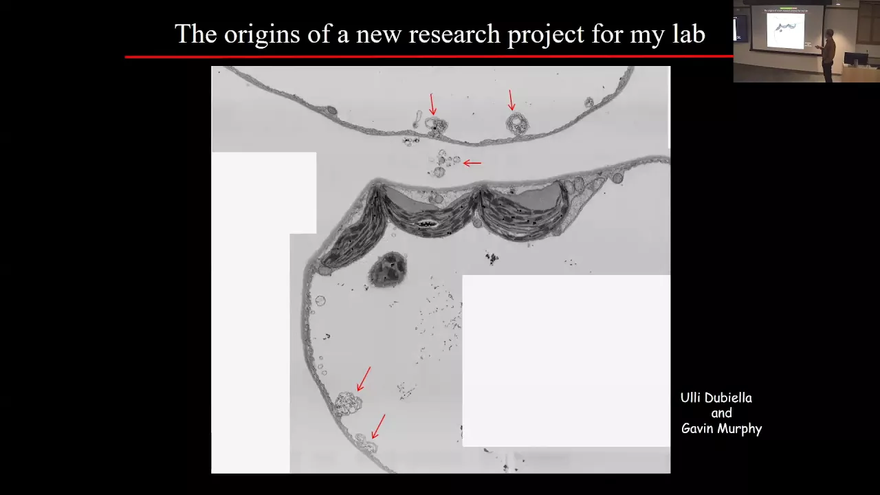
Thank you very much. I'll share my slides first. Okay, thank you very much for the introduction. And thanks for giving me this opportunity to talk about my previous PhD work at Indiana University in Roger Innes' lab. And I'm going to talk about plant extracellular RNA today focusing on extracellular, the apoplastic fluid RNA content. So the apoplast is the first battlefield between plant and pathogen and it has been shown that it contains, and it has been shown that our apoplastic fluid contains different molecules from both plant and pathogen. Recently it has been shown by Roger Innes lab, that apoplastic fluid also contain extracellular vesicles which are lipid compartment that can protect the cargo from degradation. Dr. Brian Rutter at Indiana University developed a protocol to isolate and concentrate extracellular vesicle from Arabidopsis leaves. Based on Brian studies, EVs contain anti-microbial proteins which might play a crucial role in plant-pathogen interactions The presence of EVs in apoplastic fluids raises the question of whether EVs play a role in molecule transformation, especially RNA transmission between plant and pathogen during plant pathogen interaction, especially host-induced gene silencing. To answer this question, first, we investigated the EVs associated with small RNA which resulted in identifying several small RNA and newly described tiny RNA associated with EV. Tiny RNA are RNA between 10 to 17 base-pair nucleotides that are enriched in the extracellular space and co-pelletted with the EV. Since this report, there have been a lot of other reports indicating the possible role of EVs in RNA transmission between plant and pathogen. So we decided to focus more on EVs associated or indeed encapsulated RNA. Our goal was to distinguish between EV's encapsulated RNA vurses extracellular RNA present in apoplastic fluid that might be in the shape of free RNA or bound to RNA binding protein. To distinguish between EV's encapsulated RNA versus other RNA in apoplastic fluid, we treated our EVs pelleted RNAs in the presence and absence of detergent. The presence of detergent will affect the membrane of the EVs and expose EV's cargo to RNAs. That way we can distinguish between RNA that are inside EVsand those RNA that are located outside EVs. When we treated the EV pellet with only RNase, most of the RNA were unaffected, which makes sense because we had this hypothesis that EVs encapsulate a lot of RNA, but when we added detergent to this reaction, still we have a lot of RNA unaffected. However, we in disrupted the membrane by adding detergent, that means something else other than EVs are protecting this RNA, probably RNA binding protein. So we decided to add protease to remove and digest RNA binding protein here, we added trypsin to these reactions first, and then when you have detergent, all RNA binding proteins are removed from the reaction without affecting the extracellular vesicle and extracelluar vesicles are still intact. After that, we added RNAs to remove the free RNA. But interestingly, just treating with the trypsin plus RNase that removes all RNA from in this pellet, we did not affect RNA, we didn't add detergent to this reaction, only treated with the trypsin pluss RNAse. That means that all of this RNA in this pellet is bound to RNA binding protein, and they were not inside the EV. This result indicate that EVmight not contain RNA, or they contain a very little amount of RNA.
We follow the experiment with the sequencing. Consistent with our results in a previous slide, our small RNA sequencing showed that RNA are highly affected by trypsin plus RNase treatment, indicating that they are located outside EVs, and protected by RNA binding protein. I have to mention that Trypsin pluse RNAs treatment would not affect the integrity of EVs, and they will be intact and would not affect the EVs cargo. Looking at RNA sequencing data showed that different categories of extracellular RNA were affected by trypsin plus RNase. We decided to look at some of these different categories of small RNA that were affected by trypsin plus RNase, including microRNAs. From almost 70 microRNAs that we found in apoplastic fluid, only seven microRNA were protected inside the EVs, the rest of microRNA plus small RNAs as well as tasiRNA in the apoplastic fluid were digested by trypsin plus RNAs, indicating they were located outside the EV and protected by RNA binding protein outside the EV. Besides the small RNA that we found in the apoplastic fluid that were outside the EVs, we also noticed that our apoplastic fluid contained larger RNA and this larger RNA can be detected besides the small RNA and tiny RNA in apoplastic fluid. So, we had to question here What are these RNA and whether they are from cell damage and like contamination from EV's isolation process. So, to examine the cell for RNA contamination in our apoplastic fluid, and we decided to look for the mRNA contamination, Dr. Patricia Baldridge in Donald Danforth Plant Science Centre, she performed the polyA purification and library preparation for the apoplastic fluid RNA. Since there were no polyadenylated RNA, we could not obtain any library, and that means we do not have mRNA contamination in our apoplastic fluid RNA. So still we have this question What are these larger RNA that are not mRNA contamination from cell damage. We had this concern whether they are ribosomal RNA still contamination from cell damage.
So we decided to do rRNA depletion first, on our applastic fluid RNA, and then perform the library preparation.
So we do not expect to have any RNA that are from ribosomal RNA.
Interestingly, after this process, she was able to have a library of our larger RNA. So now we know that we have a set of larger RNA in apoplastic fluid, they are not from mRNA contamination they are not rRNA. So the next question is what are these RNA? We did this RNA sequencing. Most of these RNA outside the cell were from intergenic region or coding DNA sequences. And that will indicate that they although they are not mRNA, they are still from coding DNA sequences and intergenic region. And most of these RNA were located outside the EV and protected by RNA binding protein. And only a very tiny proportion of them were located inside the EV. Based on this data that most of these RNA were not mRNA and ribosomal RNA, but still they were from intergenic region and coding DNA sequences, we hypothesised that large extracellur RNA might be the byproduct of alternative splicing or back splicing, which includes circular RNA, so we decided to test the presence of the extracellular circular RNA. To do that, we treated the extracellular RNA with RNAse R, which digests all kinds of linear RNA except circular RNA. Interestingly, we observed that accesso RNA contains circular RNA too.
We already know by all of these experiment that extracellular RNA are mostly protected by RNA binding protein, not EVs. So we were looking for RNA binding proteins in our apoplastic fluid. We identified two RNA binding protein in our apoplastic fluid, AGO2, the Argonaut 2, and glycine-rich RNA binding protein 7 seven, or GRP7. Interestingly, both these RNA binding proteins in apoplastic fluid were located outside the EVs. Because AGO2 is known as a RNA binding protein that binds to small RNAs, we expected that the AGO2 was mostly responsible for the presence of a small RNAs in the apoplastic fluid, while GRB seven as a key player in mRNAs splicing was a candidate to be responsible for the presence of circular RNAs of apoplastic fluid. We looked for the presence of the circular RNA and the percentage of a circular RNA in both AGO2 and GRP7 mutants, in apoplastic free RNA. And interestingly we we had this result that ago2 also, mutant in ago2 or knockout mutant ago2 also affect the presence of and the percentage of circular RNA in the apoplastic fluid. So, that means, that AGO2 itself directly or indirectly through microRNA or a small RNA can bind to circular RNA and is responsible for the presence of a portion of circular RNA in apoplastic fluid. And as we expected, for GRP7 as a player in the mRNA splicing GRP7 and was also responsible for the presence of the large proportion of circular RNA in the extracellular space. Based on growing evidence circRNA are enriched in m6a modification which plays critical role in circular RNA production and degradation and function. So, we had this hypothesis that extracellular circular RNA might also contain m6a modification. Beside that, when we identified tiny RNA in apoplastic fluid, we, in our in our in silico analysis showed that tiny RNA families also are predicted to be m6a modified in their core sequences, 10 base pair core sequences. So both these piece of evidence suggests that extracellular RNA enriched in m6a modification. To test this hypothesis, we did a very simple, we performed a very simple experiment. And it was RNA dot-blots blood using m6a antibody. We observed that apoplastic fluid RNA, in apoplastic fluid or we call them extracelluluar RNA, are enriched in the m6a modification. Whether these m6a modification works and function as a marker for the RNA secretion we don't know yet. So in summary, we found that apoplastic fluid not only contained small RNA, it also contained large non-coding RNA and circularRNA. We found that most of the RNA in the apoplastic fluid are not inside EVs, they're outside EVs and protected by RNA binding protein including AGO2 and GRP7. We also found that m6a modification is enriched in the apoplastic fluid RNA and it might play a critical role in RNA secretion. All of these experiment in all of these experiments, the key player or the key experiment was using trypsin plus RNase treatment to distinguish between EVs encapsulated RNA and free RNA or protein bound RNA in apoplasmic fluid. What we are looking for next is to see what is the function of this RNA, extracellular RNA, especially circular RNA tinyRNA in plant immunity, whether m6a modification play a role in RNA secretion and what is the function of AGO2 and GRP7 in RNA secretion and RNA transmission between plant and pathoge. And at the end, I would like to thank my PhD advisor, Dr. Roger Innes at Indiana University, his lab members and all coauthors and collaborators. Thank you very much. And I will take any questions you might have.
Okay, thank you so much Hana, for an excellent talk. So just a reminder, please put your questions in the q&a in the chat. And I will read them out. Just to start things off, I'm actually wondering, so how exactly does secretion of the ribonucleoprotein complexes work? Do you actually know that?
No, not yet. We have some evidence and ideas but whether they, it's hypothesis, but we are not sure how, and what is the mechanism of secretion.
Okay, and I guess another question that's probably going to be we're not sure yet, but do you have any idea what the potential functions of these long non coding RNAs might be in the apoplast.
So, there are these long non coding RNAs not like not just in plants, they have been studied in the mammalian system in the extracellular space of mammalian system, especially circular RNA.
And there are other a lot of different ideas. Some of them is like they bound, they are in the extracellular space, for example, circular RNA, they can function as a sponge for the microRNA that coming through from the pathogen, and eliminate them from doing their function or whatever is that in the plant cells, or they can also bind to like their circular RNA can also function as a sponge for the RNA binding protein, and somehow regulate their function just bind to RNA binding protein.
They are there whenever the plants need them in the extracellular space to do their job transfer RNA between pathogen, plant and pathogen, they will be released to do their function. What is exactly the function of them? We don't know yet. But these are their hypotheses, an idea about the role of long non coding RNA in the extracellular space.
Thanks. We have one question in the chat. Are you aware of any mechanistic study about what is the function of circular RNA in plants? So I guess, generally speaking, The function of circular RNA in plants? Yeah, that's, I can say that circular RNA and function and production is like a new kind of study that just like now, the scientists are interested in. I don't have much information about the plant. I know, there was a couple of papers recently published that they were looking for a function in the plant response to pathogen and plant immunity, but most of the information we have about the circular RNA function are coming from the mammalian system. And they also do not have a solid answer for these.
Some of them, they said that even circular RNA can be translated. And they are regulatory RNA that can regulate the small RNA, regulate the translation in the mammalian system, but about plant system, I don't recall any specific function, but I'm guessing it should be similar.
Okay, we have one one last question. It's a two parter.
Could the extracellular RNA be released from vesicles that move from the nucleus to the cell membrane? And, related,are the lipids in the vesicles similar to those in the nucleus or another membrane system?
So I don't know about the lipid and extracellular vesicle are similar to the nucleus. I know that the Roger Innes lab they are doing some experiments to characterise this extracellular vesicle lipids, but I don't have any answer for that. But for what was the other question? Sorry, I forgot.
I think you actually did answer both. And we'll do one, one quick last question, I swear. This is also something I was wondering about. Do you have any idea about the dynamics of extracellular vesicle contents? Or extracellular RNA is generally in response to pathogens etc. Yeah, so Brian, in Roger and his lab, he did a couple of experiments to see whether these extracellular vesicle protein change or extracellular vesicle content change in response to pathogen, he observed that this the antimicrobial molecule associated with the extracellular vesicle changes in response to pathogen infection, we also see some change in the RNA content of the apoplastic fluid in response to salicylic acid and pathogen infection. So, somehow they are dynamic.
Because we are mostly focusing on the plant-pathogen interaction, we have seen the change in the apoplastic RNA or extracellular RNA in plant-pathogen interaction.
Okay, thanks very much once again. So, we'll now move on to our second speaker of the day. This is Tesfaye Mengiste.
Tesfaye is a professor of botany at Purdue University in Indiana.
He did his studies in Ethiopia, England and here in Switzerland.
And his lab has been studying the immune system of not only Arabidopsis but several important crops such as tomato and what he'll discuss today, which I believe is sorghum. So please Tesfaye, take it away.
Hi, everyone. Thank you for the invitation and I'm happy to be part of this presentation. And my lab.... trying to move my slide, but it doesn't move.... Can you see my slide now? Okay, looks good. Wonderful. Okay, so my lab has been working on mechanisms of plant responses to Botrytis and other fungal pathogens, and try to understand the mechanisms underlying host responses. In this current paper, we have a project that focused on exploring natural variation to identify fungal resistance genes in sorghum and with the ultimate goal of improving crop disease resistance.
So, as you all know, pests and pathogens limit crop productivity. And if you've come to the Midwest in the US, you would recognise a disease called the tarspot. That's what you see here on my slides. This is a disease that has been observed in 2015, and was considered relatively mild but has increasingly become threatening. My point here is disease are everywhere. And it's not something we can solve it once and then gone. So we'd like to our research basically focuses on expanding knowledge on plant disease resistance mechanisms, and ultimately to reduce losses to pathogens. And especially these days, I think advances in genetic technologies and sequencing approaches will expedite the, you know, give us a strong handle to discover new genes and genomic regions. So, we believe that disease resistance could be improved by exploring genetic variation and also leveraging basic knowledge in plant biology. So, in today's topic, I will focus on anthracnose resistance. Sorghum is as staple crop in many countries around the world, especially in the poorer regions of the world. And it's also a relatively less studied model system. Among the most important constraints or challenge to sorghum productivity are fungal diseases. Grain mold is the number one disease around the world. And followed by that is anthracnose disease which is called by caused by a species of Colletotrichum. These two pathogens are the most widespread diseases in sorghum growing regions. What you see on this slide is basically a sorghum cultivar that does not have any disease resistance genes, at least resistance gene for anthracnose. What you see here on the left is just the fungus and some of the disease symptoms as well, and the fungal growth. If a material doesn't have a resistance gene, it is completely blighted. And the grain filling stage is completely impaired. Therefore, leading to significant reduction in crop losses... crop gains. Genetic resistance is a key for disease management, especially in a lot of sorghum-growing regions that cannot afford to buy fungicides.
So, this project was initiated to identify resistance genes in sorghum resistance germ plasm, and identify genomic regions and genes for resistance. And to do that, we've taken two major approaches. One is whole genome resequencing of biparental mapping populations generated by crossing natural variants that are resistant or susceptible. We have also taken genome-wide association studies using landraces collected from a wide area wide different regions around the world and try to link resistance loci identify resistance loci by linking sequence variation and resistance phenotypes. Sorghum is a beautiful model for exploring natural variation. If you take any trait, you'd find genetic variation for those traits. And that's actually very important for crop improvement.
In this example, you see is for example, plant height with very tall plants, or shorter plants. If you look at grain colour, red seed, green grain colour, black ones, and white ones. Any trait you consider you will find variation that's very attractive for breeders and also for people studying molecular mechanisms underlying any trade of interest. So, obviously, my lab is interested in resistance to anthracnose, that's focus of this conversation today. And additionally, we've gathered a large collection of land races and challenged them in the greenhouse for identifying resistance materials, and also conducted a huge amount of field-based phenotyping for disease resistance, and identified variants, natural variants that showed hugely resistance and unaffected growth as you see here, in this leaves that's marked our you'd only see kind of hypersensitive response or a susceptible material here where the fungal grows and these bottom leaves actually drop inoculated leave, that does not allow any fungal growth. The yellow highlighted material in the table here called SC283 was initially the one we identified to be the resistant material, resistant to many, many different strains of this fungus, also different diseases. And this incidentally comes from a region in Tanzania, where there is a high incidence of disease and also this material was selected to be grown on highly acidic soils, which is a problem in tropical areas. So this was the basis for our paper that was published in this special issue. What you see here is a field test in Ethiopia, in a disease prone region, you see all the materials in the surrounding are actually diseased of various fungal diseases. But this SC283 actually survives, although it doesn't appear to be agronomically very interesting.
It stayed green in the field throughout the season. What you see on the right is in the greenhouse, that was challenging the plants with the fungal strains. After about 10 days of disease, the susceptible material is completely gone.
Whereas SC283, the very first isolated natural variant is actually very, very resistant. This material is resistant to anthracnose class, another disease called target spot, and the rest of this is caused by Puccinia purpurea.
So in order to identify the region, or at least the genes that are responsible for disease resistance, we developed an experimental population, recombinant inbred lines. We've identified many different resistance lines you see here and each of these were crossed to a common susceptible parent, called TAM428, which is very susceptible to anthracnose. And we have selfed the very first generation, and advanced that through a single descent to an F6 generation that produced relatively fixed recombinant inbred lines or population. In the current project, we use the SC283. by TAM428 cross recombinant inbred lines, we've taken about 300 or so of this individual RILs, tested them with different strains of anthracnose, and categorised them into resistant and susceptible recombinant inbred lines. What is good about the system, the system is that the phenotypes are resistant or susceptible, it is clear cut. It is not quantitative resistance.
What we have done is we've extracted DNA from all resistant and susceptible RILs, and the parental lines. And then made four different pools of DNA, all the resistant pools, all the susceptible pools, and the parental pools and these were sequenced. And once the whole genome sequence came back, the sequencing came back, we followed what's called the QTL-seq protocol, basically trying to associate the SNP sequence variation to the genotype, what you see here on the left is sequence SNP index for resistant bulk, SNP index for the susceptible bulk, and what is called the Delta SNP index. And the region here we've shaded a little bit, you will see that in the resistance, there is an upward tick, and the susceptible a downward tick. And when this is subtracted, actually it gives you what's called the SNP index that marks the region responsible for the disease resistance in this background. And essentially, this region on chromosome seven, defines what we call the Anthracnose resistance gene locus. Once we had this larger resistance locus, then we followed it by RNA-Seq, and then try to narrow to a region thats manageable. So the rough mapping was basically done through this approach, the QTL-seq approach. And then we have done other classical mapping approaches for fine mapping purposes. This is what you see at recombination based mapping here. This is a region of interest. And we've done recombination analysis, and arrived at a region here where there is no recombination between markers. And this region contains around 20 genes here.
And among these by doing additional selection of polymorphisms, we have come to two genes that actually occupy the same genomic region, as I will explain further here. This is a part that really gave us a break here. And so what you see here in the upper panel is the SNP variation in the resistant parent, and the resistant bulk sequencing, which is very, very similar and which was very good for us with our sequencing was okay. And then the susceptible parent and the susceptible bulk was also very similar in terms of their sequences. And then we zoomed at that region, only to discover that there are actually two genes as I indicated earlier.
So there is one gene that is transcribed from left to right, and you see that there are introns and exons. And there is another gene sitting here in the middle of an intron. And this gene happened to be NBs-LRR gene. So we thought this is our ARG1 gene. And this is transcribed in opposite direction. So the initial polymorphism we identified actually is a deletion here, in the five prime region of this gene that's transcribed in the opposite direction. So this gene ended up to be a natural antisense RNA. And this is a resistance gene. So we call this ARG1 and we call these genes the CARRIER OF ARG1, or CARG. So the very initial mutation we identified or at least the QTL-Seq identified, is a delete issue in CARG that correlated with resistance. Subsequently, we identified actually, the ARG1 gene carries a stop codon. In all the susceptible lines here you see here, the very original one was in the TAM428, which we chose to make the RIL. And then after a lot of work, we identified two natural variants that actually carry a different SNP or a different mutation. So, these two mutations are actually the primary genetic lesions that were responsible for, for loss of disease resistance in the susceptible materials, and the resistance materials are all intact in this region. And this deletion here in CARG always correlated, but it is not supposed to be the causal for the phenotype.
So, then, we narrowed down this region, we are already clear where we are and then we conduct an RNA-Seq to be confident. So this is what happens. Susceptible material carries CARG gene, in the middle here is a resistance gene. And when you do RNA-Seq, you will see that the ARG1 gene, as well as the CARG gene are actually equally expressed. This just shows the transcript counts in this region. And this is the scale is zero to 20. And in addition to that, this region carries MITES these are transposabl-like element flanking both the CARG gene and ARG1, this is what happens in the susceptible material. In the resistant material, you will see that there are transcripts corresponding to the resistance gene, but the transcript is corresponding to the CARG gene are actually absent and if you look at the scale here, this is zero to 200 transcript count, and suggests that the ARG1 is hugely upregulated in SC283, in the resistant parent, but the CARG gene is completely not expressed. So we confirmed this through qPCR. You will see that in all the resistant materials,.
the ARG1 gene is highly expressed, and it is further induced after infection with Colletotrichum. In susceptible cultivars or genotypes, you see that the ARG1 gene is expressed at a low level and that is not induced that much significantly.
Whereas the CARG which is an antisense, natural antisense transcript is actually highly expressed in the susceptible cultivars, as you see here, actually is also induced. On the other hand, CARG is not expressed in all the resistance materials, not in SC283, which was the original parental line, as well as independent materials that carry different alleles. So this confirmed the RNA-seq data and added some additional dimension. Here's what you see, in terms of disease reactions.
In the susceptible parent here, in a resistant parent here, as well as representative recombinant inbred lines that came out of this cross. And these are just markers. The lesions that we've designed that 100% correlate with the phenotype. This is also true as I already alluded to, in all other independent alleles, these materials from here on SC283 to this line actually carry an intact ARG1. Whereas TAM to the end here carry a susceptible allele susceptible version or loss of function allele of ARG1 and when we quantify fungal growth in this material using fungal primer that bind to fungal DNA, this is what you see that the susceptible parent accumulates a lot of fungal growth. And you will also notice that there is a big variation in terms of both disease symptoms and the fungal growth in the various genetic backgrounds and this is to be expected. And then we did the gene expression using typical traditional RT PCR. And what we saw is in the resistant material that carries a loss of function CARG transcript, you see that the gene is increasing in response to infection here, but there is one transcript. Whereas in the susceptible one, you will see that there is ARG1 transcript but also another transcript of a smaller size, so, the susceptible material actually produces two transcripts. Both of these transcripts actually lead to a truncated protein. Whereas in the resistant background, you see an ARG1 fully full ORF, in the susceptible materials, you will see that this is a truncated protein without the LRR domain. And this is consistently true in all the resistant materials, independent genotypes. And also in all the susceptible materials, the plants produce two transcripts which do not make a full ORF.
You will also notice that there are MITEs flanking this locus, this ARG1 locus, there's one MITE here in the susceptible background in the resistant background that is another MITE, this MITE although they are both MITEs are actually polymorphic, they are very different. And also on the other side there is a MITE here, which is lacking in the resistant material. So, this is polymorphic. There is a third MITE in the intron but these are not polymorphic. So, we didn't study them that way. So, we were wondering what is the relevance of these MITEs.
So, what we did is we took these MITEs, fused them to a reporter, the classical report of fusion assay. And we took this from resistant materials and the susceptible materials. So, the very first one is ARG1 promoter from the susceptible material that carries that corresponding MITE. The second one is from the resistant material, ARG1 promoter with a different MITE.
And what you clearly see is expression of GUS is driven by the ARG1 promoter from that carries the MITE in the resistant parent is highly expressing GUS, but that is also further induced when we treat it with chitin which is a PAMP for fungus. On the other hand, this other MITE does not from the suitable parent, which drives the CARG promoter did not lead to any increased gene expression. And this is also true in whole plants. If you take the TAM428 susceptible parent and the resistant parent and if you treat these plants with mock or chitin, you will see that in the resistant material 48 hours after treatment you will see ARG1 is highly induced, whereas in the TAM428, that there is not a significant difference. So this experiment here proved what we observed with the reporter fusion assays. So all these data confirm that ARG1 is a gene that's responsible for resistance and the MITES flanking ARG1 actually the one in the ARG1 promoter confer induced gene expression response to chitin also confer high basal expression even in the absence of any treatment.
So in summary, before I conclude, through a relatively short number of years, we identified a lot of natural variants that are showing disease resistance and sorghum actually is rich of these distance of materials. The problem is not so many people work on sorghum. So resistance genes specifically were not identified. A lot of resistance germplasm were well known. A subset of this germplasm show broad spectrum resistance. spatially ARG1 confers a multi pathogen and broad spectrum resistance, which is unusual for NBs-LRR genes. We still figured out trying to figure out why It is a broad spectrum resistance gene. So we've developed an extensive amount of experimental populations, recombinant inbred lines, by crossing all these resistant lines to a susceptible material that will give us a lot of handle for future studies.
Obviously, this is an NBs-LRR there is nothing unique about it, except that at least in the sorghum case, it is embedded in the natural antisense RNA. And we really don't understand fully how this co-regulation happens, how the regulators, the ARG1.
And that's what we are trying to figure out, now, although sorghum is really not a good subject for that, relatively recalcitrant. So that's the future in terms of application in addition to the mechanism in terms of application. You know, our primary goal is to try to see how we can incorporate this into sorghum cultivars that are adapted and also are high yielding. And this work was supported, not for the basic biology rather for its application. So that's what we are trying to do. Stack this with Striga tolerance and acid soil tolerance, which are major bottlenecks in crop production where sorghum is used as as a food crop. I think that's all I wanted to say. And before I stop, I would like to thank the people who directly contributed to this work that is Sanghun Lee and Fuyou Fu and Chao-Jan Liao and Demeke Mewa. And as well as my collaborators, Gebisa Ejeta, and Adedayo from Purdue. Damon Lisch was a major contributor driving the MITE story and sequence analysis, and also a lot of collaborators from the national and regional research institutes in Ethiopia. And I will stop here. Thank you so much, and I'm ready to take questions. Thank you.
Thank you very much Tesfaye for nice talk. We have time for maybe a couple of quick questions. Remember to put them in the q&a chat. Just to start off. So is the ARG1 NLR. Do you see that conserved in other species? Or is that sorghum specific?
The ARG1 gene actually is conserved, it's nothing special.
The one that is not conserved, if you take the CARG gene and BLAST it, you will find nothing at all. And that we that's how we arrived initially that it's not making any ORF, but ARG1 is really not special. Yeah, it's Yeah. Okay.
And for the resistant lines, do you see differences in sort of basal immune responses? Is there any autoimmunity Do you see elevated, you know, SA or anything like that in the resistant lines?
So we don't know in terms of SA because I don't think anybody measured even studied SA for in sorghum, and nobody even knows if it has a role in sorghum in terms of disease resistance. But we see after inoculation, we will see this HR kind of stuff but really not without infection.
There's one more question just quickly, in many cases, in a lot of promoters do not have TEs and the expression of LRR is not induced against pathogen infection. The case of ARG1 is the MITE in ARG1 promoter responsible for this transcriptional induction by by PAMPs.
Yes, so we tried chitin you will see that the MITE confers a higher basal expression. And it's further induced when we spray chitin. That assay was done on whole plants as well as in protoplasts. Yeah.
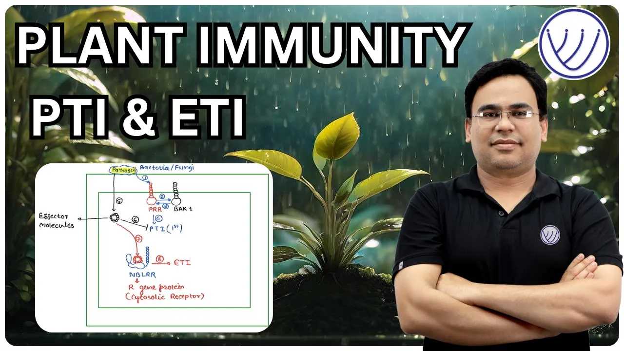
Okay, thank you very much for this excellent talk. We'll now move on to our final speaker of the day. Zhongshou Wu who is currently a postdoc in Steve Jacobson's lab at UCLA, where he's working on epigenetic modifications and DNA replication, repair and transcription. He did studies at Northwestern A&F University in China before going to the lab of Xin Li at UBC for his PhD where he worked on immune signalling NLRs and their their regulation homeostasis, which is what I believe we'll hear about today and so please take it away Okay, great, so thanks for the nice introduction and also all the organisers to provide me this opportunity to present my work here. So, all the work was done at the University of British Columbia [unclear]. How plant immune system works plama membrane localised receptors can recognise the conserved patterns of fungus termed the PAMPs, examples are flagella of bacteria chitin of fungus and the triggers PAMP-triggered immunity, termed the PTI response. However, successful pathogens can deliver effectors to the cell to dampen the PTI response. In turn plant have evolved resistant proteins to recognise the effector and trigger effector triggered immunity, termed the ETI response. The majority of the resistant proteins that belong to nuclear binding leucine rich repeat proteins and can be further classified as CNL, TNL, and RNL based on the difference at the N terminus. Both CNL and the TNL are fast evolving, while RNLs are relatively conserved across the plant species. With the structural biologists help, in recent years we have better understanding how NLR protein works. By example CNL ZAR1 one can form a pentamer upon the recognition of the effector and relocalise to the plasma membrane to serve as a calcium channel. TNL ROQ1 forms a tetrameric enzyme which contains NADase activity there and produce the chemicals to trigger the downstream immune signals.
The RPW8 domain in RNL is similar with the CC domain in structure. And Dr. Jeff Dangl group will help provide the evidence that the RNLs can form as a calcium channel on plasma membrane as well.
Interestingly, the autoactivited NLRs give us autoimmune mutants.
For example, here snc1 is autoimmune mutant. The size of the plant is smaller and it contains curly leaves. It contains a point mutation on a TNL called SNC1 which leading to amino acid substitution between NB and LRR domain. The snc1 autoimmune mutants also show enhanced disease resistance to the Pseudomonas syringae, that's a bacterial pathogen, and oomycete pathogen, Noco2. You can see there's no spots can produce on snc1 autoimmune mutants. At the molecular level, pathogen-related genes are up regulated in snc1 autoimmune the background. The autoimmune mutants are good tools to study the defence response. The reason is the size of the plant is inversely correlated with the defence response. In other words, the smaller the plant, the more resistant it is to the pathogens. So we can use the size as a an indicator of the defence response. In Arabidopsis, there are seven RNLs forming two phylogenetic clades. One clade is ADR1 clade containing three full lengths ADR1 family member and also N- terminal truncated ADR1-L3. NRG clade has two full length NRG and also N-terminal truncated, NRG1C.
So the evolution of the RNL genes is interesting. So RNL how arisen before the divergence of the gymnosperms and angiosperms.
Distinct NRG and ADR emerged within the angiosperm and the NRG genes are lost in monocot and the some dicots. The interesting evolution is that the TNL are absent from those species lacking NRG genes and so indicating a functional correlation between NRG and a TNL. Dr. Xin Li lab will try to understand how actived TNL transduce signals downstream. So Dr. Jane Parker group will help provide the evidence that the lipase-like protein, EDS1 can form two distinct complex with PAD4 or SAG101 that's required for the TNL signal transduction.
So to understand the function of the RNL in TNL pathways, we generated the higher order mutation in the snc1 autoimmune background. So first, we knocking out eds1 or pad4 in this snc1 background. The plant is wild-type like indicating fully suppression of snc1 growth effect. However, when knocking out sag101 gene in the snc1 background, only partial suppression phenotype was observed. So the plant is intermediate and still contains curly leaves. When knocking out to the three full lengths ADR1 members the in the snc1 plant, the plant is wild-type like, showing the fully suppression phenotype. However, knocking out two full lengths NRG members in snc1 background showing the partial suppression. So since knocking out ADR1 or NRG, showing the snc1 suppression phenotype, which indicated the RNL function downstream of the TNL pathway, so now we also call RNLs as helper NLRs. And also based on the genetic data here, we can conclude that the ADR1 helper NLRs work closely with the EDS1/PAD4 heterodimer. NRG helper NLRs work together with EDS1/SAG101 heterodimer. So there are two distinct modules that function downstream of the TNL pathways. So then we are wondering whether they are formal complex or not that contan the helper NLR and the lipase dimers.
So with the help of two graduate students in Dr. Xin Li lab, Lei Tian and Xeuru Liu, we used the IP based labelling, biotin labelling assay to test. In its native state without TNL activation we saw TurboID tagged ADR1-L1 successfully labelled both EDS1 and PAD4. When we co-infiltrate TNL RBA1 in the system we saw more EDS1 and PAD4 are labelled by the turbo-ID tagged ADR1-L1. So that's the EDS1 PAD4 ADR1 module, Dr. Jane Parker's group pursued a similar idea with the EDS1 SAG101 NRG modules.
Without TNL activation, the saw NRG1 successfully pulled down both EDS1 and SAG101. And with the TNL activation, they also saw more EDS1 and SAG101 were being pulled down by the NRGs.
So to summarise the observation from two groups, we found that TIR signals can introduce the complex formation which contains the lipase dimers and also the helper NLRs. However, when you look at the phylogenetic tree, we found that each clade that they are truncated NLRs present, so ADR1-L3 and NRG1C then we're wondering what's the biological function of this truncated helper NLRs. Are they pseudogenes in the genome.
However when we look at the evolution or the NRG1C we found the NRG1C is quite conserved in the Brassicaceae species. And the transcriptome analysis found the NRG1C can be induced by the pathogens. So this observation also proved by our qRT PCR, we found an NRG1C can be induced by the Pseudomonas syringae DC3000 infections.
So this other this data indicated NRG1C might be immune related. To further understand the biological functions of the NRG1C we overexpressed the NRG1C in the snc1 background. So the double mutant is slightly larger than the parent snc1 and also showing the similar suppression level with the knockout sag101 or nrg in the snc1 background. However when NRG1C was overexpressed on top of the snc1 nrg triple or snc1 sag101 background, we didn't see any further enhancement or suppression there. So indicating NRG1C antagonise the EDS1 SAG101 gene modules. And when NRG1C was knocked out in the snc1 background, the plant is even more smaller than the parent snc1, and more PR genes were detected in the double mutant background when compared to the snc1 autoimmune background.
So knocking out NRG1C showing opposite phenotype versus the overexpression of NRG1C. Then how about truncated one in the ADR1 clade that's ADR1-L3? Unfortunately, ADR1-L3 is not conserved across the plant species. And the overexpression ADR1-L3 in the snc1 background, we didn't see any immunophenotype there. However, if you look at the amino acid of ADR1-L3, we found ADR1-L3 contains a full-length NB domain which is different with NRG1C, so NRG1C [unclear] dominant. So well one thing if we truncated ADR1 as NRG1C length, what's the phenotype. So overexpression, the ADR1 truncated one into the snc1 background, we also see a suppression phenotype, so the plant is actually larger than the snc1.
So here's the summary about the truncated helper NLR. So these truncated helper NLR showing expected help to keep the activation in check to avoide the overactivation of the defence response. So here's our truncated helper NLR group.
Again, Xueru and Lei and also PhD student Weijie in Dr. Yuelin Zhang's group. Okay, I would like to say thanks to my PhD supervisor, Dr. Xin Li. And also other members in Xin Li lab.
Special thanks to Xueru and Lei. And also thanks, Dr. Yuelin Zhang at UBC. And also Dr. Oliver Xiaoou Dong to initiate this project. Definitely I would also thanks my funding support, NSERC and CFI. That's all for my presentation. I'm happy to take any questions.
Thank you for the excellent talk. We're just waiting for the questions to come into the q&a chat. Just a reminder, please place them in there. Really enjoyed this talk. I was just wondering. So you showed that if you truncate the ADR1 L3, it turns into a similar negative regulator to NRG1C. Have you guys ever tried sort of the reverse? If you can transplant the NRG1C end terminus from NRG1-1 onto NRG1C to see if you sort of restore it to a fully functional version.
We're haven't tried that way. But to we think probably also showing the similar function there. Because we think these truncated NLRs are helping our [unclear]. Later on we found that the mechanism is this truncated helper NLRs are associated with [unclear]. So it's kind of blocking the association between the heterodimers with the full-length helpers. So that's my prediction it also can occur the immune pathway.
Okay, we have a question from the chat. What is the advantage of these complexes in TIR-mediated signalling?
I think the question is about the EDS1 heterodimer with the helper NLR complex. So we think because there are multiple TIR around 218 TIR genes in Arabidopsis. So if for the transfer signal downstream or separately gonna be cost a lot, but if they merge the two the helper NLR are and lipase dimers together, so they can converge all the signals together to bolster this immune signals.
Okay, thank you. And then one last question. You showed that the NRG1C truncated helper is conserved in the Brassicaceae.
Do you see it outside of the Brassicaceae, more broadly? Or or No?
Actually, we did find that in Nicotiana benthamiana. There's also a truncated NRG. So, we also test the function of this truncated in the Brassicaceae species, we also see the similar function there. So we think the function with these truncated helper NLR is quite conserved across different species.
Okay, and then one last question before we close for the day.
This question is any crosstalk between TIR and CC NLR signalling? So I guess within the context of these helpers, Right, actually, it is so we know that a long long time ago like as a helper NLR are clade ATR1 clade that actually required the for the CC signal as well. However, we don't exactly know why they need it. Since the CC, they actually can from the calcium channel by itself. We are not sure why still need this for the functions, or the ADA, there is a cost out there.
Okay, thanks very much. So thanks again to all three of our speakers for their excellent talks. Thanks to all the audience for attending. And to Cris and Jayson for organising.
I'll now turn things back over, I guess to Cris in reverse order. Thank you very much.
Yes. So thanks to all the speakers that thanks to Thomas for moderating this webinar. The talks are great. It was really nice to see everybody and on behalf of Plant Cell the full editorial board. Thanks so much, everybody for joining us today.
Thanks, everyone, for participating and thank you to our speaker. Just want to remind you all I know there's a question about the certificate, but um, you guys will be getting certificate and will be receiving an email very soon. So keep an eye out for that and then stay hopefully we'll catch you into our next webinar. Have a great day, everyone. Bye, everybody. Thank you. Bye bye.
Up Next

Systemic Acquired Resistance and Induced Systemic Resistance in Plants
@lizbrauer3951
6.6K views•2021-09-02

Bacterial Communication: Quorum Sensing and Biofilm Formation Explained
@JHUAAP
12.6K views•2011-06-28

Enteric Nervous System Explained: The Gut's Brain | Neurobiology Lecture
@alumniu6029
438 views•2018-09-12

Bacteriophages: Earth's Deadliest Killers and Future Antibiotics
@kurzgesagt
34.6M views•2018-05-13
Related Study Plans & Knowledge Roadmaps
Structured learning paths in Biology