




















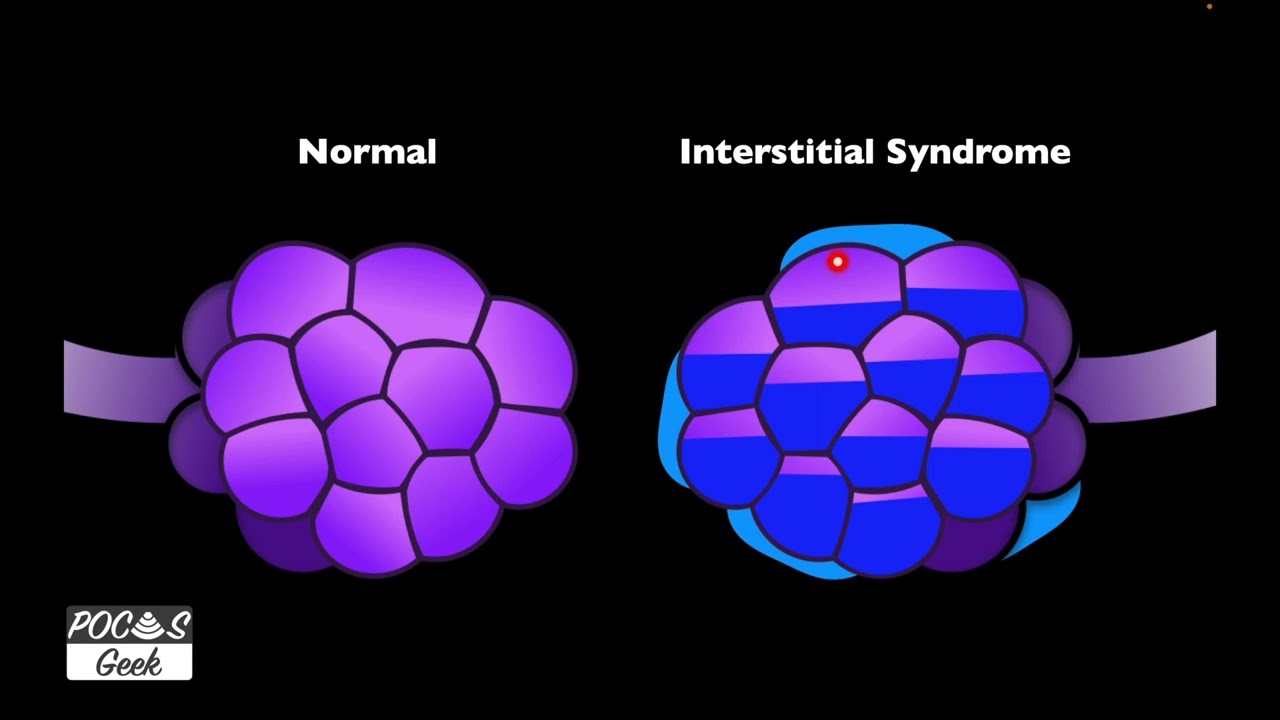
Kerley B-Lines are horizontal, perpendicular lines seen on chest X-rays that indicate pulmonary venous congestion and are a key radiographic sign of heart failure; they appear as white lines extending from the pleural surface at approximately 90 degrees to the chest wall, resulting from fluid accumulation in the interlobular septa due to elevated pulmonary venous pressure from cardiac dysfunction.
Kerley B Lines on Chest X-Ray: Heart Failure Signs Explained for Medical Students
Added:Basic radiological anatomy of the chest, including the boundaries of the cardiac silhouette and normal pulmonary vasculature.
The pathophysiology of congestive heart failure (CHF), specifically how left ventricular dysfunction leads to pulmonary venous hypertension.
Fundamental principles of chest X-ray (CXR) interpretation, including understanding standard projections (PA and Lateral) and basic tissue densities.
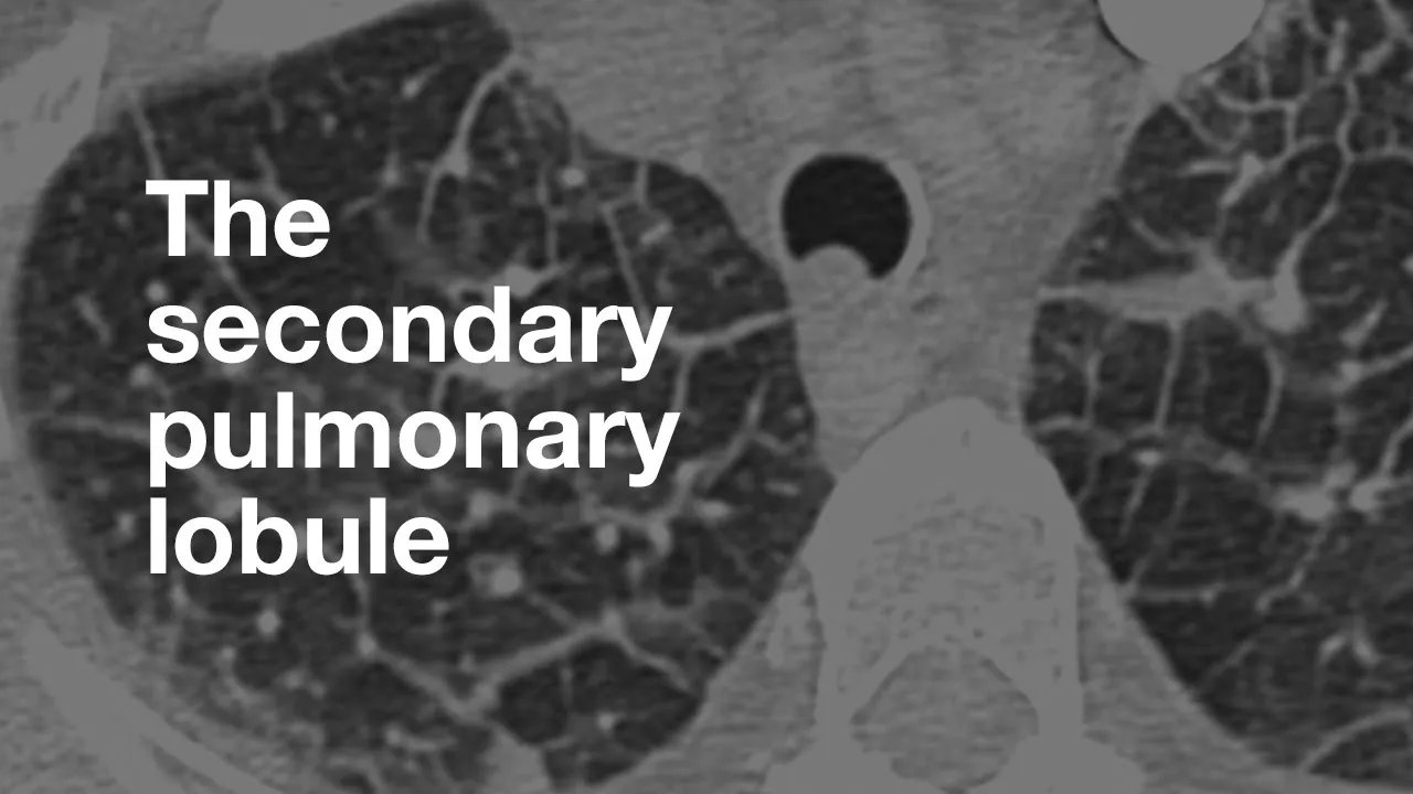
Anatomy of the pulmonary interstitium and lymphatic system, specifically the structure of the secondary pulmonary lobule.
Distinguishing between the progressive stages of congestive heart failure on CXR, from pulmonary venous congestion to interstitial edema and alveolar flooding.
Formulating a differential diagnosis for Kerley B lines, including non-cardiac etiologies such as lymphangitic carcinomatosis, sarcoidosis, and interstitial lung disease.
Clinical management of acute decompensated heart failure, correlating radiological findings with physical exam signs and appropriate diuretic therapy.
Comparing chest X-ray findings with point-of-care lung ultrasound (POCUS) to identify sonographic 'B-lines' in acute dyspneic patients.
23.7K views794likes9:02Original Release: 2020-04-07
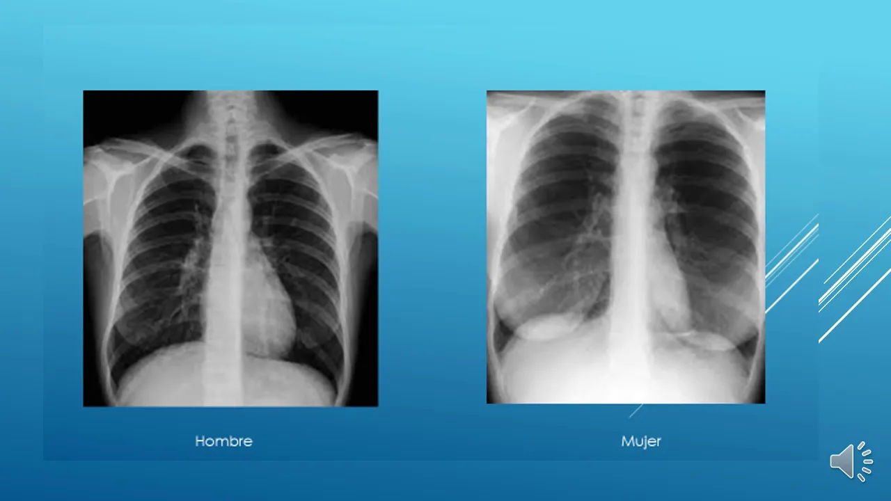
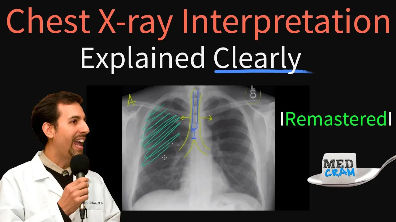
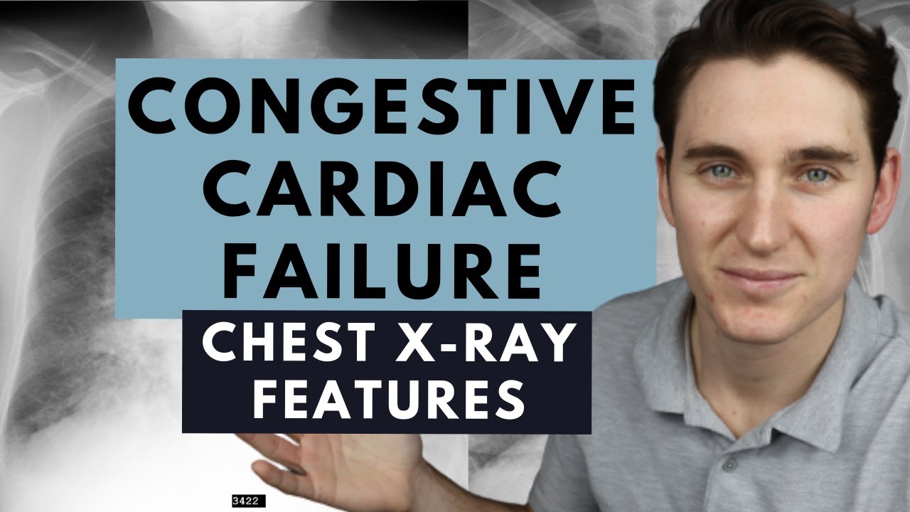
hey you guys I'm dr. Sharma I'm radiologists which means I like to interpret images that's pretty much my job and that's the specialty that I chose after graduating from medical school so I want to explain to you what curly beelines look like and not only that but essentially show you what when I'm looking at a chest x-ray what things I'm looking for what pathologies I see in anatomical structures that I'll assess to start to think hey I think this person has heart failure I think this person's got some of the findings that we would see in sort of vascular congestion that sort of thing so in my previous lectures I sort of go through not only the normal findings normal Anatomy but also some of the pathologies that you should be able to see whether you're a medical student whether you're a nurse whether you're whatever specialty or just watching this for fun some of the things sort of take you in the mind of like what I would look for and if you're gonna be looking at chest x-rays yourself no matter what specialty that you're in what you should also be looking for so first let's start with this chest x-ray which is now a frontal we just have a frontal view of the chest and the clinical history is that this patients coming in with sort of shortness of breath they're not able to breathe as well and that's really all the history that we get so right off the bat I'm gonna just give you a couple seconds or you can pause the video just to go ahead and look at what findings we trying to sort of trying to look at trying to look for I've got some arrows over here on the right and I've also got some things outlined on the radiograph itself but even then you should it's sort of you should be able to look at this and say okay I think these things are wrong what I love to do and what I tell people all the time when I'm sort of teaching some of these topics is to pull up a normal chest x-ray when you're able to pull up something and compare something to normal no matter what it is when you're maybe like scrolling through social media and there's like oh can you tell the difference from this fish to that picture they're not just giving you just the wrong picture they're giving you normal they're making they're making you compare two pictures and saying hey what's wrong with what's going on so over here we have a very nice normal check start you're going to actually go through this in detail in my prior videos that we have the heart which is nice and normally sized and then case and we have the vasculature that we can see but they sort of taper off they're not too prominent the vast feature is not very prominent we can sort of see this here and we see the Carina and the main bronchi as well as the trachea and again the hearts really normally sized that was a little bit off here on the pathological chest x-ray meaning the chest or that we think something is wrong we've got these lines that are drawn here okay and we're obviously trying to take now not obviously but what we're doing here is we're measuring the heart size we measure the heart size in comparison to the the chest no matter what patient it is because every patient is different every person's heart size is gonna be different their chest is gonna be different therefore as just an eyeball measure and sometimes we even measure this on the computer we'll take the measurement or just eyeball the measurement of the thorax the widest point of the thorax if the heart is 50% or less than that we tend to call that normal okay if the heart is greater than that which is definitely in this case if it's greater than 50% of the the widest point of the of the chest we now call that cardiomegaly so we are now concerned that this patient's heart is enlarged for some reason when you look at this nice clear space the nice clear area of the lungs it's nice and black that means that there's a lot of air there isn't too much gunk in there the intra lot of your slept are probably nice and clear there's nothing no really congestion going on that's what this looks like on the normal here I'm sure you can already say there's kind of more like white stuff sort of this just like gunk that's going on I could sort of tell the lines are going further to the end of the of the chest ORS here they sort of yeah they sort of you see them here but they taper off and it just becomes black and you can't really see it beyond that that's sort of just deep more prominent interstitial markings of the lungs in fact someone in some of these areas you can actually see there's sort of consolidate of areas that are starting to develop it's a little bit early but you can start to call these sort of Pachi Pachi consolidations so sort of centrally in addition to that once you start to look at sort of the upper portions of the lungs you see these vessels that are here that sort of look more prominent we do we can kind of see them here but they're not really as obvious this is called cephalization of the of the pulmonary vessels okay so sucessful ization this is not very good at that drawing settle is and you get the point cephalization of the pulmonary markings and finally the most important thing that we are here to learn about is actually let's go through the findings first is these lines right here so these arrows point these out in these lines see this white line here it's very horizontal it's very horizontal and perpendicular to the chest wall you can literally do a 90 degree more or less perpendicular to the chest wall and that's you don't see that in this chest x-ray you don't when you go out to the this chest x-ray it's at the chest wall you don't see these perpendicular lines that are sort of more prominent you're just seeing these out of nowhere like what could really be going on here so let's pull up our question what what most huh so the most likely abnormal labrum normality okay I'm not really sure why I said typed it like that I must have been typing this late or something here was in a rush but a is a white blood cell count is that abnormal is the BNP the brain natural peptide is that abnormal the ACE inhibitor level or a rheumatoid factor so in this case we've got a lot of findings that are sort of pointing towards something we've got cardiomegaly we've got interstitial prominence and sort of these patchy consolidations throughout and cephalization of the pulmonary vasculature and those lines that we were talking about those curly B lines going to explain the pathology sort of the picture of what those are essentially so this is going to be heart failure and therefore this patients most likely going to have an elevated B and P most likely sometimes in severe heart failure chronic heart failure really large cardiomegaly sometimes they start to their heart is just training so much that their BMP starts to clear and sometimes we have like a normal BNP but that's more detail than you need to know so we've cardiomegaly alveolar and history social pattern of lung markings that appear more prominent and then curly B lines so curly B lines so the alveoli are very important and they're all actually organized in a unit called a secondary pulmonary lobule a secondary pulmonary lobule contains the central arterial and then around it is gonna be the venous structures it's actually around the secondary pulmonary nodule that we have the venous structures within the inter lobular septa these are septa that split apart the the secondary pulmonary lobules okay so we have these units within the units are the alveoli okay we have the venous structures which align the outside of this said unit and what happens is in heart failure the heart starts to fail it's not able to pump blood out into the body as well it starts to get bigger and engorged itself and it starts to get full because it's like I really cannot pump blood out the blood starts to pool and starts to increase pressure there's a lot of increased pressure here and starts to pull backwards okay so these are the pulmonary veins and like we said the pulmonary veins are here trying to drain this way but really quite can't to the heart these start to engorge the pulmonary veins start to encourage and then these inter lobular septa start to get filled with fluid and they start to get bigger and bigger until they become prominent filled with fluid and then as you know fluid and those types of mediums were able to see more on a chest x-ray it comes up more prominently and that's where we get these curly curly B lines it's the prominence of the inter lobular septa because of venous congestion that's specifically what that is so if you're seeing these little perpendicular lines along the chest wall think karlie Butte lines let me know if you guys have any questions if there's something I didn't explain too well on this and yeah good luck
Up Next
13:04
Chest X-Ray Atlas: 5 Essential Cases for Medical Students
@Med4VL
28.5K views•2020-07-24

48:47
Graphic Medicine: Comics for Collaborative Healthcare Communication
@nationalpatientadvocate
189 views•2023-12-04

10:28
Neuroanatomy: Central and Peripheral Nervous System Divisions Explained
@AKLECTURES
136.2K views•2014-09-20

3:05
Stages of Labor and Vaginal Birth | Childbirth Animation
@nucleusmedicalmedia
52.1M views•2017-08-18
Related Study Plans & Knowledge Roadmaps
Structured learning paths in Medicine
medicine
Abnormal LFTs: Diagnostic Workup & Pathology
01Liver Anatomy and Basic Physiology
02Introduction to Liver Function Tests (LFTs)
03Hepatocellular vs. Cholestatic Patterns
+ 3 more learning modules
12h6 modules
Explore Roadmap
medicine
Analyzing Blood Smears: RBC, WBC & Platelets
01Introduction to Blood Components & Smear Preparation
02Normal Morphology of Blood Cells
03Erythrocyte Pathologies & Morphological Variants
+ 2 more learning modules
12h5 modules
Explore Roadmap
medicine
Mechanical Ventilation: Waveforms & Alarms
01Anatomy and Physiology of Breathing
02Introduction to Mechanical Ventilation & Core Modes
03Ventilator Settings and Waveform Interpretation
+ 2 more learning modules
12h5 modules
Explore Roadmap
medicine
Neuro Exam: Localization & Stroke Syndromes
01Foundations of Neuroanatomy and Vascular Supply
02The Neurological Examination: Step-by-Step
03Principles of Neurological Localization
+ 3 more learning modules
18h6 modules
Explore Roadmap