



























This video teaches five essential chest X-ray findings for medical students: (1) Right middle lobe pneumonia shows consolidation with air bronchograms and silhouette sign while preserving the hemidiaphragm; (2) Right lower lobe atelectasis presents with triangular density, silhouette sign, and decreased lung volume signs including tracheal deviation and elevated hemidiaphragm; (3) Congestive heart failure demonstrates cardiomegaly, vascular redistribution, pulmonary venous congestion, interstitial edema (curly B-lines, peribronchial cuffing, fissure thickening), pleural effusions, and potentially alveolar edema; (4) Simple pneumothorax appears as a thin visceral pleural line with absent bronchovascular markings and no mediastinal shift; (5) Skin folds can mimic pneumothorax but show bronchovascular markings extending to the periphery.
Chest X-Ray Atlas: 5 Essential Cases for Medical Students
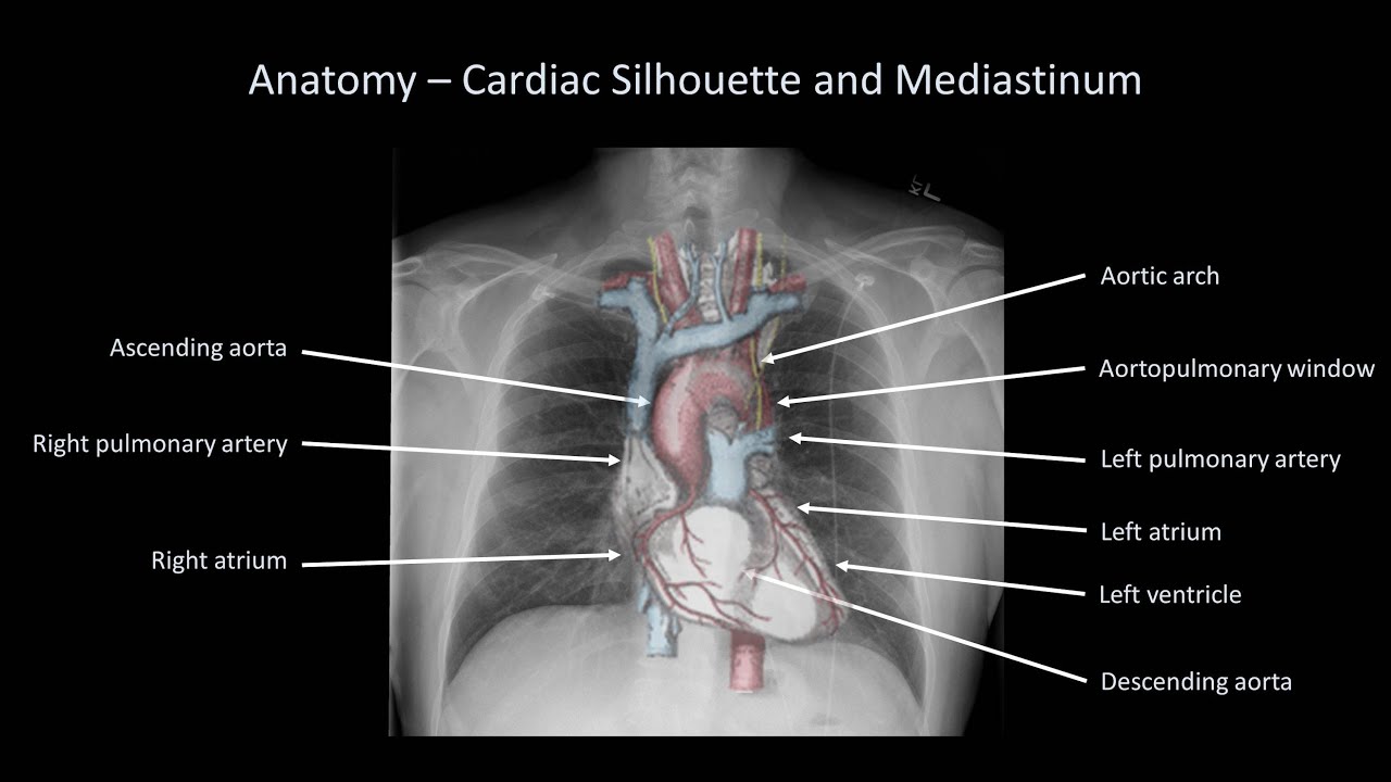
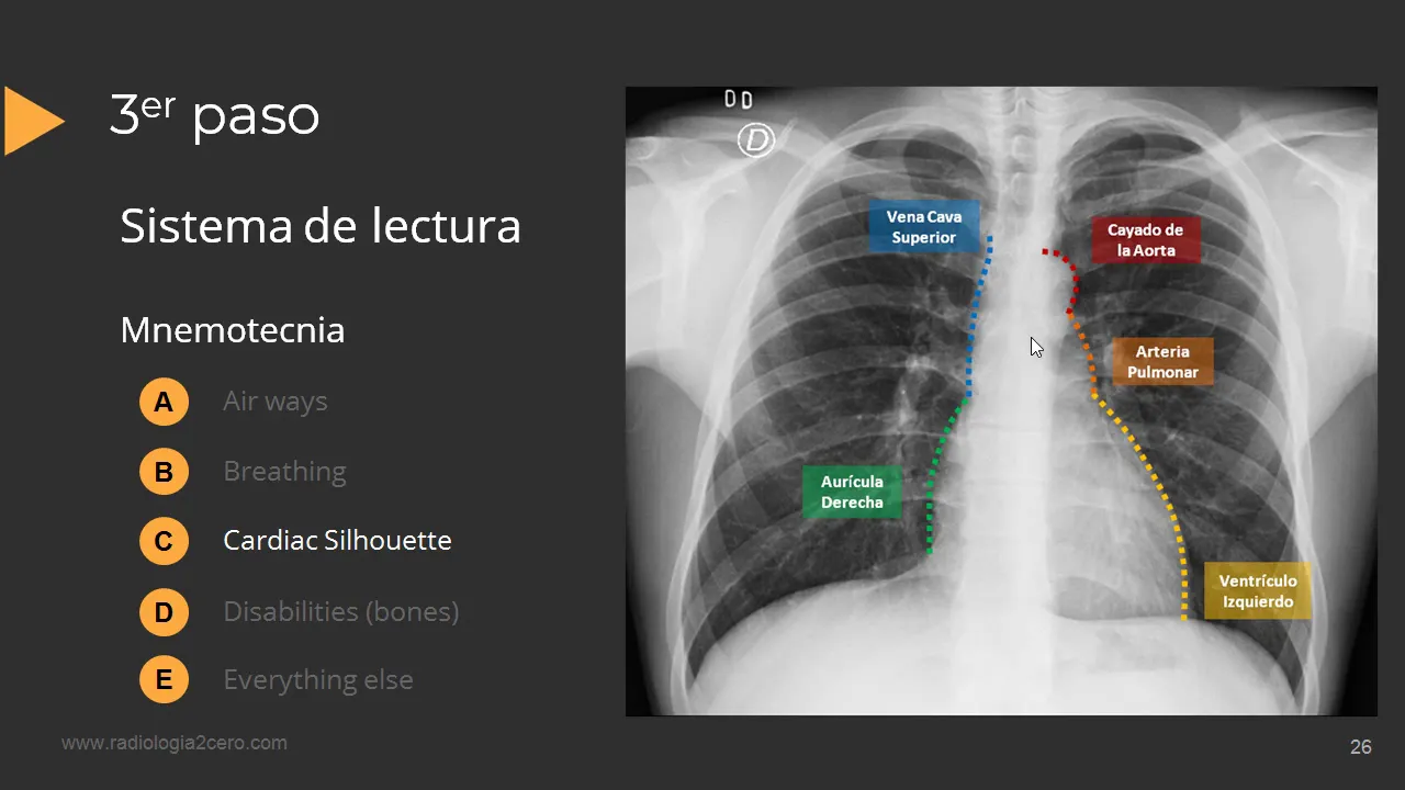
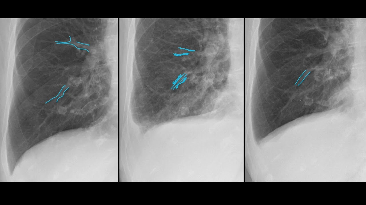
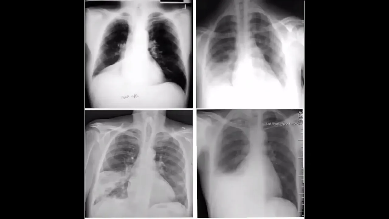
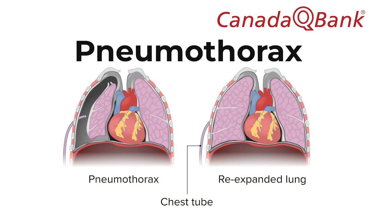
Added:[Music] when presented with an x-ray of a patient with right middle lobe pneumonia the first finding that you may notice is an area of consolidation the presence of fluid density material within the right middle lobe results in a wedge-shaped opacity in the right lower zone adjacent to the heart another finding that is a little more difficult to appreciate is the presence of air bronchograms air filled bronchi that are surrounded by alveolar infiltrates can appear as dark radiolucent branching columns within the area of a pacification consolidation of the right middle lobe results in a placement of the right heart border obscuring the interface between the lung and the heart this is referred to as the silhouette sign lastly note that the superior border of the right hemi diaphragm is clearly defined this helps to differentiate our right middle low pneumonia from right lower low pneumonia in which case the right hemidiaphragm would be obscured so to quickly recap on an x-ray depicting right middle of pneumonia we would expect to see right lower zone pacification with air bronchograms and an indistinct red heart border but with a clearly demarcated right hemi diaphragm i'll also add that unless there is coincidental lectures there is usually no loss of lung volume as well keep in mind that radiographic evidence of pneumonia can worsen during the first few days of treatment and take several weeks to completely resolve when presented with an x-ray of a patient with right lower low betalectusis the first finding that you may notice is an area of increased pacification atelectasis of the right lower lobe results in a triangular density immediately at the right lung base collapse of the right lower lobe results in effacement of the right heart border obscuring the interface between the lung and the heart this is referred to as the silhouette sign note that the interface between the lung and the medial aspect of the right hemidiaphragm is also obscured adolectasis may also result in signs of decreased lung volume which can include ipsilateral tracheal deviation depression of the minor fissure and elevation of the right hemidiaphragm a radiographic sign that is referred to as tentine [Music] so to quickly recap on an x-ray depicting right lower low batalectasis we might expect to see right arizona pacification with a placement of the borders between the lung and both the heart and the medial aspect of the right hemidiaphragm and signs of decreased lung volume such as ipsilateral tracheal deviation and diaphragmatic tinting congestive heart failure can produce a moderative findings on plane chest radiography which findings are present depends on the severity of the patient's condition on an erect posterior anterior chest x-ray suggestive findings include cardiomegaly which does not necessarily need to be present vascular redistribution that is the syphilization of blood flow pulmonary venous congestion pulmonary interstitial edema pleural effusions and in more severe disease alveolar edema which is not clearly evident on this radiograph cardiomegaly particularly in concert with other findings is a suggestive feature of congestive heart failure however keep in mind that congestive heart feeler can occur in the presence of a normal-sized heart cardiomegaly is said to be present when the cardiothoracic ratio is greater than 0.5 that is when the largest transverse distance between the left and right heart borders of the cardiac silhouette is greater than half the width of the thorax in this image the cardiothoracic ratio is approximately 0.6 now you should also be aware that on ontario posterior films the termination of heart size is unreliable due to magnification vascular redistribution is one of the first signs of congestive heart failure increased blood flow to pulmonary vessels in the upper lung zones results in an increase in size relative to blood vessels in the lower lung zones this caudal to cranial redistribution of blood flow should only be inspected for an erect x-ray since equalization of blood flow may occur in the supine position the finding of upper low blood diversion however can be difficult to appreciate on a plane radiograph one other way that it can be identified is by the discovery of a superior lobe artery with a greater diameter than its accompanying bronchus now since it is hard to be certain on this radiograph this example should be taken for illustrative purposes the bronchus is the ring-like structure with a hollow center while the artery is filled in normally the upper lobe artery to bronchus ratio is less than one to one here the reverse is seen with a larger artery than bronchus you may have also noticed the prominence of the hyaluro region and the widening of the vascular particle in this radiograph the fullness of the right hilum is particularly evident whereas the left eye element is predominantly obscured by an enlarged cardiac silhouette unlike with lymph node enlargement in which case the highlight can appear as a large lumpy mass the hilar region in this x-ray has irregular borders due to thickened outward branching vessels the increase in hilar size as well as the vascular pedicle widening in this x-ray are due to pulmonary venous congestion an edematous interstitium is another important feature of congestive heart failure three signs of thickened interstitial tissue include curly beelines peri-bronchial coughing and interlobal fissure thickening curly b lines are fine linear opacities that are only one to two millimeters in width they are typically located peripherally in the lower long fields near the costophrenic angles when viewed close up they can be seen to extend perpendicularly in words from the pleura and are up to three centimeters in length these opacities can also be seen head-on in which case they refer to as curly sea lines or radiating outward from the hilah in which case they are called curly a-lines peri-bronchial cuffing is another discrete finding which occurs due to edema of the bronchial wall when viewed head-on it appears as a donut or a ring when viewed tangentially that is from the side it appears as two parallel lines which to some extent resemble tram tracks note that perry bronchial coughing can occur in other conditions as well such as chronic bronchitis also evident in this image is a thickened minor that is horizontal fissure this finding is easier to detect on a lateral radiograph where both the oblique and horizontal figures may be visible collectively interstitial edema results in widespread blurring of long markings the loss of definition results in a hazy appearance of the long fields in highland bilaterally now let's turn our attention to the bottom right corner of the radiograph note that there is a decrease in definition of the left costophrenic angle a finding that is consistent with a pleural effusion here is another x-ray of congestive heart failure in this case there are bilateral plural fusions which can be identified by the loss of costs for neck angles and obscured hemidiaphragms below the left lung we see the meniscus sign in which the opacity has a concave upper border and is higher laterally than medially alveolar edema is a more severe sign of congestive heart feeler and is not clearly evident in this radiograph that we've thus far been examining in this image many of the aforementioned findings of congestive heart failure are present plus alveolar edema which classically results in central and symmetrical aerospace disease the outer third of the lung is frequently spared resulting in a characteristic batwing configuration however the patterns of pulmonary pacification are variable and can be more or less diffuse and asymmetric or patchy to quickly recap the plane radiographic findings that can occur with congested heart failure include cardiomegaly signs of vascular redistribution such as cephalization and increased artery to bronchus ratio in superior segments signs of pulmonary vascular congestion such as higher enlargement and a white and vascular pedicle signs of pulmonary interstitial edema such as curly beelines peri-bronchial coughing and interlobal fissure thickening pleural effusions and in more severe cases alveolar edema animal thorax refers to the presence of gas or air in the pleural space it is considered a simple pneumothorax when there isn't any mediational shift to the contralateral side and the patient is stable in regards to detecting a pneumothorax the most important aspect is simply looking for it the x-ray may be almost entirely normal except for a small crescent of lucency next to the lung which can be easy to miss can you see a crescent of increased trans radiance in this x-ray you may need to look closely this area of increased blackness represents free air notice the lack of bronchovisible markings within this region a very helpful sign of a pneumothorax is the demarcation of the visceral pleural on the x-ray which is identified by a thin sharp white line this fine hairline opacity occurs between the lateral border of the lung and free air in the pleural space again notice the lack of pulmonary vascular markings in the area beyond the pleural line now if the lung is consolidated then the visceral pleura won't be distinguishable as a separate line but rather as the edge of the consolidation within the confines of the visceral pleural lines you can see long tissue which can be identified by the presence of lung markings the contralateral along however may appear to have more prominent lung markings due to increased vascular flow as mentioned earlier with a simple pneumothorax the heart and mediational structures remain in the natural positions for example notice the central position of the trachea in this x-ray here's a more obvious x-ray of a pneumothorax no lung markings are visible beyond the border of the right lung which has collapsed notice the increased opacity of the collapsed right lung in comparison to the normal left lung as well the right heart border cannot be identified because the collapsed lung is indistinguishable from the heart this is referred to as the silhouette sign and the trachea essential as mediating shift has not occurred normally the chest x-ray is performed with a patient on inspiration can you detect the pneumothorax on the inspiratory x-ray now how about on the expiratory x-ray occasionally the air between the longer the pleural space is easier to detect with an expiratory image to quickly recap with a pneumothorax a thin sharp white line the visceral pleura separates the lung in the free air which is void of bronchovesicular markings as well in the case of a simple thorax there will not be any signs of mediastinal shift now although most x-rays are taking the eurex position before the purpose of detecting a pneumothorax anyway this weight air collects superiorly and laterally and fortunately not all patients can stand so when the radiograph is taken with the patient in the supine position they are actually collected basically which may seem counterintuitive but in that position the anterior and lateral cosmetic sulci are actually the highest points in which free pleural air can collect when air collects in this location it may result in a deep sulcus sign which is an indirect sign of pneumothorax spine x-rays however lacks sensitivity for detecting an orthorex and should not be relied upon if negative if the patient cannot stand then a lateral decubitus x-ray should be taken with the affected lung facing upwards a skin fold on the chest can result in an abrupt drop off an opacity thereby mimicking the radiographic findings of a pneumothorax a skin fold however has a dark band opposed to a light at its lateral contour as well in the case of skin folds bronchovascicular markings extend to the periphery of the lung whereas with animal thorax they do not keep in mind however that apical bronchovisicular markings may be difficult to appreciate [Music] you
Up Next

Chest X-Ray Interpretation for Beginners: A Systematic Guide
@DrJamesGill
66.1K views•2022-04-10

Graphic Medicine: Comics for Collaborative Healthcare Communication
@nationalpatientadvocate
189 views•2023-12-04

Neuroanatomy: Central and Peripheral Nervous System Divisions Explained
@AKLECTURES
136.2K views•2014-09-20

Stages of Labor and Vaginal Birth | Childbirth Animation
@nucleusmedicalmedia
52.1M views•2017-08-18
Related Study Plans & Knowledge Roadmaps
Structured learning paths in Medicine