![[Neurociencias] Procesamiento del Lenguaje (Area de Broca y Wernicke)](https://i.ytimg.com/vi/PdhFGq96eg8/maxresdefault.jpg)










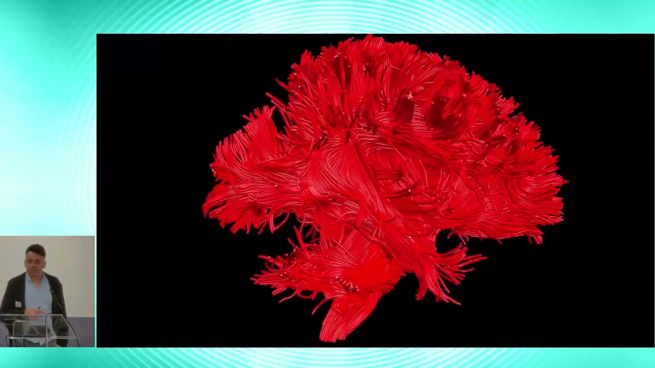

![dr. M. Němý: Co prozradí difuze vody o mozku [Fyz. čtvrtek]](https://i.ytimg.com/vi/WKd2mSnLXLs/maxresdefault.jpg)







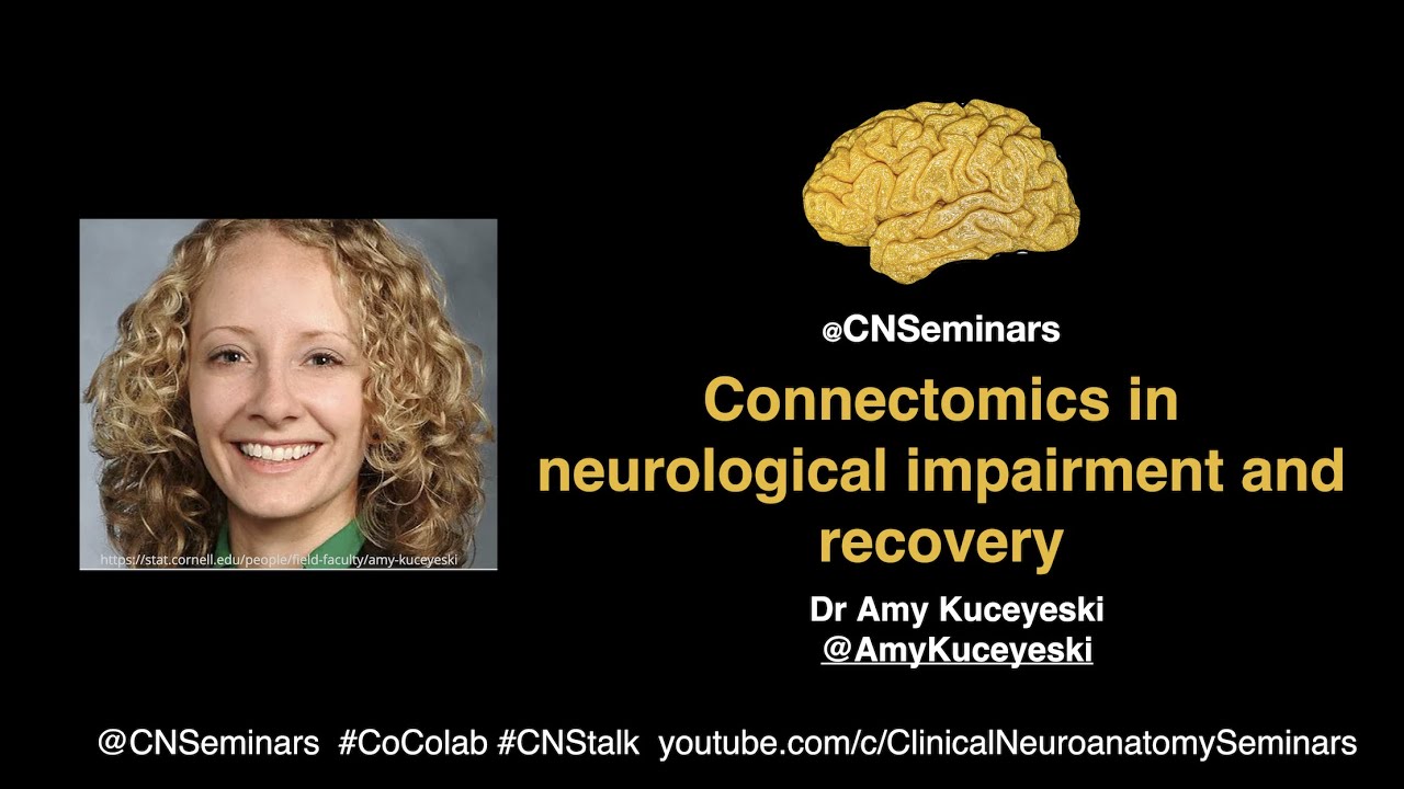
This lecture explores how studying white matter connectivity (the 'disconnectome') provides more accurate predictions of long-term language recovery than traditional lesion analysis. Dr. Forkel demonstrates that individual anatomical variability in white matter tracts—particularly the arcuate fasciculus and its indirect segments—significantly influences recovery outcomes after stroke, with accounting for this variability nearly doubling the explained variance in recovery predictions compared to clinical and demographic data alone. The research reveals that language is an emergent property of distributed brain networks rather than residing in specific cortical regions, challenging classical localizationist models and demonstrating the clinical value of multivariate, multimodal approaches in neuroscience.
Beyond Lesions: Language Disconnectome for Long-Term Predictions
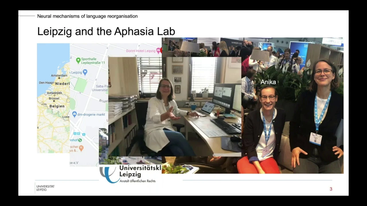
Added:it is actually 11:30 on the east coast of the US OFA so it's sear time welcome everybody in the room Welcome to our online audience um we're really happy to um to welcome our Our Guest uh speaker today coming all the way from the other side of the Atlantic but before I introduce Dr Stephanie forco uh I want to alert you to our meeting into two weeks time on March 7th we'll be listening to Danielle fee who graduated from our very own University of South Carolina and she'll be talking about syntactic processing in bilingual Aphasia so please again uh join us for that one today however uh we are joined by Stephanie forl um Dr forl is an associate professor of psycholinguistics at the rbout University in Nan where she also heads the clinical neuroanatomy and language research group and she's a principal investigator and leader for the language and communication theme spanning Across The ders Institute for brain cognition and behavior the max Blan Institute for psycholinguistics and the center for language studies at rbout University Nan in the Netherlands um Dr FCO got her PhD from King's College in London in 19 in 19 in 2013 and after that she spent uh she was a global citizen uh working in a lot of places around the globe in France in Germany in the UK and also in the United States before landing in a beautiful Nan her research centers on neuro variability and its impact on cognition across different brain States including health and disease and by utilizing Advanced neuroimaging Techniques her goal is to better understand the emerging properties of the brain's connectivity before I hand it over to uh Stephanie um everybody should check out her beautiful website which is apply called www.star.com uh with a lot of information that is of interest to most of our audience uh Stephanie is also the uh the organizer and the I think the principle instigator of the clinical neuroanatomy seminars uh which one can find on YouTube uh so she's a passionate advocate for science communication uh with that I'm going to give the floor to Dr Stephanie farle uh for her talk Beyond lesions unlocking the language disconnect home for long-term predictions it's all yours Stephanie thank you very much thanks for the wonderful introduction uh lovely to meet you all uh in the room and also online and also to see some familiar names in the group um whoops let me TI that works now so um as you heard today we're going to talk about um the language disconnector and what we can learn in terms of predicting um um symptoms long term and you already heard that I'm running a YouTube channel so every now and then you will see a symbol at the bottom of the slide and that means that even though I'm rushing over it to give you an overview of the research of the lab there is a dedicated talk for the slides that have the YouTube symbol now before we uh dive deep I just want to make sure that we're all on the same page and that means starting with Anatomy now um there is not just one type of anatomy there's many types of anatomy and that is partially driven by how we look at the brain so we can look at the surface look at the gy and the susai we can look at section Anatomy so that means either cutting through an actual brain or using MRI images and slicing through to see the subcortical structures the basic ganglia for example but also the gray white differentiation we can look at the connection in the brain and we're going to talk a lot about those uh in the course of the next hour and we can obviously ask participants to perform a task or do resting state functional Imaging and get the um colorful blops on the cortex now I work a lot with um neurosurgeons and I learned they have a different type of Anatomy on top of that which is everything is upside down now the reason it is important to be aware of this is because every type of anatomy may have terminology that differs but they refer to similar brain areas or structures so that is just something to be um aware of when we look at the brain now um a simple question usually this is quite interactive so I'm I can't see you at the moment so I'm just going to give you a little bit of time to choose your number um to the question how how many loes does the brain have and you can see the numbers from one to seven and I hope that each of you there's usually a bit of discussion starting in the audience when I ask that question um but just choose your number for now and I have a feeling that I may know roughly where you uh land on the scale and that is that most of you probably don't choose the numbers one two three but the first hands would go up for four uh five six some for seven and some even need uh the number eight now the reason that we have this discrepancy is because the way we Define the loopes has changed over time so if we look at it if you thought it was four loopes then you are with the original definition from the 19th century where you have the frontal the parietal the oipal and the temporal Lo those of you who said five uh in in their mind uh probably already included the insula lobe and have reached the 70s and those of you who said six uh are in the '90s including the limic lobe and then there is a controversy whether or not there is an additional lob uh referred to as the central L now this is important to be aware of when we discuss anatomical findings just to make sure that we Define what we classify um in our papers to make sure that we agree on some of the principles in the end now what is interesting is that um as we disagree in the field and across the fields um so do the um AI techniques that we use uh do as well so a very simple method of investigating that question is that I asked Google and Google thinks it is for loopes I am quite persistent so I asked it again a couple of months later and Google changed its mind and that is mainly because someone uh published a paper now uh around that time chpt came about so I obviously as everyone else played around with it to see how good chat's Anatomy is turns out uh it is as good as you want it to be if you just keep asking it the same question so this is literally just me hitting repeat on the same question and the first time Chad GPD said there's five loopes then I repeated the question it said four loes and then I repeat it again and then it started apologizing uh and said it is six loopes so um this just shows you that when it comes to Anatomy there is a disagreement in the field on the basic principles of the organization of the brain and that is also reflected in some of the tools that we use now um I hope we're all on the same page now so let's dive into the Beyond delion aspect of this talk so here are a couple of famous cases that I'm sure toas audience do not need an introduction and they're all famous because they presented with a lesion and a clinical presentation that was then matched in a clinical anatomical correlational way whereby for fineas Gau we had the uh frontal L for patients we had the posterior inferior frontal gyrus and for patient hm on the right um we had the bilateral U medial temporal lob for memory so it's personality language articulation and memory mapped onto cortical areas uh in a different L now this is what uh a lot of our field is based on a lot of the brain uh cognition or brain Behavior associations that we know these days are based on initially single case observations and they do have their um validation these days they're still driving our field um but one of the things that quite interesting is if we take exactly the same patients and this is work that Michelle has done about 10 years ago now um so he took the legions from these three famous cases instead of looking at the cortical damage they caused he looked uh using a disconnection uh Approach at the white matter connections that were affected given the presence of the lesion in uh the area they were described and what you can see is that for fia's gate even though it's quite a localized Legion in the frontal L is quite an extensive disconnection that was caused by this lesion where tracts in the frontal L were disconnected but also the onate facus that connects the frontal lobe to the anterior temper L same when we look at broker's patient so the the lesion and Nina drus has beautifully shown that in her MRI study of that patient um Extended into the white matter behind the posterior inferior frontal gyrus and when you look at the white matter that is running underneath that gyrus then you can see that leion disconnected most likely the front asan tract obviously the AR facus the long segment but also the frontal um parital segment of the archit facus and a few more connections that you can see here now for patient hm we can see that the entire limbic network was affected by the surgical lesions that um were affected in his brain now what this shows you is that by losing using the exact same input data we can come to very different conclusions about um the correlation between brain structure and clinical presentation now I want to spend a little bit of time to show you how we can actually study the white matter because there's only two methods that we have available in the human brain and the first one is postmortem so that is called Kingler postmortem dissection and that is a method where um you have a specimen and you go through it I'm going to show you the steps in a minute um but that is obviously a very destructive method now luckily for us uh not too long ago so we were born in the same year um inv Vio tractography uh became available and that now lets us study the white matter Connections in the living human brain now tractography I'm happy to discuss this a little bit has many limitations as do all the Neuro Imaging methods that we have available to us but it is the only method available where we can actually look at the white met Connections in the living human brain and is not destructive which is a big Advantage so I promised uh I show you a little bit of how cling La morm dissections work so this is not how we do it particularly but given the human tissue act I'm can't show you a recording of our own dissection so I took this from a book uh very good book and recommend it references here but what you see is that you start by pulling away or pushing away the gray mattera and in order to do that you have to prepare the specimen in quite a laborious way which ideally lasts about 3 month where the brain gets Frozen and thought in a repetitive cycle and that makes the gray mattera a bit more fragile and you can then peel it away and you can see that you can expose those beautiful white matter connections underneath and you can see them with uh the naked eye as you can see in this example now what does it take to do a cling Laos morm dissection well it takes a good specimen as I just said but also quite a bit of patience it's not a quick and easy method to learn or to master um so we usually take about half a day to go lateral to medial and half half a day to go medial to lateral in the end you end up with a very thin slap of uh white matter what you can see is that the instruments you need are quite straightforward and everyone who does Kingler postmortem dissections has their own way of um preparing their spatula for example so some like it a bit thinner some like it a bit thicker personal preference but you get used to it um and then ideally but not necess necessarily the use of a microscope is quite helpful so uh you start from the surface of the brain and as always you begin with the anatomy so I give you a second to identify a couple of structures on this brain um as you can see is not a textbook brain even though this brain had no um um brain pathology at the time of death so this is just normal variability that we can see and if I now give you couple of pointers here so we have the syan Fisher or the lateral Fisher underneath the superior Tempa garist and the superior Temple Circus this is where we usually start in the depth of the circus to peel away the cortex and then you go around and do a SE shape around the entire brain and there's the big and a small sea along the sorai and when you do that you end up with the specimen that looks like this now what you can already appreciate is that finding your way around gets harder and harder as you go further so I'm just going to give you a couple of pointers of where we are obviously we can still see the lateral fissure quite nicely but the rest of the landmarks are getting a little bit harder to see now in terms of the white matter that you can see first the white matter is nicely layered in the brain so the first one that we can see is the archid fulus that is arching around the syvan fisser and we can see some u-shaped fibers that interconnect neighboring gy now in The Next Step I'm going to cut the archet fulus and g go one step deeper and now you can see that we exposed the ventral Network as it's often referred to that includes the inferior front otive fulus the onate facus we can see the beginning of the internal capsule and then also towards the occipital L you can see quite nicely into specimen the vertical occipital facus now we do this uh every year in in a white mattera dissection course and the Beautiful advantage that that gives you is that you see at least 15 brains in one go and what you can appreciate is that when you look at the brains they all look different so here's one example of two randomly chosen brains and if we just look at the um lateral fissure and the super marginal gyrus that sits at the end of it you can see that the two right hemispheres I'm showing you here have quite a degree of variability both in the fissure as in the gyrus now this is not um unheard of or uncommon and I'm going to show you a couple more examples so you may have heard about the uh inverted Omega the hand knob area so again giving you a second to find your way around we're looking from the top at the brain here and I'm just going to show you where the central circus is and both uh um examples here and you can already appreciate that there's variability between the two brains but also between the left and the right Hemisphere and if we look at the hand knob area or the classically called inverted Omega you can see that in the far left it is actually a textbook in Ed Omega but that is not the case um for both Hemisphere or between people now another beautiful example uh that I was lucky enough to get from our colleague Pascal Belin and France is the um anatomy of the superior temporal circus here so you can appreciate the variability in the structure but also he mapped The Voice selective areas in the auditory cortex so you can see that there is a huge degree of structural variability but also a huge degree of functional variability now the auditory cortex is not the only primary cortex that shows such a degree of variability if we look at the visual cortex for example using a different method so this cytoarchitectonic mapping now you can see that um there's quite a degree of variability between the left and the right hemisphere in the same brain but also across uh people and similarly we see that also for um the areas 44 and 45 in the posterior inere frontal gyrus now I could go on and give you many many more examples of this so this is just to say that this is not the exception but the norm and that led um killas and Katan Ammons to conclude that variability is not noise but rather that it is actually an invaluable um principle in the brain to understand the principles of brain Evolution and development and for interpreting statistical maps in task-based functional Imaging studies now we don't account for variability in your Imaging as much as we probably should and there's various reasons for it and the field is moving more and more into capturing this variability and trying to statistically account for it but I want to give you a toy example that hopefully makes it uh a bit more tangible of um what the problem is when we look at group studies rather than variability so here is a uh slide of a couple of women anyone in the room or online knows who these women are no no one brave enough to speak up fair enough um so what you see here is uh an overview of all the women who won a Noel prize for uh physiology and medicine and every year I'm hoping to update it um but it hasn't happened uh this year so we'll we'll see if we get lucky next year um but yeah so these are all the women that won the Nobel price and I asked my student to create an average face so we know what you have to look for uh or look like when you uh want to win the Noel prize as a woman and here we go this is the average face now uh what you can see is a couple of things here number one is the algorithm worked we have an average face fantastic two eyes a nose and mouth roughly in the same place um so that is a good uh sanity check but what we can also see is that the face in the middle doesn't actually look like any of the women around it nor do any of the individual women around it look like the average face in the middle now while this is extremely obvious when we look at a face it is a lot less obvious when we look at the average brain so this is the m& template and we can see areas here that are very sharp and crisp so for example the cordate or the basic glia um they have sharp boundaries um and that means that there is less variability so we tend to be more alike whereas areas where we tend to be more variable when you look for example at the tempal um cortex here the image becomes a bit more blurry now I've already shown you um examples of the variability in the cortex so this is the inverted Omega example again but the same variability or the same degree of variability can also be seen in the white matter so here are two randomly drawn healthy participants from a local data set here in n and we mapped the extended language Network and we can see that when we look at the onate for example in the top image is barely visible whereas in the bottom image it's extremely prominent and similarly when we look at the anterior segment of the archid for example there's quite a degree of variability now these are obviously handpicked examples so we wanted to know what that looks like on a group level and this is work that polar croxen was leading on um where we mapped the variability of the white matter across the population and what we can see is that variability is not homogeneous across the brain so you see this beautiful gradient here where areas that are deeper inside the brain or sort of more older parts of the brain have a colder color and that means there is less variability or if you flip it around there's more similarity between us and as we go out there is more and more variability now this is important for various reasons this is important for example if we use an atlas so if you use an atlas in the center of the brain it's fine because we're more alike but as you go out to the surface of the brain it becomes more and more difficult to rely on an atlas approach and this is in the healthy brain and obviously in the pathological brain this is exaggerated so here I'm giving you one example of um one of our tumor patients so in green you see the brain tumor and we wanted to know where the cortical spinal tract is so we did a um um invivo dissection using tography that's what you see in blue and you can nicely see the Fanning of the cortical spinal tract and then it consolidates towards the internal capsule now when we used an atlas and plotted the atlas onto the patient brain what you can see is and read the outline here that the atlas would have completely missed where the actual cortical spinal tract is so this is quite important to be aware of especially in a clinical setting now as I mentioned um um we only have those two methods available to us to look at the white matter of the brain but there's many other methods available in other species and when you apply them you can see that our understanding of uh other behavioral and cognitive systems like the visual system here or the auditory system are extremely detailed so here every single box is a cortical area and every line is a connection and you can see especially for the visual system that is a very dense plot now when you open most the textbooks to look at what language looks like in the brain then most of textbooks certainly still have this uh rather outdated image with two cortical areas and one white matter Connection in between now tractography has done a fantastic job in mapping a more extensive white meta Network beginning with a validation of the original description so here on the far left you see the arch phicus the long segment as it's called or the frontal temporal connection so the direct connection between the frontal and the temporal uh cortices and then the early 2000s Marco katani used tractography to show this indirect segment that uh relays in the inferior parial and these two branches are either referred to as anterior and posterior segment or horizontal and vertical connections now since uh the early 2000s many more connections have been implicated to be relevant for language functions and that is through um other means of testing the function for example during brain surgery using direct cortical stimulation now what we did uh recently is we dissected the extended language Network into two large data sets and then we were interested in the cortical projections sorry of these white meta tracts and when you do that uh number one is that these tracks are all bilateral so we find them in the left and the right Hemisphere and when you project them onto the cortex you can see that again there is a degree of variability so you have some hotspot areas where they very reliably project to shown in darker colors here so in red and areas where there is slightly more variability between people sorry now what you can also see is that all the white matter that has been associated with various language processes is projecting onto most of the cortex in the left and the right hemisphere so not just the classical definitions of Brokers and veric a fasia uh area and that is so much so that in the beginning we thought we just plot them all on one brain and it was so busy that we couldn't do that so we had to split it up for um each segment um in the language Network now as I mentioned they are bilateral tract so we were also interested in looking at the asymmetry and what you can see is that there is no consistent pattern of uh asymmetry across the language Network so some tracks tend to be more right lateralized others are more bilateral and others again are more left lateralized and what we were also interested in and this is work done by Lil Doan in the lab is to look at the variability overall so not just um per tract but also which side of the brain when it comes to the language network is actually more bearable and is that specific to certain tracts so this is work that Lil did and this is the result of it so what you see here is that overall there is a difference between the left and the right hemisphere so the left hemisphere tends to be more variable than the right and also this variability can be measured in a tract specific way now Peter haard and I recently uh triy to put that all together and come up with a uh new model anatomically based where all these tracks that you can see here have been implicated to be involved in some kind of language processing and here we're applying a very broad definition of language in the brain so the vertical exhibitor facus for example in green um towards the right of the brain has been implicated in reading the archid in uh learning a new language and motor phic mapping um the front asan tract in initiation um and sequencing so there is many many tracts that have been implicated for some or another um language process and on the right you see pretty much the same image but we try to show uh the anatomical areas that are involved so again the boxes are are the cortical areas then in pink you have the subcortical structures that have been implicated and then the lines are all the white met connections that you see in the middle here of the image so this is the anatomical part um we now have a fairly good understanding of the extended network of language in the brain and we know that there is a huge degree of variability both at the cortical level but also the sub iCal level and then the next question is does it matter is it relevant that we are actually different when it comes to our brains so we um embarked on a meta analysis of 326 studies that took uh as quite a while to review but we learned a great deal and the first thing that we learned is that most of what we know about the function of the white matter we actually know from Clinical population so the majority of studies were conducted in neurological and Neurosurgical patients and about 29% in psychiatric patients and only 25% in healthy participants the other thing that we learned is that there are certain tracks that are superstars in the field like the cortical spinal tract and other tracts that are quite um neglected like the aumo frontal pathway for example now there is obvious reasons why you have this discrepancy and that relates back to to um how prominent these tracts are so the cortical spine tract is obviously much larger than the frontal pathway and therefore also slightly easier to dissect now the other thing that we learned is that these tracts tend to be prominent in one field more than the other so the cortical spine tract is extremely prominent in neurology and neurosurgery um for obvious reasons because neurosurgeons obviously want to try and avoid causing any motor deficits so they always need to know where the cortical spinal tract is but um if we look at the inferior longitudinal phicus for example that is primarily studied in healthy participants especially when it comes to reading now as a Next Step what we did is we looked at the functional correlations for all of the tracts that we looked at and in total there was uh 36 tracks I believe so has quite a substantial uh supplementary material in this paper but I'm just going to zoom in to my favorite tracks which are the arids and what we can see is that the Arid does correlate reliably with language but not exclusively so you can also see correlations with sleep memory executive functions auditory functions attention and motor functions and that is a pattern that we have seen pretty much across all the tracts that we looked at so there is uh no one track one function Association um which again brings us back to the idea of localization ISM in the brain so initially stating um when we look at the famous cases that I opened with that one cortical area is involved in one um function and we're moving away from that on the cortical level and we started in a similar space with the white mattera where one tra like the arit is the language tract but in fact when we look at it it looks like that the white matter is recruited for multiple functions but partially specialized in certain functions so at this point we know that we are different in terms of our anatomy we know that this is also relevant when it comes to mapping functions and now the question was um if we can use this information in a clinically meaningful way so what we did in this particular study is we uh recruited acute stroke patients who had a lesion to the left hemisphere and we recruited them in the acute stage for Imaging and Ne psychological assessments and then we followed them up six month and also a year after symptom onet and what you see plotted here is on the x-axis the size of the archid facus in the healthy right hemisphere so the leion was on the left and we looked at the archit on the right plotted against a recovery 6 month after symptom onset now the blue box on top is where you are above the cut off line so these uh three patients that you can see there are officially recovered back to normal 6 month after symptom onet and I'm just going to highlight three patients for you here so patient number one you can see on the far left didn't really recover well 6 months after symptom onet and when we look at the archit phus in the right hemisphere it is rather thin and small and as we go up to Patient number three you can see that the archid is very thick and strong in patient number three now what is interesting about this study is that when you only look at clinical and demographic data we can predict about depending on the studies you're looking at between 30 to 40% of the variance in recovery Now by simply adding the variability in anatomy in the archid phus in this case we can nearly double that um explained variance so that is uh quite a strong indication that looking and accounting for variability is actually um quite useful in a clinical setting now another um study and type of a fasia in particular that we were interested in was conduction of fasia so conduction of fasia is um characterized by fluent articulation intact comprehension but one of the things that doesn't work for example is repeating what you heard so the information that is processed in the um auditory cortex cannot be sent towards the frontal L for articulation and this is one of the classical disconnection syndromes disconnection because it was hypothesized that the frontal infer frontal gyus is intact that the superior Temple gyus is intact but the communication between the two particular via the archid forculus is interrupted now with the emergence of CT and MRI imaging that narrative shifted slightly towards the inferior prial lob as studies showed that you can have leion to the infer Ral lobe that is not affecting the arute fasiculus and still have um a clinical presentation of conduction of fasia so we wanted to look at that um in a multimodal Imaging uh Paradigm so we had tractography data but also cortical morphometry data in patients with primary Progressive aasia and the sister cohort um from the Chicago team and we looked at the a uh not asymmetry the the atrophy In classical language areas and also the three segments of the archid phicus now the first thing that we saw quite to our surprise is that the size of the Arid forculus so classical long segment did not correlate with repetition deficits at all but the indirect segment so the um anterior and posterior segments did correlate with repetition deficits when we looked at the cortical level across all these regions that you can see on the top here the only one that survived multiple comparison correction was in fact the posterior super marginal gyus or the inferior parial Lobo now what that meant is that after 150 years of the archid fulus uh or disconnection of the archid fulus leading to conduction and fasia we took the arch phicus out of the equation and just kept the indirect segment and proposed a new model whereby lesions can occur anywhere in this network including the inferior frontal gyus the inferior parial or the temporal and the connections in between to cause repetition deficits now obviously depending on where in this network the lesion will occur the flavor of the repetition deficit will be slightly different now um a couple of years ago our I presented this at a conference and one of the neurosurgeons in the audience was extremely excited about it because he actually observed a similar pattern in his uh patients when he stimulated the uh inferior parial lobe leading to um repetition deficits so I'm going to show you the video very briefly and I hope the audio Works um but for those of you who are a bit squeamish if you want to close your eyes close your ears for about a minute um now is the time to do so and then just get someone next to you to pinch you when it's over for everyone else uh what you're going to see in a second is a Neurosurgical intervention where they try to map language deficits uh across the cortex and um as they stimulate the cortical areas they ask the patient to um repeat what they heard so here we go today is a nice day today is a nice day I like watching movies TR there's there's she's fluently having a problem there yeah no EGS hands or buts okay so actually get one you put that's on four yes four just right repetition that's DCS yeah yes house okay let's try again today is nice day nice day bit okay so what you can see here obviously finding your way around anatomically is a bit difficult but if when they stimulated the inferior parial lobe that patient quite consistently had repetition deficits now what I've shown you so far is that we can look at individual uh brains and particular Health uh individual um patients and dissect the white matter that we're interested in but we can also take a different approach that is known as the disconnect them where you delineate the lesion bring it into m& so a standard space and then plot that lesion onto X numbers of um healthy connectomes or attractor stams so these uh are taken from data sets like the human connectum project and as you bring the lesion into each and every single one of these normative connectomes what you then can see is the estimated um reconstruction of the streamlines that are affected by the lesion now we can then binarize this per uh individual connecto and come up with a disc connector map that gives you the probability of these white matter connections to be um affected by a given lesion now this is particular uh interesting to do when uh you have patient data where you don't have the diffusion weighted um Imaging available so you can't do tractography in the indidual patient but you always will have a CT or a T1 scan that you can use for this uh disconnector analysis now we um applied this to data of conduction of phasia so first taking all delions and plotting delions to see where the heat map indicates the area of most overlay and not surprisingly we see the posterior temporal inferior prior region here and when we do the disconnector however we can see that the white matter that is affected is extending far beyond that particular region um even into the the right hemisphere in quite a few of these um patients now this is uh an example dedicated to one clinical presentation but Leah tootsi recently did this beautiful work where she took over 1,000 stroke patient lesions and mapped the disconnection of all of them and then used an embedding dimensional reduction approach to Cluster delions or the the connections disconnection profiles for every uh stroke patient so every dot you see here is an individual stroke patient and the pattern of disconnections here are clustered based on similarity so if they're closer together they tend to be more alike and if they're further apart they tend to be more uh distant now we were then able to combine this embedded space with neuropsychological data that was provided by Macio caretta and map the um different neuros psychological assessments in the same space and this allows us to relate the um pattern of disconnection caused by a lesion with the neuropsychological assessment of the neur neuropsychological cognitive behavior battery that was applied and in total there was over 80 assessments available so this is quite a substantial data set and what this allowed us to do is to predict long-term recovery meaning that uh given leion of a patient you can map the disconnection the pattern of disconnection you project that into this embedding space and that allows you to get a prediction of where that patient most likely will be in a year's time now all of this we uh made available on a website called the disconnector symptom Discoverer and the video shows you how it works so you see the space that I've just explained to you where you have the different uh dimensions and every single dot is one of the patients and then you have for every test available in the battery you have a description of how it was assessed and what you have and it's quite as I mentioned a substantive list of neuros pychological assessments now on the website you can just upload a leion mask it will calculate the disc connection so the all the white matter that is most likely affected by this lesion and then you let it run and this video is in real time so this is uh how long it takes you so it's quite fast it will give you the prediction of where that patient is likely going to be in about a years time time across all these neuropsychological scores and you can just export this as a Excel fire and take that further for your statistics now this approach was recently um validated out of sample by Tom hope and Kathy Price from UCL that used the same approach on the independent plur data to predict language recovery long term now the other thing that we can do with this approach is we can actually map a neuros pychological white meta Atlas so the map in the middle is probably slightly unfamiliar to most of you but it's a crosssection um through the brain so it's an axial slide looked at from the top and in the middle on the top for example you see the Corpus colome and then for each of these connections you have the most uh prominent Association of the neurosc logical scores and this is a dynamic map so I'm obviously just showing you one slap of the atlas but you can scroll through and see how these networks change um throughout the brain now if we put this all together uh which is what Michelle and I recently did is is leading us to uh an understanding of language being an emergent property of the connected brain um and I'm going to spend the rest of the time that I have to slightly disentangle what we mean by that so language is by no means the only emerging property in the brain but probably one of the most elegant ones that we have now what we did in this uh review is that we looked at the classical um neurobiological model where you have the uh posterior inferior frontal gyus the temporal gyus and uh also the inferior parial and you have this Association a classical localizationist uh view of one region one function now when you do that model with three regions the computational flexibility that this model provides is quite limited so our complex and dynamic interaction uh style that we have when we use language uh wouldn't really be explained by just having those three regions available similarly when we connected them in the uh early uh 2000 model where you have the direct segment of the archit and then the indirect route again the flexibility in the system is too limited to explain language processing as we know it so what we proposed um and as you can see in this toy model it's not the proper anatomy of the model that we think it is but we're still in the process of mapping the actual Anatomy to it is that language is an integrative function of the brain or IM mergent property of the brain whereby cortical regions and white matter connections get dynamically recruited for different linguistic processes as they are needed and together they produce what we refer to as language now I still find this quite abs ract so I came up again with a toy example that hopefully makes this slightly more tangible and the best example that I could find is this one so if you ask me where language is in the brain that question is equally hard um to answer than asking where music is in the orchestra because music is not in the individual uh musician it's not in the conductor it's not in the instrument it's not in the notes it's in all of them coming together in a timely organized fashion that music emerges and do as similar to what I believe language in the brain is like and with that I would like to thank my team and look forward to your questions still there yes hello Stephanie I'm so sorry I was just late so you didn't hear the Applause you got from our room but I think um if you have your cam on you could I don't know what what kind of view you have you would be able to see our room so I see the room yes thank you so much for your talk um let's start with uh questions from the room while people uh online uh please feel free to start typing your uh your questions thank you stanie this is uh wonderful as always um and very interesting uh to especially see the neuros pychological aspects of all this so I you know I I appreciate the point you make about individual variability right and your example of the average face not reflecting any of the individuals but it actually does depending on what you're interested in right so the average face has two eyes it has a nose it has a mouth and those are very important essential functions of a face you know to produce speech or to breathe or to you you know look at people Etc right so I mean I think isn't it so I guess the question is like how you know relevant is that critique if you're trying to understand certain essential functions that are shared like if you're looking at the brain yes of course there's variability but how many people lack a superior temporal gyus right or how many people lack an arch fulus altogether and so I guess my question I want to push you a little bit on that is is there a can you and obviously the individual variability is relevant for things are recovery which you show very well but if you're trying to understand basic architecture or functionality is that a problem to ignore that variability um very good question and obviously I have thought about this quite a bit um and the answer is it depends on what we look at in terms of the processes and what the aim is if we want to have a general understanding of roughly where things happen in the brain then you can look at the group level there's absolutely nothing wrong with that and then the inter individual variability doesn't matter as much but what we see is that a lot of the models that we have available are based on for example fmy average data and when we bring that back to the individual especially in the clinical setting then these Maps don't inform us where language necessarily is and while if we do a healthy control um student study where we just want to see where certain processes light up it doesn't matter to get it wrong within a like margin of error but if you come to a neuros surgon then you better get it right so it depends on what we're interested in and what the aim of knowing is in the end um but depending on where you are you can use the full spectrum of the methods available and they all have their Merit and their limitations obviously a technical question um you show kind of a standard tractography um where you imported or overlaid like the lesion on top of the the maps um is that from a specific tool that uh you can access or like where where is that found um so that's on the bcbl lab.com website or on my website under the open data I think the writer is called uh you can find the toolbox it's part of the BCB toolkit it's called the disconnector maps um and the data so the individual connect homes that I've shown um are taken from the human connecton project and they're also individually available on the website so all of this is uh openly and publicly available so yeah B have more question I probably keep talking about this for hours so this is a testament to how interesting your talk is but um how do you distinguish between complex and emergent right so you talk about language as an emergent function but why not just say it's complex there are multiple underlying components you have to integrate them all together to understand the whole picture what makes it emergent as opposed to Simply complex um very good question there's lot of people uh trying to figure this out at the moment um the difference that I would see as the most prominent difference is that if it's complex we could potentially decode it and explain the variance as we go along oops you're moving but the emergence is in the fact that we can't map the emergent function per se it's in the inter direction of processes and that to me is the difference if it was complex we could at some point with enough data time and resources and sophisticated tools we could map the individual components of a complex function but the emerging function is in the interaction and therefore a lot harder to capture thanks Stephanie I want to go to some of the online questions to give them a chance as well uh we got a question from uh Peter prman or Peter prman given the association of repetition the phological loop and working memory can you please further discuss the specificity of the described conduction ofas Network for language versus working memory more generally yeah so um there's the as I said there's there's different uh linguistic processes that have been mapped and the classical study that Marco has done for example looking at the archid facular as a language pathway was using the California verbal learning test which has this verbal working memory component to it um that was mapped onto the uh long segment and we're still in the process of really disentangling all the individual segments um of the archid facus but also the extended white matter Network in terms of which processes um rely on which connections so uh the concrete answer is still to be seen um but we are getting closer to mapping um more comprehensive functional structural Network when it comes to the white matter great thank you very much and we have a question from Dorian pustina who actually also recognized one of your Nobel Prize winners so he was actually able fantastic excellent work Stephanie have you noticed any discrepancy between the virtual track reconstructions and the tracks found in the sections in in terms of presence or consistency very good question um as a bit chicken and egg question so um when we started out with tractography cling Laos morm dsection was seen as the gold standard to validate the white meta connections that we see in the living human brain using tractography and what we have learned over the years is that postmortem dissections and invivo tractography gra y by enlarge suffer similar limitations in their methodology so to give you one example and you've seen that in in the beginning of the talk if you want to dissect out the white mattera using um cling Laos morm dissection you start off by taking away the cortex and you go layer by layer deeper into the brain to expose the white matter underneath now that means that by virtue of the method is very hard to say where the white matter is terminating similarly with tractography the closer you get to the cortex the harder it is to uh track the streamlines now this is improving as we go along on both accounts so in the white mattera world there is now beautiful work done where you get a digital reconstruction and you take images at every step of the dissection and then afterwards you can reconstruct it in 3D and try to um scroll through and see where the white meta most likely terminated in the cortex and also on the invivo side we're getting closer and closer to cortical areas um but there is several limitations that make it difficult to uh cross validate the two methods now in terms of um the difference that we see it's limited in a sense that if if you look at the core of the tract you find a similar reconstruction in postmortem and in Vivo dissections but when we look at the archit for example um if you use a um DTI algorithm compared to a spartic convolution algorithm where that track projects to varies a great deal so with classical DTI it goes to the ventro premotor and the posterior inferior frontal gyus with a more advanced tractography algorithm it goes all the way nearly to the frontal Pole now there is no way in knowing where those connections actually terminate if it is the tractography based on uh DTI or SD or in fact somewhere in between we can't tell based from tractography but also Kingler poort section doesn't help us to disentangle that so the similarity in the core is given but as we go closer to the cortical regions it becomes harder and harder um to be sure that you can validate it now another thing to bear in mind is that there's different types of tractography so some use a methods driven approach and others like the ones that we are using are actually validated for the last 20 years on postmortem dissection so they're in engineered to show you reliably what we know from postmortem n sections thank you very much and we have a question from kak Neu um following up on the response to Will's last question emerging versus complex is this a suggestion then that we should not be ascribing any more specific processes to specific cortical regions good question um so as with many things the truth is probably somewhere in between um certainly it's not a one Function One cortical region um Association across the entire brain there are certainly cortical areas that are specialized or primarily serving a given function um but doesn't apply to the entire brain now on the flip side I'm not saying let's get rid of all cortical functional Association and every part of the brain can just chip in and they're all equal in their um functioning and relevance that's not the case we know there are certain patterns and for example depending on where a lesion roughly occurs in the brain will have an impact on the types of aases that we can see so um yeah the the truth is somewhere in the middle that not every cortical area is engineered towards one function but also it's not the entire brain that is completely completely flexible and plastic and can do everything thank you Stephanie I think that's a good note on which to uh end our uh your talk and our discussion for today thank you so much and thanks to the online audience for attending and we hope to see you next time thank you bye
Up Next

Broca's Aphasia Explained: Stroke Survivor's Experience
@Tactustherapy
2M views•2017-06-14

Deciphering Neural Circuits Controlling Anorexia in Mice | Richard Palmiter
@AllenInstitute
2.3K views•2011-10-10

Vagus Nerve (CN X): Anatomy, Nuclei & Functions Explained
@Alilamedicalmedia
305.2K views•2022-10-31

How Exercise Benefits Your Brain: Science Explained
@TED
11.4M views•2018-03-21
Related Study Plans & Knowledge Roadmaps
Structured learning paths in Neuroscience