Broca's aphasia, also known as non-fluent aphasia, is a communication disorder caused by damage to the Broca's area of the brain, resulting in difficulty producing speech while comprehension remains relatively intact; individuals with this condition often experience frustration with their inability to express thoughts verbally despite understanding language, as illustrated by stroke survivor Mike Caputo who founded Voices of Hope Aphasia, a support program offering structured rehabilitation sessions to help those with aphasia regain communication abilities.
Broca's Aphasia Explained: Stroke Survivor's Experience
Added:Deep Dive
Basic neuroanatomy of the human brain, specifically the location of the frontal lobe and the typical left-hemisphere dominance for language functions.
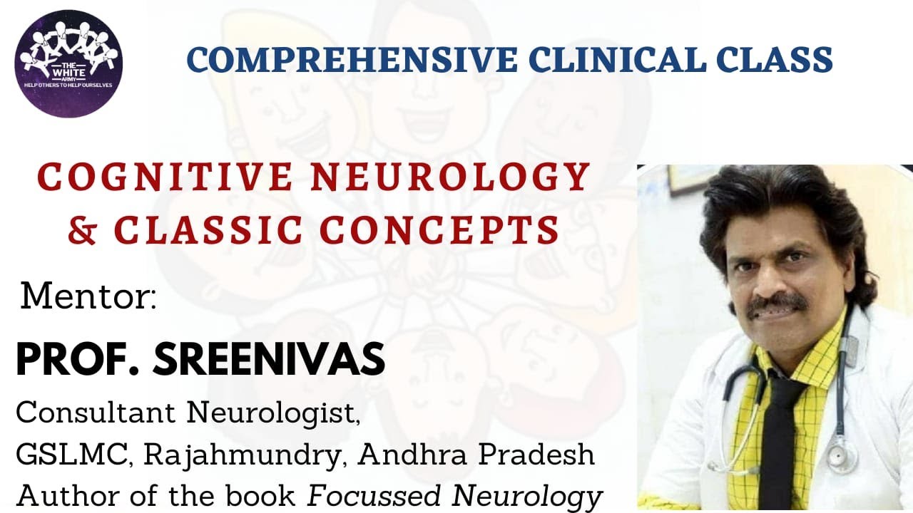
Cognitive Neurology and Classic Concepts
2.4K
Cerebral lateralization refers to the specialization of functions in one hemisphere. The left hemisphere is dominant for speech and language functions (Broca's area, Wernicke's area), while the right hemisphere is dominant for spatial orientation and non-verbal functions. For most right-handed individuals (>90%), language areas are located on the left side, making it the dominant cortex. This lateralization allows humans to occupy a commanding position in the animal world through language, intelligence, and manual dexterity. The frontal lobe lies anterior to the central sulcus and superior to the sylvian fissure, supplied by the anterior cerebral artery (medial parts) and the superior division of the middle cerebral artery (convexity).
Click to watch →
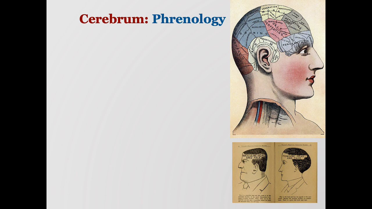
Annotated Brain Physiology 3
137
Cerebral dominance describes how one hemisphere becomes more specialized for certain functions. About 90% of humans are right-handed, indicating left hemisphere dominance, with this preference present from fetal development. Language areas are typically left-hemisphere dominant: Broca's area (frontal lobe) controls speech production, while Wernicke's area (parietal-temporal junction) controls comprehension. Despite handedness variations, 95% of right-handers and 70% of left-handers have left-hemisphere language dominance. This lateralization has survival value, as language processing is critical for communication and social interaction.
Click to watch →

Neurocientista: Suas Ações Não Estão No Seu Controle! - Dr. Carlos Sherman | Lutz Podcast #250
129.2K
The left hemisphere dominates language functions in most right-handed individuals, housing Broca's area (speech production) and Wernicke's area (language comprehension). Damage to these areas produces specific language deficits. This lateralization explains why left-handedness correlates with certain cognitive profiles and why language disorders often affect one hemisphere preferentially.
Click to watch →
hemisferios y lóbulos cerebrales
146.4K
The brain consists of two mirror-image hemispheres connected by the corpus callosum, a white fiber bundle transferring information between sides. The left hemisphere is dominant in most individuals and specializes in verbal functions. It contains Broca's area (speech production, motor control for language) and Wernicke's area (language comprehension). The left hemisphere is characterized as verbal, analytical, symbolic, temporal, rational, digital, and linear in its processing style. Each hemisphere is divided into four lobes separated by the Rolandic, Sylvian, and parieto-occipital fissures.
Click to watch →
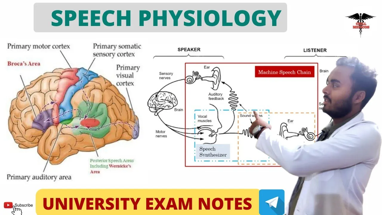
Speech physiology || Broca Area | Wernicke Area | CNS Physiology
24.9K
The dominant hemisphere concept explains that approximately 95% of right-handed individuals have left-hemisphere dominance for speech, while left-handed individuals may show different patterns. The frontal lobe contains multiple speech-related areas: Broca's area (area 44, lower lateral surface) controls speech articulation; the prefrontal cortex (anterior to Broca's area) manages complex speech movements; and the frontal motor area (medial surface) controls writing movements. These areas work hierarchically to plan, sequence, and execute speech-related motor patterns.
Click to watch →
Deep Dive
The physiological definition of a stroke (cerebrovascular accident) and how localized brain tissue damage occurs due to disrupted blood flow.

Stroke & Cerebrovascular Accident (CVA) - Medical-Surgical - Nervous System | @LevelUpRN
244.9K
A stroke, also known as a cerebrovascular accident (CVA), is defined as the sudden interruption of blood flow to the brain. This interruption deprives the brain of oxygen and nutrients, leading to brain tissue damage and necrosis. Understanding this fundamental mechanism is essential for recognizing stroke symptoms and initiating timely treatment.
Click to watch →

Nervous System - CVA/Stroke | For NORCET | ESIC | RRB | DSSSB | RPSC | CHO Exams |By Raju Sir
65K
Stroke (Cerebrovascular Accident) is a cerebrovascular event where blood supply to the brain is interrupted. The brain receives blood supply primarily through the carotid arteries. When blood vessels become blocked or rupture, blood flow and oxygen delivery to brain tissue is disrupted. Ischemic stroke (85% of cases) occurs when blood clots block vessels, while hemorrhagic stroke (15%) occurs when vessels rupture. Both types cause brain tissue necrosis due to oxygen deprivation. Transient Ischemic Attack (TIA) is a warning sign where blood supply interruption lasts less than 24 hours without permanent damage.
Click to watch →
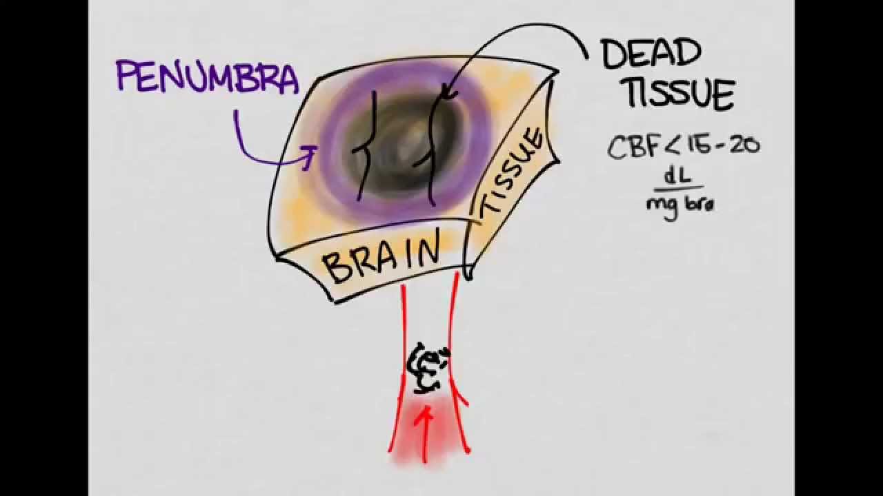
Ischemic Stroke 01: Introduction
31.3K
Ischemic stroke creates two distinct tissue zones: the infarct core (dead tissue receiving no blood flow) and the penumbra (surrounding tissue receiving reduced but insufficient blood flow). Brain tissue dies when cerebral blood flow falls below 15-20 deciliters per milligram of brain tissue per minute. The penumbra remains viable temporarily and can be salvaged if blood flow is restored. Cerebral autoregulation normally maintains constant blood flow despite systemic pressure changes between 50-150 mmHg. During stroke, this curve shifts rightward, requiring higher blood pressure to maintain adequate cerebral perfusion. This compensatory hypertension should not be immediately treated, as it represents the body's attempt to preserve blood flow to ischemic brain tissue.
Click to watch →

Cerebrovascular Accident (CVA)- Brain Stroke ( Nursing Lectures in Hindi MSN 2 )
196.9K
Cerebrovascular accident (CVA), commonly known as stroke, is a condition characterized by the death of brain tissue due to insufficient blood supply to specific areas of the brain. The brain tissue dies when blood flow is interrupted, leading to tissue damage in particular brain regions.
Click to watch →

Niña Amada Mía - Capítulo 096 (Parte 2)
375.3K
A stroke, or cerebrovascular accident, occurs when blood flow to part of the brain is interrupted, depriving brain tissue of oxygen and nutrients. This can happen through two main mechanisms: ischemia (blockage of blood vessels by clots) or hemorrhage (rupture of blood vessels causing bleeding into brain tissue). The result is damage to brain cells in the affected area, which can lead to various neurological deficits depending on the location and extent of the damage. Immediate medical intervention is critical, as strokes are time-sensitive emergencies where rapid treatment can minimize brain damage and improve outcomes.
Click to watch →
Deep Dive
The fundamental distinction between expressive language (producing speech and writing) and receptive language (comprehending speech and reading).
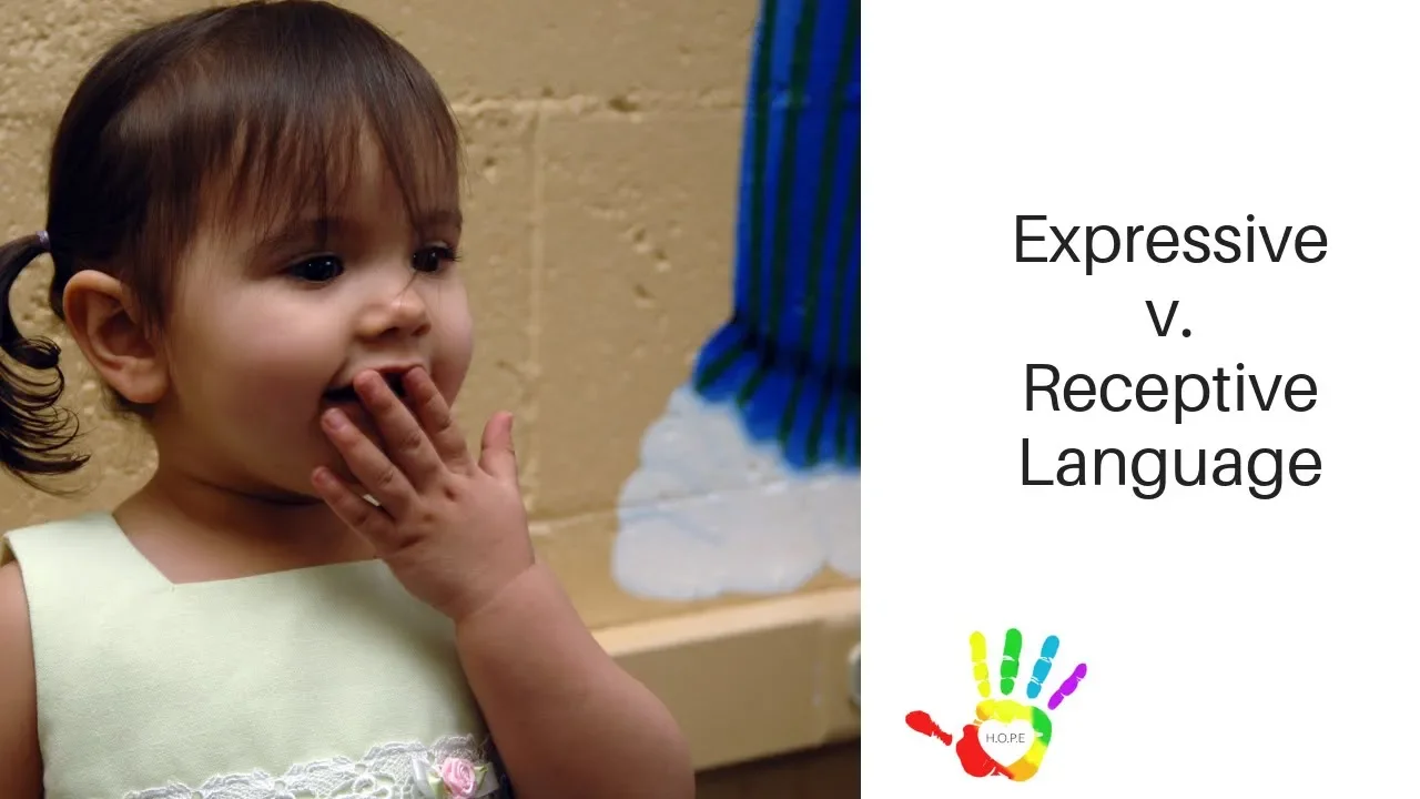
Receptive versus Expressive Language
18.6K
Receptive language refers to the ability to understand and follow another person's language (such as selecting a card when asked 'which one says ribbit' or bringing a pencil when told 'go get me a pencil'), while expressive language refers to the ability to respond or give a vocal response using one's own language (such as naming an object when shown a picture or describing its color).
Click to watch →

CTET 31 JAN पास करने के लिए सभी विषयो का निचोड, इनसे होगा पेपर पास✌️
312.9K
Expressive language skills (productive skills) involve producing language through speaking and writing. Receptive language skills (receptive skills) involve understanding language through listening and reading. Both types of skills are essential for effective communication.
Click to watch →

CTET FEB 2026 | सम्पूर्ण हिन्दी मैराथन Live Class | माध्यमिक स्तर | एक क्लास में पूरा हिन्दी | 4 PM
34.2K
Listening and reading are receptive skills (ग्रहणात्मक कौशल) where learners comprehend language. Speaking and writing are expressive skills (अभिव्यक्तात्मक कौशल) where learners produce language. This fundamental distinction is essential for understanding language teaching methodology.
Click to watch →

CTET 7 JULY 2024 भाषा कौशल by Sachin choudhary live 8pm
634K
Language skills are divided into two categories: Expressive skills (उत्पादक कौशल/प्रोडक्टिव स्किल) - skills used to express one's own thoughts to others, which include speaking and writing. Receptive skills (गणनात्मक कौशल/रिसेप्टिव स्किल) - skills used to receive and comprehend others' thoughts, which include listening and reading. Language skills are not learned in a fixed sequence (like listening first, then speaking, then reading, then writing). Research shows children can learn these skills in any order. The skills can be learned integrally rather than sequentially.
Click to watch →

HTET 2025-26 Hindi / English Pedagogy | Complete Quick Revision | Important Highlights of Pedagogy
9.5K
Language skills are categorized into two types: (1) Expressive skills (अभिव्यक्तात्मक कौशल) - skills used to express oneself, which include speaking (वाचन) and writing (लेखन); (2) Receptive skills (गृहीत कौशल) - skills used to receive and understand language, which include listening (श्रवण) and reading (पठन). This distinction is important for effective language teaching as it helps teachers understand how students process and produce language differently.
Click to watch →
Deep Dive
An introductory understanding of neuroplasticity, which is the brain's capacity to reorganize neural pathways in response to learning or rehabilitation after injury.

Introduction to Neuroscience - Neurorehabilitation
14.8K
Neuroplasticity is the brain's capacity to reorganize neural pathways through experience or exercise, underlying all learning including skill acquisition in adulthood. Recovery after injury involves learning compensatory movements using alternative strategies. Two types exist: functional plasticity (shifting functions to undamaged areas) and structural plasticity (growing new axons and synapses). Currently, clinical efforts primarily target functional plasticity since structural manipulation remains technically challenging. Understanding these mechanisms allows rehabilitation to be viewed as applied neuroplasticity.
Click to watch →
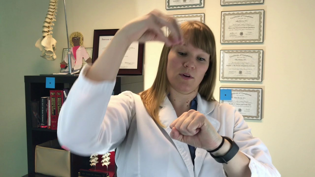
WHAT IS NEUROPLASTICITY?
194
Neuroplasticity is the brain's ability to form and recognize synaptic connections in response to learning, experience, or injury, allowing the brain to reorganize itself by strengthening good neural pathways and weakening inefficient ones through consistent practice and stimulation.
Click to watch →

The Changing Brain - Thursday 17 March 2016
263
Neuroplasticity (brain plasticity) is the brain's ability to reorganize itself by forming new neural connections throughout life. The term combines 'neuro' (neurons) and 'plastic' (changeable). This allows neurons to compensate for injury and disease like stroke and neurodegenerative conditions, and to adapt to learning experiences and environmental changes. The brain stores information in connections between neurons, which are continuously updated. Connections strengthen with use and weaken with disuse. This dynamic nature enables recovery after brain damage and is the foundation for rehabilitation approaches.
Click to watch →
Ramazan Baythak Neuroplasticity Sahil Adeem 250420
17.4K
Neuroplasticity is the brain's ability to reorganize itself by forming new neural connections throughout life. The brain contains numerous neural pathways that function like roads and routes, strengthened through repeated use and mental activities. Neurotransmitters facilitate communication between neurons, and the flexibility of these connections can be enhanced through practice and learning. The brain's capacity to adapt and change in response to learning, experience, or injury is fundamental to all learning processes and cognitive development.
Click to watch →

Workshop - Movimento Humano e Neurociências - Aula 06
235
Neuroplasticity is the brain's ability to reorganize itself by forming new neural connections throughout life. Learning and memory can occur at any age, though there is a critical period from birth to age 4 when the brain has greater predisposition for learning. During initial skill acquisition, many brain regions are activated, but with practice, the brain becomes more efficient, requiring less neural activity. Neural pathways are formed through repeated practice and become strengthened with continued use. The brain can reorganize after injury by activating previously latent pathways and forming new connections. This adaptive capacity is the foundation for rehabilitation after neurological injury.
Click to watch →
Prerequisite Knowledge
- Concept 01Basic neuroanatomy of the human brain, specifically the location of the frontal lobe and the typical left-hemisphere dominance for language functions.
- Concept 02The physiological definition of a stroke (cerebrovascular accident) and how localized brain tissue damage occurs due to disrupted blood flow.
- Concept 03The fundamental distinction between expressive language (producing speech and writing) and receptive language (comprehending speech and reading).
- Concept 04An introductory understanding of neuroplasticity, which is the brain's capacity to reorganize neural pathways in response to learning or rehabilitation after injury.
Subsequent Learning
- Step 01A comparative analysis of other key communication disorders, particularly Wernicke's (receptive) aphasia and Global aphasia.
- Step 02An exploration of clinical therapeutic approaches used in speech-language pathology, such as Melodic Intonation Therapy (MIT) or Constraint-Induced Language Therapy (CILT).
- Step 03The psychological, emotional, and social impacts of chronic communication deficits on a patient's quality of life and family dynamics.
- Step 04The application of advanced neuroimaging techniques (such as fMRI and diffusion tensor imaging) in mapping language networks and tracking post-stroke recovery.
Aphasia Journey
Living With Aphasia
Aphasia Journey
0:09Playing Section
- 1
Stroke seven years ago, formerly worldwide sales at Autodesk.
- 2
Wife assists with speech, condition identified as aphasia.
- 3
Co-founded Voices of Hope with Dr. Hinckley in St. Petersburg.
Challenging the Classic Localizationist View of Broca's Aphasia
While traditional models view Broca's aphasia as a localized, purely expressive speech deficit caused by damage to Broca's area, modern cognitive neuroscience offers a critical counter-perspective. Contemporary research demonstrates that language processing relies on highly distributed, dynamic neural networks rather than isolated brain regions. Studies show that damage confined strictly to Broca's area rarely causes persistent Broca's aphasia; instead, the syndrome typically requires more extensive damage to surrounding white matter pathways and cortical networks. Furthermore, psycholinguistic research has revealed that individuals with Broca's aphasia do not have entirely 'intact' comprehension, as they frequently struggle with understanding complex syntactic structures (such as passive sentences). This shift from a strict localizationist framework to a network-based model suggests that the disorder is not merely a motor speech impairment but a complex deficit in syntactic processing, reshaping how clinicians approach diagnosis and rehabilitation.
Deep Dive
A comparative analysis of other key communication disorders, particularly Wernicke's (receptive) aphasia and Global aphasia.
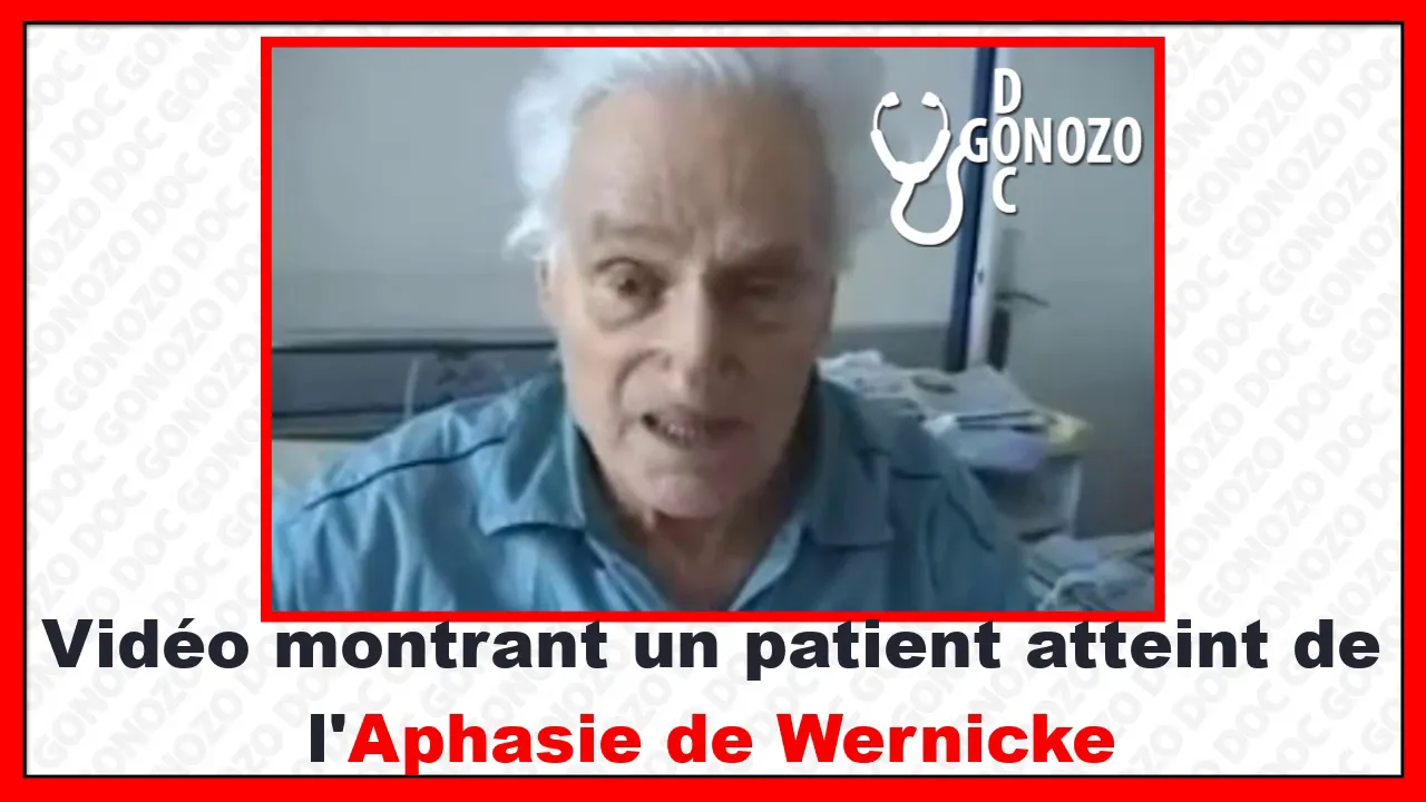
comparaison entre l'aphasie de BROCA et l'aphasie de WERNICKE
57.4K
Broca's aphasia is an expressive (non-fluent) language disorder caused by damage to Broca's area in the inferior frontal gyrus of the dominant hemisphere, characterized by difficulty producing speech and finding words (anomia), while Wernicke's aphasia is a receptive (fluent) language disorder caused by damage to Wernicke's area in the posterior superior temporal gyrus, characterized by fluent but incomprehensible speech and the patient's false belief that they are being understood.
Click to watch →

This person doesn't belong in the Shivering Isles
417
Receptive aphasia (Wernicke's aphasia) results from damage to Wernicke's area, causing fluent but meaningless speech. Patients speak grammatically correctly but cannot comprehend language, often producing word salad. Jargon aphasia is a subtype where patients substitute common words with obscure synonyms used in different contexts (e.g., 'commune' for 'speak'). This use of low-frequency words is unique to jargon aphasia. Global aphasia results from damage to both Broca's and Wernicke's areas, causing severe impairment in both comprehension and production. Statistics show global aphasia is most common immediately after stroke, but less severe types become more common a year later as patients recover.
Click to watch →

Aphasia: Broca's, Wernicke's and More | OT Miri
48.9K
Aphasia is an acquired language disorder caused by brain injury, typically in the left hemisphere, affecting speech production, comprehension, reading, and writing; Broca's aphasia (expressive) involves impaired speech production with slow, labored, telegraphic speech but relatively good comprehension and patient awareness; Wernicke's aphasia (receptive) involves fluent but nonsensical speech with poor comprehension and lack of awareness of errors; anomic aphasia is the mildest form characterized primarily by word-finding difficulties while maintaining fluent, meaningful speech; global aphasia represents the most severe form with widespread impairment across all language modalities.
Click to watch →
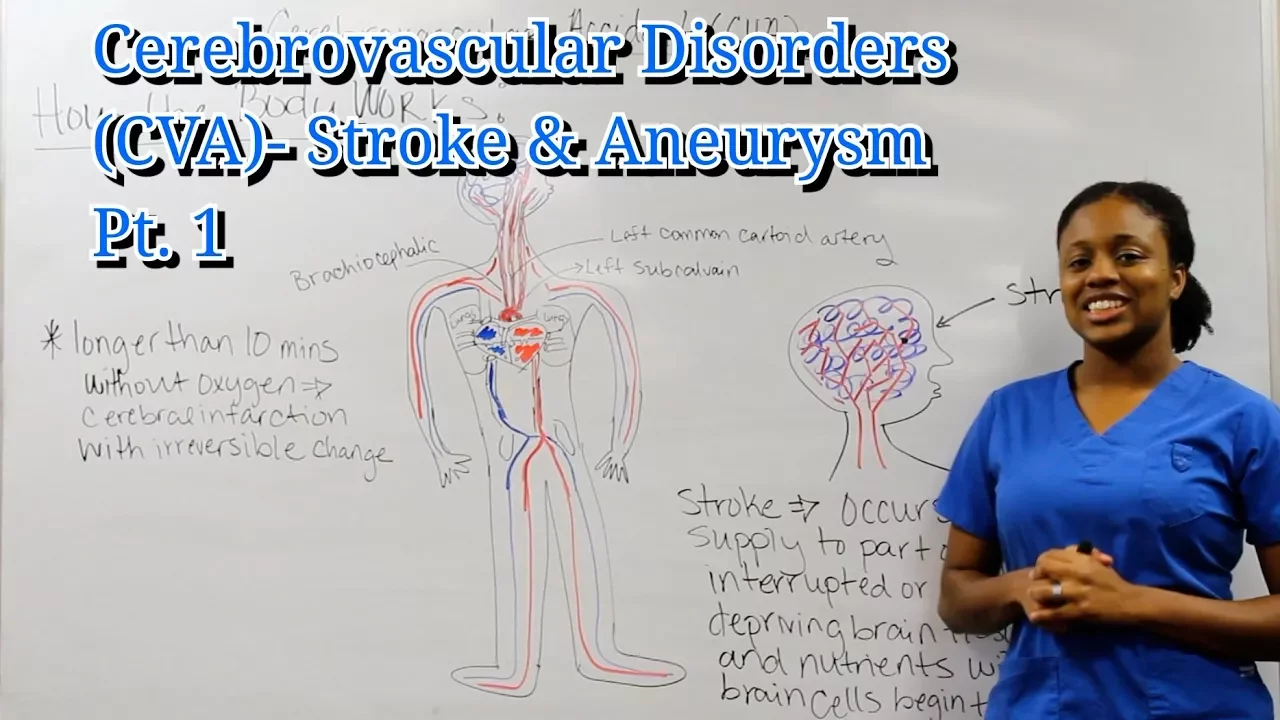
Medical Surgical Neurological System: Cerebrovascular Disorders Pt. 1
8.6K
Communication deficits in stroke include aphasia types: (1) Expressive aphasia (Broca's area) - comprehension intact but inability to express oneself, characterized by effortful, non-fluent speech; (2) Receptive aphasia (Wernicke's area) - literal interpretation of language, fluent but incomprehensible speech; (3) Global aphasia - combination of both expressive and receptive deficits, representing the most severe communication impairment.
Click to watch →
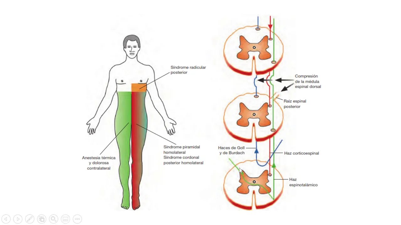
Fisiopatología - Alteraciones de la vía motora principal y la vía sensitiva (Parte 2)
8.8K
Language function requires comprehension, naming, and production. Wernicke's area (posterior temporal lobe) handles comprehension - lesions cause receptive aphasia. Broca's area (inferior frontal lobe) handles production - lesions cause expressive aphasia. Conduction aphasia affects repetition with preserved comprehension and production. Global aphasia affects all language functions. Motor aphasia preserves comprehension but impairs production. Assessment evaluates comprehension, naming, and repetition. Patients with global aphasia have the poorest prognosis.
Click to watch →
Deep Dive
An exploration of clinical therapeutic approaches used in speech-language pathology, such as Melodic Intonation Therapy (MIT) or Constraint-Induced Language Therapy (CILT).
Melodic Intonation Therapy
12.4K
Melodic Intonation Therapy (MIT) is a speech-language therapy technique that helps individuals with expressive aphasia (difficulty producing words) by using melody and rhythm to activate the right hemisphere of the brain, which can reroute speech pathways from damaged left-side brain areas; the therapy involves six steps: therapist-guided left-hand tapping and humming, client listening and rehearsal, humming and singing together, fading therapist support, alternating turns, and finally independent speech production, starting with simple phrases like names and building to more complex sentences.
Click to watch →
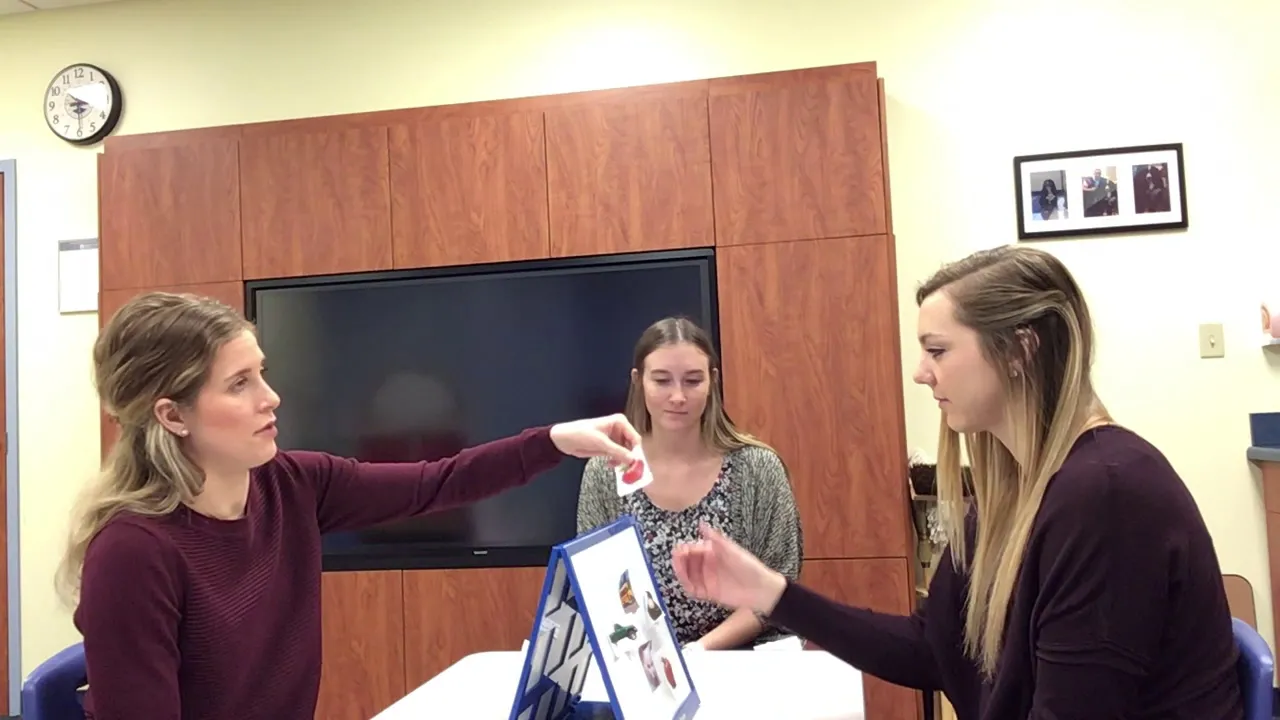
Constraint Induced Language Therapy for individuals with Aphasia
4.3K
Constraint Induced Language Therapy (CILT) is an intensive rehabilitation approach for individuals with aphasia that uses three core principles—constraint (limiting communication to verbal output only), forced use (requiring verbal requests to obtain items), and mass practice (3-4 hours daily for 10 consecutive days)—to promote verbal language recovery through structured, progressively complex communication tasks.
Click to watch →

Melodic Intonation Therapy Example
74.8K
Melodic Intonation Therapy is a speech-language pathology intervention where clinicians model target phrases, clients join in by humming or saying the phrase together, and clinicians gradually fade out their participation while clients produce phrases independently; phrases are considered correct when each word is produced intelligibly and accurately, with the overall objective being for clients to produce grammatically correct sentences with minimal verbal queuing eight out of ten times.
Click to watch →

Melodic Intonation Therapy
63.2K
Melodic Intonation Therapy (MIT) is a music-based rehabilitation technique that helps patients with stroke, dementia, Alzheimer's, or acquired brain injury recover speech by using singing to bypass damaged speech centers in the left hemisphere of the brain and engage the right hemisphere's compensatory mechanisms; the therapy involves singing short songs, repeating phrases, and gradually transitioning from singing to speaking, which has been shown to help patients rebuild vocabulary and communication abilities even after being told they will not speak again.
Click to watch →

Integrative Health For Treating Aphasia with Abbe Simon | No Plateau Podcast - Season 2 Episode 4
1.2K
Constraint-Induced Language Therapy applies CIMT principles to aphasia by constraining alternative communication forms to force use of targeted language skills. Techniques include: (1) Restricting single-word responses to encourage multi-word utterances; (2) Using barriers to prevent gesturing or writing when describing pictures; (3) Limiting obvious vocabulary to promote creative expression. While scientifically sound, CILT is less popular than CIMT for motor recovery because speech therapy traditionally focuses on life participation and functional communication across multiple modalities.
Click to watch →
Deep Dive
The psychological, emotional, and social impacts of chronic communication deficits on a patient's quality of life and family dynamics.
Communication Difficulties Post stroke
696
Communication disorders adversely impact patient quality of life and personal safety by causing trouble expressing basic needs and preferences, social withdrawal, work return difficulties, and learning challenges during therapy sessions. Families face challenges understanding loved ones' wants, while healthcare professionals struggle with patient compliance and rehabilitation progress assessment. Discharge arrangements must consider cognitive and communication status.
Click to watch →

CRID
2.4K
Communication difficulties create significant limitations for people with disabilities, resulting in profound psychological, social, and emotional consequences. These barriers prevent individuals from expressing their thoughts, emotions, and feelings, which can lead to depression. Communication equipment and resources help optimize communication abilities, enabling people with disabilities to achieve better quality of life and develop their cognitive capabilities to levels comparable to others.
Click to watch →

1º Congresso Brasileiro de Análise do Comportamento e Autismo da FME
2.3K
Communication deficits create significant stress for families, who may struggle to understand what their loved one wants, needs, or is experiencing. This uncertainty can lead to frustration, anxiety, and difficulty meeting the individual's needs. Effective communication intervention addresses these challenges by providing families with reliable ways to understand and respond to their loved one's needs, improving family quality of life.
Click to watch →
E.S. Meyers Lecture 2016 University of Queensland
503
Communication failures create lasting family distress and inappropriate medical interventions. Patients who expressed desire to leave hospital soon become surrounded by machines in ICU. Families receive calls wanting 'everything done' while doctors deliver futile interventions. Patients develop hospital-acquired complications like pneumonia. Families are heartbroken when they realize staff always knew the prognosis but never communicated it until death was imminent.
Click to watch →

2019 Evening for Healers - Martha Wilson
681
The loss of ability to communicate creates significant psychological distress for patients and their families. Patients may experience fear that their condition is permanent, while family members struggle with the emotional burden of not hearing their loved one's voice. Understanding this psychological impact is essential for providing comprehensive care that addresses both physical and emotional needs.
Click to watch →
Deep Dive
The application of advanced neuroimaging techniques (such as fMRI and diffusion tensor imaging) in mapping language networks and tracking post-stroke recovery.

Dynamics of language reorganization after left temporo-parietal and frontal stroke
1.4K
This longitudinal fMRI study reveals that language recovery after left hemisphere stroke follows a sequential pattern: initial global network disturbance (diaschisis) in areas distant from the lesion, followed by reactivation of domain-general networks (dorsal lateral prefrontal cortex and insula), then gradual restoration of preserved left hemisphere language networks, with lesion-homologous activation occurring only in frontal stroke patients and not in temporal-parietal stroke patients.
Click to watch →
Neurocon 23 Professor Gesa Hartwigsen
187
Longitudinal fMRI studies of stroke patients (temporal parietal or frontal lesions) across acute, subacute, and chronic phases reveal critical roles for domain-general networks in recovery. Early increases in task-related activity in bilateral prefrontal regions (insula, DLPFC, supplementary motor areas)—belonging to multiple demand networks for cognitive control—correlate with better language improvement. Effective connectivity analysis shows early facilitatory connectivity from domain-general multiple demand cortex regions to perilesional temporal cortex. This early recruitment of domain-general networks to support language-specific regions predicts better preserved performance and initial recovery. Common mechanisms of flexible neural network recruitment appear across healthy young, aging, and lesioned brains: domain-general networks serve compensatory roles in all contexts. The key insight is that neural networks must remain flexible to adapt to challenges, and domain-general contributions represent fundamental mechanisms underlying cognitive resilience. These findings suggest therapeutic strategies targeting network interactions rather than single areas.
Click to watch →
Николай Павлов - Неврология и речь
21.7K
Resting state networks are brain regions showing correlated activity fluctuations even during rest, suggesting functional connectivity between distant regions. These networks are important for understanding speech processing. Resting state fMRI can measure correlations between speech-related brain regions in aphasia patients. Increased correlation over time after stroke suggests successful neural reorganization and recovery potential. Stroke recovery occurs through recruitment of homologous regions in the opposite hemisphere or perilesional regions surrounding damage. Modern rehabilitation uses TMS to stimulate homologous regions and promote the more favorable recovery pathway.
Click to watch →

Neuro Connect - Functional Plasticity in the Language Network by Gesa Hartwigsen
192
The human brain demonstrates remarkable functional plasticity in the language network, where domain-general cognitive systems (attention, cognitive control, working memory) compensate for disruptions or challenges to specialized language areas through increased connectivity and recruitment; this compensatory mechanism operates across the adult lifespan, from healthy aging to post-stroke recovery, and can be studied using combined neuroimaging and neurostimulation approaches.
Click to watch →

#CNStalk - Probing flexible neural network recruitment with neurostimulation - Gesa Hartwigsen
482
Language recovery after stroke requires changes in the dynamics of network interactions, not just the language-specific network. Research shows that in the acute phase after stroke, there is a strong increase in facilitatory drive from domain general areas to perilesional language areas. Stronger increases in this facilitatory influence in the early phase are associated with better preserved language performance in patients with temporal-parietal lesions. This suggests domain general networks contribute to recovery, potentially from the earliest stages.
Click to watch →
Aphasia Journey
Living With Aphasia
Aphasia Journey
0:09- 1
Stroke seven years ago, formerly worldwide sales at Autodesk.
- 2
Wife assists with speech, condition identified as aphasia.
- 3
Co-founded Voices of Hope with Dr. Hinckley in St. Petersburg.
Challenging the Classic Localizationist View of Broca's Aphasia
While traditional models view Broca's aphasia as a localized, purely expressive speech deficit caused by damage to Broca's area, modern cognitive neuroscience offers a critical counter-perspective. Contemporary research demonstrates that language processing relies on highly distributed, dynamic neural networks rather than isolated brain regions. Studies show that damage confined strictly to Broca's area rarely causes persistent Broca's aphasia; instead, the syndrome typically requires more extensive damage to surrounding white matter pathways and cortical networks. Furthermore, psycholinguistic research has revealed that individuals with Broca's aphasia do not have entirely 'intact' comprehension, as they frequently struggle with understanding complex syntactic structures (such as passive sentences). This shift from a strict localizationist framework to a network-based model suggests that the disorder is not merely a motor speech impairment but a complex deficit in syntactic processing, reshaping how clinicians approach diagnosis and rehabilitation.
Basic neuroanatomy of the human brain, specifically the location of the frontal lobe and the typical left-hemisphere dominance for language functions.
The physiological definition of a stroke (cerebrovascular accident) and how localized brain tissue damage occurs due to disrupted blood flow.
The fundamental distinction between expressive language (producing speech and writing) and receptive language (comprehending speech and reading).
An introductory understanding of neuroplasticity, which is the brain's capacity to reorganize neural pathways in response to learning or rehabilitation after injury.
A comparative analysis of other key communication disorders, particularly Wernicke's (receptive) aphasia and Global aphasia.
An exploration of clinical therapeutic approaches used in speech-language pathology, such as Melodic Intonation Therapy (MIT) or Constraint-Induced Language Therapy (CILT).
The psychological, emotional, and social impacts of chronic communication deficits on a patient's quality of life and family dynamics.
The application of advanced neuroimaging techniques (such as fMRI and diffusion tensor imaging) in mapping language networks and tracking post-stroke recovery.
2M views19.7Klikes3:59@TactustherapyOriginal Release: 2017-06-14
Megan: Can you tell us your name?
Mike: I’m Mike Caputo.
Megan: And Mike, when was your stroke?
Mike: I was um, seven years ago.
Megan: And what did you used to do?
Mike: Um, well, um, worked, um, Autodesk.
Um.
Seven, seven, (cued “s”) Sales. Sales.
Worldwide.
And very good, yeah.
Megan: And who are you looking at over there?
When you turn your head?
Mike: That’s my wife.
Megan: Okay, and why is she helping you to talk?
Mike: Um, she’s...Speech.
Um.
Megan: So you have trouble with your speech?
Mike: Yeah.
Megan: What's that called?
Mike: Aphasia.
Megan: And so why don't you work now?
Mike: Um, I, I, well I do!
Megan: What do you do now?
Mike: Voices of Hope Aphasia.
Megan: What is Voices of Hope?
Mike: Um, Peterburg, um Peterberg. (St. Petersburg, Florida) Um, and um, Dr Hinckley and um, and um, myself, um, founder.
Founder for me.
And um, I, I um, members, um, members, um, the, the uh, members, probably seven-, six zero people.
Megan: So 60 people are part of Voices of Hope, which is an aphasia support group that you founded, and Dr. Jackie Hinckley is part of that.
Mike: Yes.
Megan: Okay. Great.
[Wife: It's not a support group.]
Mike: No, it’s programs.
It’s it's, um, three month, three days.
Um, um, Monday, Wednesday, Friday.
And the, the um, and they laugh, and and talked, and um, music, hear this this beautiful, it's... Megan: Great.
Can you tell me, what does it feel like to have aphasia?
Mike: Um it’s, it’s hard, it’s um, well it’s um, speech, it’s like, um, words that don’t understand.
Brain is good, you know, um, but it’s um, speech like um, I don’t know, it’s like um, words, yuk! [laughs] Megan: Alright, thank you so much.
Bye-bye.
Up Next

7:11
Blue Light and Circadian Rhythm: Optimize Sleep with Light Management
@HubermanLabClips
114.2K views•2022-08-11

21:55
Deciphering Neural Circuits Controlling Anorexia in Mice | Richard Palmiter
@AllenInstitute
2.3K views•2011-10-10

4:10
Vagus Nerve (CN X): Anatomy, Nuclei & Functions Explained
@Alilamedicalmedia
305.2K views•2022-10-31

13:03
How Exercise Benefits Your Brain: Science Explained
@TED
11.4M views•2018-03-21
Related Study Plans & Knowledge Roadmaps
Structured learning paths in Neuroscience
neuroscience
Appetite Regulation: Brain Circuits & Hormones
01Foundations of Endocrinology and Brain Anatomy
02Introduction to Energy Homeostasis
03Peripheral Signals: Leptin, Ghrelin, and Insulin
+ 2 more learning modules
14h5 modules
Explore Roadmap
neuroscience
Neuroeconomics: Value, Risk & Brain Mechanisms
01Foundations of Neuroscience and Brain Anatomy
02Introduction to Neuroeconomics and Subjective Value
03Dopaminergic Prediction Errors & Reinforcement Learning
+ 3 more learning modules
18h6 modules
Explore Roadmap
neuroscience
Spatial Navigation: Cells, Grids & Brain Maps
01Foundations of Brain Anatomy and Memory
02Place Cells and Head-Direction Cells
03Grid Cells: The Brain's Coordinate System
+ 2 more learning modules
12h5 modules
Explore Roadmap
neuroscience
BCIs: Neural Decoding & Prosthetic Control
01Neuroscience Foundations: How the Brain Signals Intent
02Signal Acquisition: Recording the Brain's Activity
03Neural Decoding: Translating Brain Waves into Data
+ 2 more learning modules
25h5 modules
Explore Roadmap