Density gradient centrifugation is a separation technique where particles migrate through a liquid medium with gradually increasing density (typically sucrose solutions) during centrifugation, accumulating at positions where their own density matches the surrounding medium; this method enables purification of biological samples such as ribosome subunits, isotopically labeled DNA, and Okazaki fragments in biochemistry and molecular biology research.
Density Gradient Centrifugation | Principle and Applications
Added:Deep Dive
Basic principles of centrifugation, including centrifugal force, sedimentation, and how a standard centrifuge operates.

Centrifuge | principle of centrifuge | centrifuge machine introduction
218.7K
A centrifuge is a machine device used for sedimentation of solid particles in samples. When blood samples are placed in centrifuge tubes and rotated, heavier particles like red blood cells settle at the bottom while lighter serum rises to the top. Centrifuges are classified by speed: low-speed for urine processing, high-speed for blood banking and research, and refrigerated for temperature-sensitive samples. The operating principle is centrifugal force, where particles experience outward force proportional to their mass, angular velocity squared, and distance from the center. The centrifugation force formula is F = m × ω² × r, where m is particle mass, ω is angular velocity, and r is radius of rotation.
Click to watch →

PRINCIPLES OF CENTRIFUGATION
514
Sedimentation is the natural settling of particles in liquid suspensions due to gravitational force, as seen when sand settles in water over hours. Centrifugation accelerates this process by applying centrifugal force, an outward fictitious force on objects moving in circular paths. This force overcomes opposing buoyant and frictional forces, causing denser particles to settle as pellets while lighter phases remain as supernatants. Separation efficiency depends on particle size, shape, density, medium viscosity, and rotor speed. A centrifuge consists of three components: rotor (holds sample tubes), motor (provides power), and drive shaft (connects rotor to motor). Two key formulas govern centrifugation: centrifugal force F = mω²r, and Relative Centrifugal Force (RCF) = 1.12 × 10⁻⁵ × rpm² × r.
Click to watch →

Sedimentation basic principles of centrifugation
7.2K
Sedimentation is the process by which particles in suspension settle out of a fluid under the influence of forces such as gravity or centrifugal acceleration, with the terminal velocity depending on the particle's properties and the applied force; centrifugation exploits this principle by applying high centrifugal forces (up to 980,000g in ultracentrifuges) to separate particles based on their size, density, and shape, with the relative centrifuge force calculated as RCF = m × r × ω², and analytical ultracentrifugation enabling real-time monitoring of sedimentation velocity and equilibrium to determine molecular mass and study biomolecular interactions.
Click to watch →
Centrifuge and Autoclave. | رسول ستار
201
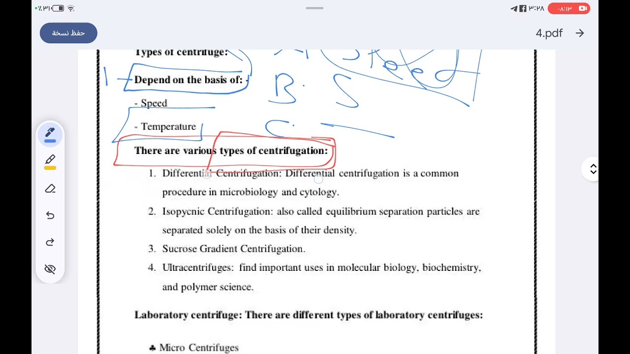
A centrifuge is a device that separates mixture components using centrifugal force based on weight and density. When spun at high speed, heavier particles sediment faster than lighter ones. Centrifugal force is an apparent force pushing objects outward from the center of rotation. The principle relies on differential sedimentation rates - heavier components settle more quickly. Four main types exist: differential centrifugation (by weight), density gradient centrifugation (by density), isopycnic centrifugation (by sugar concentration), and ultracentrifugation (for proteins at high speeds). Laboratory centrifuges include microcentrifuges, clinical centrifuges, high-speed centrifuges, and industrial centrifuges.
Click to watch →

CENTRIFUGE-WORKING PRINCIPLE | COMPONENTS | TYPES
140
A centrifuge is a laboratory instrument that uses centrifugal force to separate components from solutions. The working principle relies on sedimentation, where particles settle at different rates based on mass and density. Natural gravitational force causes slow sedimentation, but centrifugal force accelerates this process by creating high-g environments. The centrifuge generates centrifugal force perpendicular to the rotation axis, which is much stronger than normal gravity. This force overcomes or accelerates gravity, enabling rapid separation of particles that would not settle under natural conditions.
Click to watch →
Deep Dive
The physical concepts of density, buoyancy, and viscosity, and how they influence the movement of particles in a liquid medium.

مش هتنسى الدرس ده أبداً 💡 المادة وخصائصها - علوم أولى إعدادي ترم أول
1.9K
Viscosity is a physical property of liquids that describes their resistance to flow. It measures how easily a liquid flows - liquids with low viscosity flow easily (like water), while liquids with high viscosity flow slowly (like honey). There is an inverse relationship between viscosity and the speed of movement through a liquid - liquids with lower viscosity allow objects to move through them more easily and quickly. Density is a physical property defined as mass per unit volume (measured in grams per cubic centimeter). It is used to distinguish between materials - materials with density less than water (1 g/cm³) float on water, while materials with density greater than water sink.
Click to watch →

Mechanical Properties of Fluids in 45 minutes 🔥 | NEET 2025 | NEET AVATAR SERIES
4.3K
This comprehensive section covers surface phenomena, buoyancy, and viscous flow. Contact angle determines meniscus shape: θ < 90° creates concave meniscus (wetting), θ > 90° creates convex meniscus (non-wetting). Capillary rise is h = (2T cosθ)/(ρgr), inversely proportional to tube radius. Buoyant force equals weight of displaced liquid: F_b = ρVg. For hollow bodies, volume exceeds displaced liquid volume, so density is less. Stokes' Law gives viscous drag force: F = 6πμrv. Terminal velocity is v = (2r²(σ - ρ)g)/(9μ), directly proportional to r². For liquids, viscosity increases with pressure and decreases with temperature; for gases, viscosity is independent of pressure but increases with temperature. These principles explain particle motion, sedimentation, and the behavior of objects moving through viscous media.
Click to watch →

Fluida Statis dan Fluida Dinamis | Fisika Medis
194
An object floats in a fluid if its density is less than the fluid's density, sinks if greater, and remains suspended if equal. For example: blood (1,060 kg/m³) sinks in water (1,000 kg/m³), ice (0,92 × 10³ kg/m³) floats in water, ethanol (0,81 × 10³ kg/m³) floats in water. When mixing fluids, the one with lower density rises to the top while the one with higher density sinks to the bottom. Viscosity (kekentalan) measures a fluid's resistance to flow. When a glass plate moves across a liquid surface, molecules near the plate move at the same speed due to adhesion, while molecules farther away experience resistance, creating a velocity gradient. This principle is used in a viscometer to measure fluid viscosity, with applications in assessing male fertility - sperm must have appropriate viscosity to be motile enough to reach and fertilize an egg.
Click to watch →

Mechanical properties of fluid part 2 || RHT || OSSTET || OAVS @physicsfever
634
This section covers fundamental fluid properties. Density determines whether objects float or sink: objects less dense than the fluid float, while denser objects sink. The human body has density slightly greater than water, explaining why humans sink but can swim more easily in saltwater due to its higher density. Viscosity is the internal friction between fluid layers, causing resistance to flow. The velocity gradient describes how velocity changes across fluid layers, with zero velocity at stationary boundaries. These concepts form the foundation for understanding fluid behavior and motion.
Click to watch →

Class-03 PHYSICS SCIENCE || Ghatnachakre Short Notes 2026 || UPPCS 2026 || BPSC 2026 || Sharad Sir
1.3K
This section covers viscosity and buoyancy principles. Viscosity (श्यानता) is the property of liquids that resists flow between layers. Honey has the highest viscosity among common liquids, followed by glycerin, machine oil, blood, and water. For liquids, viscosity decreases as temperature increases, while for gases, viscosity increases with temperature. Buoyant force (उत्प्लावन बल) is the upward force exerted by a fluid on an immersed object, equal to the weight of the fluid displaced. Objects float if their density is less than the fluid's density. Water has maximum density at 4°C (277 Kelvin), which is why ice floats on water. This property allows aquatic life to survive in winter, as the bottom layer of water remains liquid at 4°C while the top layer freezes.
Click to watch →
Deep Dive
Fundamental structural properties of macromolecular biomolecules, specifically DNA, RNA, and proteins.

Biomolecules(जैव-अण) in 75 Minutes - NEET हिंदी 2025
138.6K
Proteins have four structural levels: primary (amino acid sequence with peptide bonds), secondary (alpha helices and beta pleated sheets with hydrogen bonds), tertiary (complex 3D folding with peptide bonds, hydrogen bonds, disulfide bonds, ionic interactions, van der Waals forces), and quaternary (multiple polypeptide chains). N-terminus has free amino group, C-terminus has free carboxyl group. Nucleic acids (DNA, RNA) are genetic materials consisting of polynucleotides. Both have sugar-phosphate backbones with phosphodiester bonds. DNA has deoxyribose (C5H10O4), RNA has ribose (C5H10O5). DNA has thymine, RNA has uracil. Base pairing: DNA (A-T 2 bonds, G-C 3 bonds), RNA (A-U 2 bonds, G-C 3 bonds). B-DNA double helix has 10 base pairs per turn, 36-degree angle, 2.0 angstrom spacing.
Click to watch →

12th Chemistry | ONESHOT Revision Vol 2 | Public Important Questions 2025 | Shravanee Ma'am
45K
DNA is double-stranded with deoxyribose and thymine, stable and found in nucleus/mitochondria. RNA is single-stranded with ribose and uracil, unstable and found in cytoplasm/ribosomes. Denaturation loses higher-order structure without affecting primary structure. Peptide bonds link amino acids via carboxyl-amino group condensation. Essential amino acids (histidine, isoleucine, leucine, lysine, methionine, phenylalanine, threonine, tryptophan, valine) must be obtained from diet.
Click to watch →

الصف الثالث الثانوي / ( فاهم حافظ ) الدرس الثاني تركيب ال DNA 🧬
46.4K
DNA (Deoxyribonucleic acid) is a biological macromolecule composed of genes, classified as a polymer made of nucleotide monomers. Each nucleotide contains three components: a sugar, a phosphate group, and a nitrogenous base. DNA and RNA are the two main types of nucleic acids. DNA stands for Deoxyribonucleic acid (حمض نووي ريبوزي منقوص الأكسجين), meaning it has one oxygen atom removed from ribose. RNA stands for Ribonucleic acid (حمض نووي ريبوزي), meaning it has complete ribose sugar. The key difference is that DNA has deoxyribose sugar while RNA has ribose sugar. A nucleotide consists of three components: a sugar molecule, a phosphate group, and a nitrogenous base. The sugar in DNA is deoxyribose (C5H10O4), while in RNA it is ribose (C5H10O5). The nitrogenous bases in DNA are adenine (A), thymine (T), cytosine (C), and guanine (G). In RNA, uracil (U) replaces thymine. Each nucleotide contains only one nitrogenous base. A gene is formed by the specific sequence of nucleotides, and different sequences create different genes despite having the same basic components.
Click to watch →

Class 12 Chemistry: 50 IMP Questions and Answer 🔥 MP Board Quarterly Exam Marathon
1.9K
This section covers biomolecule fundamentals. DNA and RNA differ in sugar (deoxyribose vs ribose), base (thymine vs uracil), and structure (double vs single strand). Proteins have four structural levels: primary (amino acid sequence), secondary (alpha-helices, beta-sheets), tertiary (3D arrangement), and quaternary (multiple polypeptide chains). The instructor emphasizes that these concepts are essential for understanding biological molecules and frequently tested in board exams. The section concludes with encouragement for students to study thoroughly and wish them success in their quarterly examinations.
Click to watch →

الدرس المفصل الوحدة 01 آليات تركيب البروتين
180.4K
DNA and RNA differ in several key structural aspects. DNA contains deoxyribose sugar and the nitrogenous base thymine, while RNA contains ribose sugar and uracil instead of thymine. DNA typically exists as a double helix with two complementary strands, whereas RNA is usually single-stranded. The carbon numbering in the sugar molecule (specifically the 2' carbon) distinguishes DNA from RNA, as the 2' carbon in deoxyribose lacks an oxygen atom, while ribose has an oxygen atom at this position. These structural differences affect the stability and function of each molecule.
Click to watch →
Deep Dive
The chemical nature of solutions, solubility, and how concentration gradients (such as sucrose gradients) are established.

141 Chapter 5 Part 1
108
In a sucrose solution, there is a concentration gradient of sucrose (50% on one side, 0% on the other). If sucrose could pass through the membrane, it would diffuse from high to low concentration. However, because sucrose cannot pass, water moves instead to compensate for the solute gradient.
Click to watch →

Módulos Específicos - EXANI-II / PreMedicina Clase 23 Parte 1 - Tipos de Transporte Membrenal 🧬🔬
355
Una solución es una sustancia homogénea compuesta por solutos (sustancias que se disuelven) y solvente (sustancia líquida que disuelve). Los solutos pueden ser sólidos, líquidos o gaseosos. El gradiente de concentración es la diferencia de concentración de una solución entre dos regiones, específicamente entre el líquido extracelular y el intracelular. La concentración está determinada por la cantidad de solutos presentes en cada región.
Click to watch →
L-5 Centrifugation | BBCCT-103 Block-1 Unit-4 | IGNOU BSc Biochemistry
114
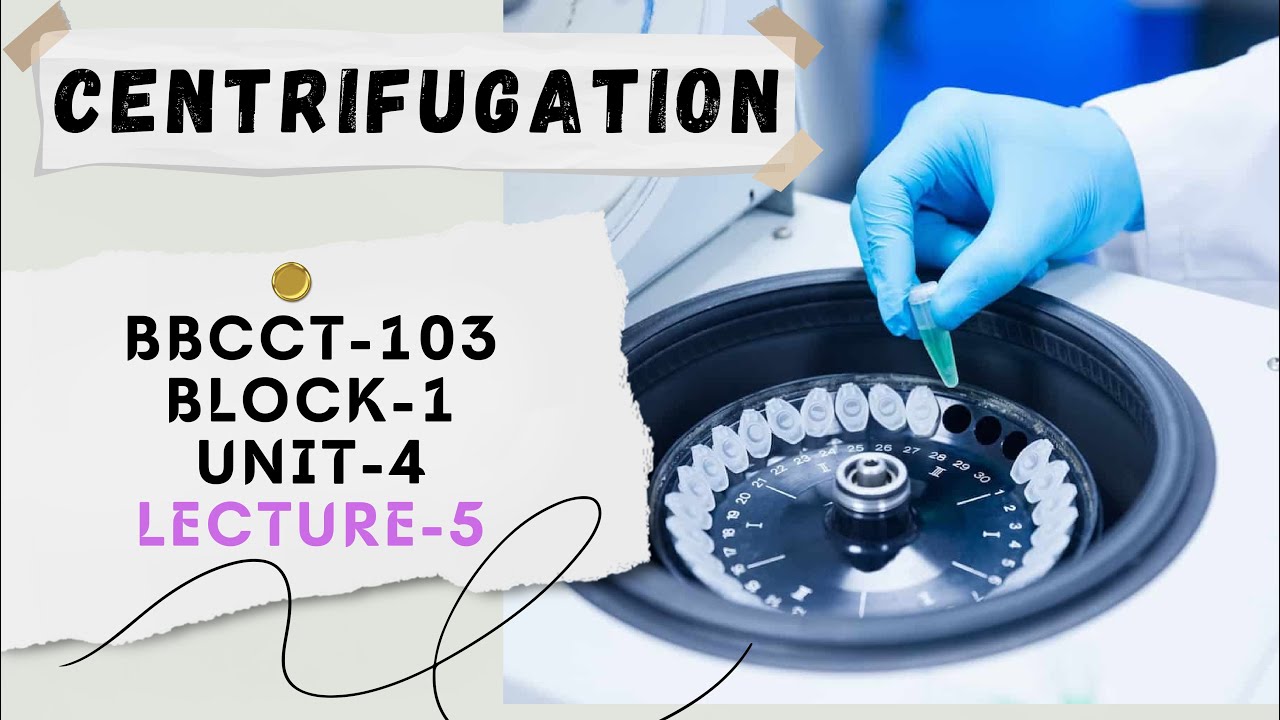
Isopycnic gradient refers to a smooth density transition allowing particles to settle at their respective densities without mixing. Sucrose solution (5-20% concentration) is commonly used as the gradient medium because it is readily available and does not interfere with biological particles. The gradient is prepared using a gradient maker to create a linear pre-formed density gradient before sample loading.
Click to watch →

Méthodes d'étude de la cellule: révision + QCS (MEB+la technique d'isolement) part 2
4.3K
A sucrose gradient is prepared by layering solutions of different sucrose concentrations (e.g., 20%, 15%, 10%, 5%) in a tube. The principle is that higher sucrose concentration creates a denser environment. Organelles migrate through the gradient until they reach a position where their density matches the surrounding sucrose solution.
Click to watch →

9700/12/Oct/Nov/2023 Q.21 to 40 (Part 2)
113
For sucrose to be loaded into phloem, there must be a relative concentration gradient. Companion cells should have the highest concentration of sucrose because sucrose is transported by active transport. Mesophyll cells should have the lowest concentration, and phloem sieve tube elements should have the second highest concentration. This gradient drives the active transport process.
Click to watch →
Prerequisite Knowledge
- Concept 01Basic principles of centrifugation, including centrifugal force, sedimentation, and how a standard centrifuge operates.
- Concept 02The physical concepts of density, buoyancy, and viscosity, and how they influence the movement of particles in a liquid medium.
- Concept 03Fundamental structural properties of macromolecular biomolecules, specifically DNA, RNA, and proteins.
- Concept 04The chemical nature of solutions, solubility, and how concentration gradients (such as sucrose gradients) are established.
Subsequent Learning
- Step 01The distinction between rate-zonal (size and shape-dependent) and isopycnic (purely density-dependent) gradient centrifugation.
- Step 02The historical significance of density gradients in molecular biology, such as the Meselson-Stahl experiment proving semi-conservative DNA replication.
- Step 03Advanced purification workflows, including the isolation of sub-cellular organelles, viral particles, and membrane proteins.
- Step 04Analytical ultracentrifugation (AUC) methods used to characterize the hydrodynamic properties and molecular weights of macromolecules.
Gradient Basics
Applications
Gradient Basics
0:02Playing Section
- 1
Explains stepwise sucrose gradient construction for density separation.
- 2
Particles migrate to layers matching their own density during spinning.
- 3
Fractionation after centrifugation enables easy sample purification.
Artifacts of Hydrostatic Pressure and Shear Stress: The Shift to Chromatographic and Gentle Biophysical Alternatives
While density gradient centrifugation is a foundational technique for separating biomolecules, it has significant limitations that have led researchers to adopt alternative methods. The intense gravitational forces (often exceeding 100,000 x g) generate extreme hydrostatic pressure and shear stress. This can cause artifactual dissociation of weakly bound macromolecular complexes, denature sensitive proteins, and damage genomic DNA or delicate replication intermediates like Okazaki fragments. Furthermore, preparing sucrose gradients is time-consuming and suffers from low throughput. To overcome these limitations, modern biochemistry increasingly relies on gentler, high-resolution alternatives. Techniques such as Size Exclusion Chromatography (SEC), Asymmetric Flow Field-Flow Fractionation (AF4), and native Mass Spectrometry (MS) allow researchers to isolate and analyze macromolecular complexes in their native states without subjecting them to destabilizing physical forces. These modern methods provide more accurate representations of in vivo molecular interactions, offering a critical counterpoint to the reliance on traditional ultracentrifugation.
Deep Dive
The distinction between rate-zonal (size and shape-dependent) and isopycnic (purely density-dependent) gradient centrifugation.

Difference Between Rate Zonal Centrifugation and Isopycnic Centrifugation
2K
Rate zonal centrifugation separates particles based on size using sedimentation rate, employs low-speed swing rotor, creates flat gradient, forms pellets, and is used for proteins, enzymes, hormones, nucleic acids, and cell organelles; whereas isopycnic centrifugation separates particles based on density using sedimentation equilibrium, employs high-speed fixed-angle or swing rotor, creates steep gradient, does not form pellets, and is used for subcellular particles like mitochondria, lysosomes, and peroxisomes.
Click to watch →
Centrifugation | Part 3: Differential; Density Gradient: Isopycnic, Rate Zonal || GATE 2022 || DBT
4.3K
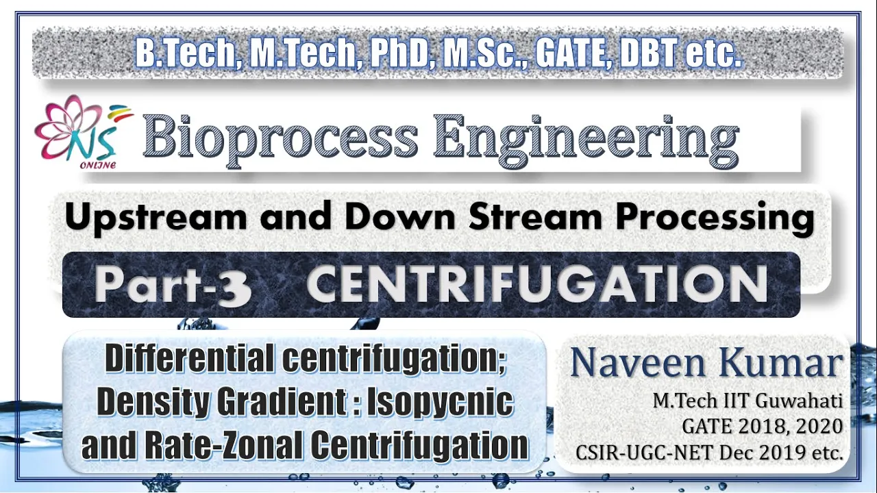
Density gradient centrifugation forms gradients before centrifugation to separate particles of similar size but different density. Isopycnic centrifugation separates based on density: particles sink until reaching their isopycnic point where particle density equals surrounding liquid density. This enables separation of particles with identical sedimentation rates but different densities. Rate zonal centrifugation separates based on sedimentation rate (size and mass) rather than density, providing better resolution than differential centrifugation. Key criteria include sample density less than gradient minimum, particle density greater than gradient maximum, sufficient path length, and appropriate run time.
Click to watch →

M-09. Rate zonal size separation & Isopynic centrifugation
608
Density gradient centrifugation separates particles based on their sedimentation velocity, governed by Stokes' equation V = (D² × (ρ_p - ρ_l) / 18μ) × G, where velocity depends on particle size, density difference from the medium, and centrifugal force; rate zonal centrifugation separates particles by both size and density using sucrose gradients with critical timing, while isopynic centrifugation separates particles solely by density using cesium chloride gradients where particles migrate to equilibrium positions where their density matches the surrounding medium, with each method having distinct advantages for different biological applications.
Click to watch →
CUET PG Life Science | Unit 1 - Techniques | CENTRIFUGATION | Gargi Jauhari |
1.4K
Fixed angle and vertical rotors have shorter run times but lower resolution. Swing-out rotors provide the best choice for high-resolution banding in rate-zonal and isopycnic centrifugation. Rate-zonal centrifugation separates particles based on size and shape, while isopycnic centrifugation separates based on density. Understanding these distinctions helps researchers select appropriate rotors for their specific separation needs. The choice depends on whether time or resolution is the priority for specific experimental requirements. Proper rotor selection ensures optimal separation efficiency and prevents damage to both the equipment and samples.
Click to watch →
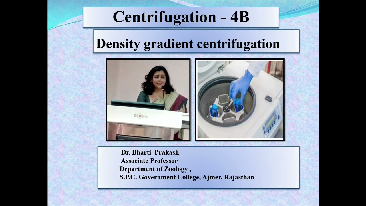
Centrifugation 4B : Density Gradient Centrifugation by Dr. Bharti Prakash
2.3K
Density gradient centrifugation is a preparative ultracentrifugation technique that separates particles based on their density or sedimentation velocity by creating a density gradient in the centrifuge tube; it includes two main types: rate zonal separation (which separates particles based on size and mass using a shallow gradient where particles stop at different positions according to their sedimentation speed) and isopycnic separation (which separates particles based on density using a steep gradient where particles migrate to their isopycnic point—the position where their density matches the surrounding medium), with applications ranging from separating cellular organelles to isolating nucleic acids and viruses.
Click to watch →
Deep Dive
The historical significance of density gradients in molecular biology, such as the Meselson-Stahl experiment proving semi-conservative DNA replication.

تضاعف الADN / تطور كمية ال ADN|ثانية ثانوي|شرح تجارب مسلسون و ستال بأبسط الطرق😍👌
98.8K
This section details the Meselson-Stahl experiment that proved DNA replication is semi-conservative. They used E. coli bacteria and nitrogen isotopes: N-14 (light) and N-15 (heavy). Density gradient centrifugation separates DNA by density: light DNA (1.65 g/cm³) floats, heavy DNA (1.80 g/cm³) sinks. Bacteria grown in N-15 produce heavy DNA. When transferred to N-14 and divide once, hybrid DNA forms (1.72 g/cm³) with one heavy and one light strand. When hybrid DNA divides again, two bands appear: light DNA and hybrid DNA. This 50:50 ratio confirms semi-conservative replication where each original strand serves as a template for a new complementary strand.
Click to watch →
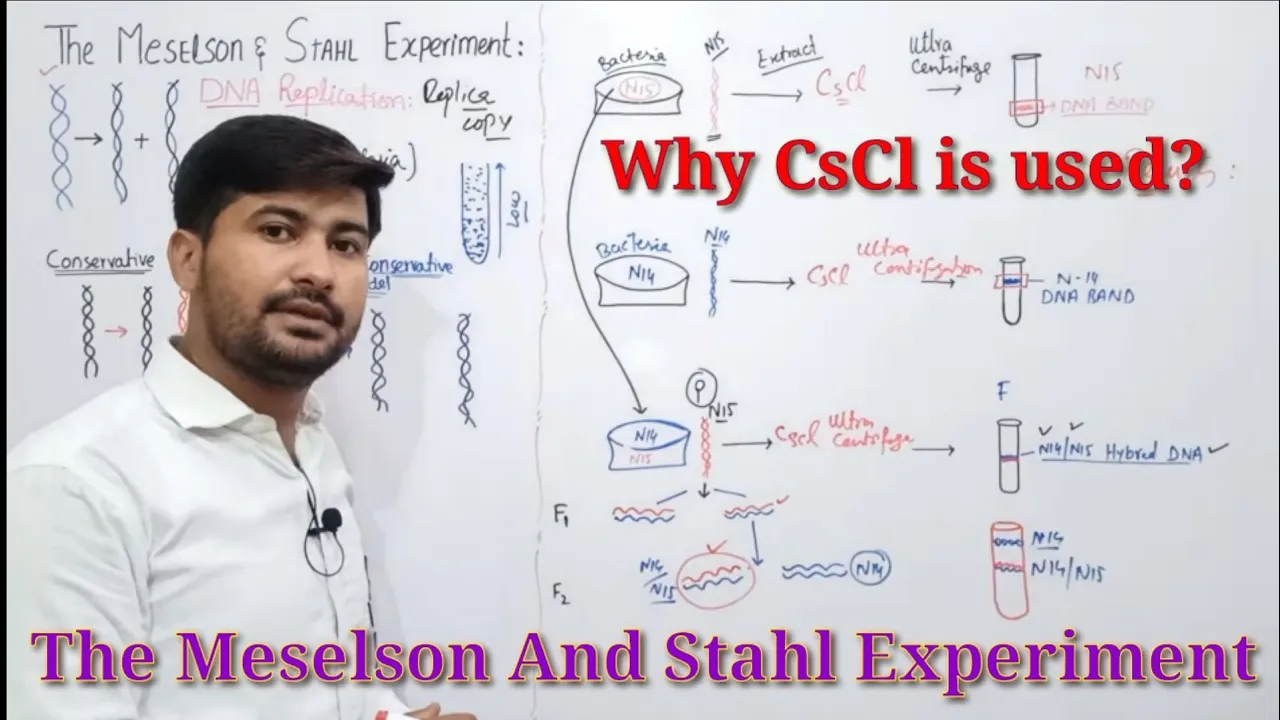
20.6 The Meselson and Stahl experiment | Models of DNA replication| fsc 2nd year Biology
66.3K
This lecture covers the Meselson-Stahl experiment that proved DNA replication is semi-conservative. Three models existed: conservative (parental DNA intact), semi-conservative (each strand serves as template), and dispersive (parental material dispersed). The experiment used E. coli bacteria grown in heavy nitrogen (N15), then transferred to light nitrogen (N14). DNA was extracted and subjected to density gradient centrifugation using cesium chloride. After one generation, a single intermediate band appeared, ruling out conservative replication. After two generations, two bands appeared (intermediate and light), confirming semi-conservative replication and ruling out dispersive model. Each daughter DNA contains one parental strand and one newly synthesized strand.
Click to watch →

DNA Replication | Molecular Basis of Inheritance | L4 | NEET 2022/23 | Komal Yadav
105.2K
The Meselson-Stahl experiment (1958) proved that DNA replication is semi-conservative. Bacteria were grown in heavy nitrogen (N-15) medium, then transferred to normal nitrogen (N-14). After one generation, all DNA was hybrid (one heavy strand, one light strand) with intermediate density. After two generations, half was hybrid and half was light. This demonstrated that each daughter DNA molecule contains one original strand and one newly synthesized strand. The experiment used density gradient centrifugation to separate DNA based on density, with heavy DNA sinking to higher density regions and light DNA rising to lower density regions.
Click to watch →

Meselson and Stahl experiment
508.2K
The Meselson-Stahl experiment (1958) demonstrated that DNA replication is semi-conservative by using density gradient centrifugation to distinguish between parent and daughter DNA strands; cells grown in heavy nitrogen-15 (N15) media were transferred to normal nitrogen-14 (N14) media, and after one generation, only an intermediate-density band appeared, proving that each original DNA strand serves as a template for synthesizing a new complementary strand.
Click to watch →

6.7 -DNA REPLICATION (SEMI CONSERVATIVE) || CHAPTER 6-CHROMOSOMES AND DNA || SECOND YEAR BIO
49.9K
The Meselson-Stahl experiment definitively proved DNA replication is semi-conservative. Scientists used nitrogen isotopes: N-14 (light, 7 protons + 7 neutrons) and N-15 (heavy, 7 protons + 8 neutrons). Bacteria were grown in N-15 medium, making their DNA heavy. After transfer to N-14 medium, DNA was analyzed using density gradient centrifugation. After one generation, a single intermediate-density band appeared, ruling out conservative replication. After two generations, two bands appeared (intermediate and light density), confirming semi-conservative replication. After multiple generations, the intermediate band decreased while the light band increased. This pattern showed that each original heavy strand serves as a template for a new light strand, producing intermediate-density molecules. Subsequent replications produce molecules with two light strands. The experiment provided definitive proof that DNA replication is semi-conservative.
Click to watch →
Deep Dive
Advanced purification workflows, including the isolation of sub-cellular organelles, viral particles, and membrane proteins.
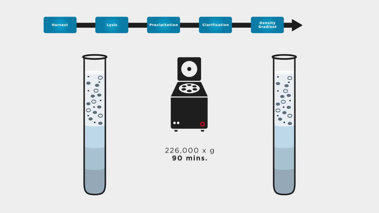
Producing and Purifying Viral Systems in the Laboratory - Beckman 2017
21.1K
Viral particle purification involves a multi-step centrifugation workflow: initial low-force centrifugation separates soluble virus from cells, followed by cell lysis and sedimentation of debris; viral precipitation using polyethylene glycol creates pellets that are clarified through washing and resuspension; finally, density gradient ultracentrifugation (using sucrose or cesium chloride gradients) isolates active virus based on size and density, achieving purification from empty particles, incomplete proteins, and cellular debris.
Click to watch →
Viral Particle Production 101 Video
5.5K
Viral particle purification involves a multi-step centrifugation workflow including initial low-force separation of virus from cells, cell lysis and sedimentation of debris, precipitation using polyethylene glycol, washing with sodium benzoate, and finally density gradient ultracentrifugation to isolate active virus based on size and density (1.14-1.24 g/mL), which separates fully loaded virus from empty particles and cellular debris.
Click to watch →

Subcellular Fractionation / Cell Fractionation - Biochemistry
17.1K
Subcellular fractionation systematically isolates organelles through sequential centrifugation at increasing speeds. The nuclear fraction (5-10 μm diameter) precipitates first at 600g for 10 minutes. Mitochondria, chloroplasts, lysosomes, and peroxisomes deposit at 15,000g for 5 minutes. The microsomal fraction (plasma membrane, ER, polyribosomes) requires ultracentrifugation at 100,000g for 60 minutes. Ribosomal subunits and enzyme complexes need even higher speeds. The cytosol remains undeposited after 300,000g for 2 hours. Fractions appear in order of decreasing density: nuclear > mitochondrial > microsomal > cytosol. Organelle purity is assessed using marker molecules (specific enzymes or biomolecules for each organelle) rather than expensive methods like electron microscopy, with DNA testing confirming nuclear isolation.
Click to watch →

Centrifuge - Principle, Rotors (Fixed Angle & Horizontal), Types of Centrifuge, Application in Hindi
78.6K
Ultracentrifuges operate at extremely high speeds ranging from 60,000 to 100,000+ RPM, far exceeding the capabilities of standard high-speed centrifuges. These powerful instruments enable separation of very small particles including proteins, nucleic acids, subcellular organelles, viral particles, and macromolecular complexes. The extreme centrifugal forces allow resolution of molecular weight differences that cannot be achieved with conventional centrifugation, making ultracentrifuges indispensable in advanced biochemistry and molecular biology research.
Click to watch →

BIOL 4610 Ch 4.3-4.4
228
Organelle isolation begins with gentle cell disruption using osmotic protectors (0.25 M sucrose) and methods like sonication or filtration. Differential centrifugation separates organelles by sedimentation rate: low-speed spins pellet nuclei; higher speeds pellet mitochondria, chloroplasts, lysosomes, and peroxisomes; ultracentrifugation pellets membranes and ER. Equilibrium density gradient centrifugation further purifies organelles by buoyant density (lysosomes ~1.12 g/cm³, mitochondria ~1.15 g/cm³, peroxisomes ~1.23 g/cm³). Antibody-based purification targets specific proteins using protein A from Staphylococcus aureus. Mass spectrometry identifies proteins through trypsin digestion, MALDI-TOF analysis, and database comparison, enabling comprehensive proteomic characterization of organelle compositions.
Click to watch →
Deep Dive
Analytical ultracentrifugation (AUC) methods used to characterize the hydrodynamic properties and molecular weights of macromolecules.

Introduction to Analytical Ultracentrifugation: AUC 101
73.3K
Analytical Ultracentrifugation (AUC) is a specialized technique using an ultracentrifuge with absorbance and interference detection optics to monitor macromolecules in solution under native conditions, capable of detecting particles at low concentrations without standards; the instrument spins samples at speeds up to 60,000 RPM while measuring optical density or refractive index along the cell radius over time, with two primary experimental approaches—sedimentation velocity (high-speed, creating pellets to calculate sedimentation coefficients, diffusion coefficients, and molecular weights) and sedimentation equilibrium (lower-speed, establishing gradients to determine molecular weight, homogeneity, association constants, and aggregation states)—enabling characterization of proteins, nanoparticles, peptides, polymers, cells, liposomes, drug conjugates, and viral payloads.
Click to watch →
CUET PG Life Science | Unit 1 - Techniques | Analytical and Preparative centrifugation |
235
Analytical ultracentrifugation uses very high acceleration (up to 250,000 g) to sediment biological macromolecules in solution. Modern systems use optical detection (absorbance and interference) to monitor concentration gradients in real-time. It operates in two modes: sedimentation velocity (measuring sedimentation rate) and sedimentation equilibrium (measuring isopycnic positions). Applications include determining relative molecular mass, oligomeric state, and hydrodynamic properties; assessing sample purity and homogeneity; studying ligand binding interactions. Advantages include working in solution without immobilization, analyzing a wide range of particle sizes, and providing absolute measurements.
Click to watch →

Introduction to Hydrodynamic Techniques
257
Analytical ultracentrifugation uses UV absorption optics (280 nm for proteins) and interference optics (for any macromolecule) to detect sedimenting molecules. Sedimentation velocity experiments measure how fast molecules sediment under centrifugal force, with the sedimentation coefficient (s) representing the rate per unit centrifugal field. Larger particles sediment faster, while asymmetric particles sediment more slowly due to increased friction. At low speeds, opposing forces of sedimentation and diffusion reach equilibrium, creating a stationary concentration gradient dependent only on molecular weight. This equilibrium state enables absolute molecular weight determination independent of calibration standards. The sedimentation coefficient relates to molecular weight through the power law equation s = K' * M^B, where B is the frictional coefficient dependent on conformation.
Click to watch →

Ultracentrifugation explained
439
Ultracentrifugation is an advanced centrifugation technique operating at 60,000-150,000 rpm to separate biological molecules that normal centrifugation cannot handle. Refrigeration prevents heat-induced degradation of DNA and RNA. The sedimentation principle states denser particles settle faster under centrifugal force, with the sedimentation coefficient (v/ω²r) characterizing particle behavior. Biological molecules are too small for gravitational separation due to thermal motion. Analytical ultracentrifugation (AUC) analyzes particle properties during centrifugation using real-time detection systems (absorbance, interference, fluorescence). The sedimentation coefficient enables determination of molecular mass, hydrodynamic properties, and characterization of size/shape changes under different experimental conditions.
Click to watch →
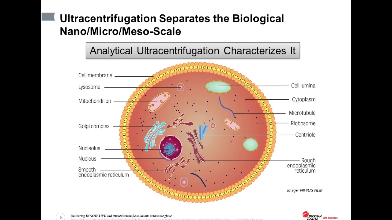
An Introduction to characterization of biomolecules using analytical ultracentrifugation
2.2K
Modern AUC instruments like the Optima AUC feature 4-hole rotors (60,000 rpm) or 8-hole rotors (50,000 rpm) with sector-shaped cells containing air gaps, buffer regions, and sample regions. Detection uses interference or absorption optics to record concentration versus radial distance over time, tracking the meniscus boundary movement. Two experimental approaches exist: sedimentation equilibrium (low speed, long duration, 6-sector centerpieces) for accurate molecular weight and binding constants; sedimentation velocity (high speed, short duration, 2-sector centerpieces) for molecular shape determination. Multi-wavelength detection enables simultaneous monitoring of multiple components within single experiments. Sample loading uses narrow-stem pipette tips, with typical volumes around 450 μL requiring absorbance 0.1-1.0 at 230nm for optimal detection.
Click to watch →
Gradient Basics
Applications
Gradient Basics
0:02- 1
Explains stepwise sucrose gradient construction for density separation.
- 2
Particles migrate to layers matching their own density during spinning.
- 3
Fractionation after centrifugation enables easy sample purification.
Artifacts of Hydrostatic Pressure and Shear Stress: The Shift to Chromatographic and Gentle Biophysical Alternatives
While density gradient centrifugation is a foundational technique for separating biomolecules, it has significant limitations that have led researchers to adopt alternative methods. The intense gravitational forces (often exceeding 100,000 x g) generate extreme hydrostatic pressure and shear stress. This can cause artifactual dissociation of weakly bound macromolecular complexes, denature sensitive proteins, and damage genomic DNA or delicate replication intermediates like Okazaki fragments. Furthermore, preparing sucrose gradients is time-consuming and suffers from low throughput. To overcome these limitations, modern biochemistry increasingly relies on gentler, high-resolution alternatives. Techniques such as Size Exclusion Chromatography (SEC), Asymmetric Flow Field-Flow Fractionation (AF4), and native Mass Spectrometry (MS) allow researchers to isolate and analyze macromolecular complexes in their native states without subjecting them to destabilizing physical forces. These modern methods provide more accurate representations of in vivo molecular interactions, offering a critical counterpoint to the reliance on traditional ultracentrifugation.
Basic principles of centrifugation, including centrifugal force, sedimentation, and how a standard centrifuge operates.
The physical concepts of density, buoyancy, and viscosity, and how they influence the movement of particles in a liquid medium.
Fundamental structural properties of macromolecular biomolecules, specifically DNA, RNA, and proteins.
The chemical nature of solutions, solubility, and how concentration gradients (such as sucrose gradients) are established.
The distinction between rate-zonal (size and shape-dependent) and isopycnic (purely density-dependent) gradient centrifugation.
The historical significance of density gradients in molecular biology, such as the Meselson-Stahl experiment proving semi-conservative DNA replication.
Advanced purification workflows, including the isolation of sub-cellular organelles, viral particles, and membrane proteins.
Analytical ultracentrifugation (AUC) methods used to characterize the hydrodynamic properties and molecular weights of macromolecules.
89.1K views1.6Klikes2:30@quickbiochemistrybasicsOriginal Release: 2022-04-29
hey guys quick back M Basics here let's talk about density gradient centrifugation the term density gradient means density of liquid and centrifuge tube keeps changing as we move from the top to the bottom of the tube one of the most common ingredient used for making density gradient is a solution of sucrose the gradient is prepared in stepwise manner for this solution of sucrose with different concentration is prepared the most concentrated one is first layered in the bottom of the tube this is followed by stepwise layering with decreasing concentration of sucrose the topmost layer is least concentrated while the bottom layer is the most concentrated one the sample which is to be analyzed is layered on the top and centrifugation is carried out the particles which are very dense will travel towards the bottom layer while the particles which are less dense will remain on the top notice this carefully here as the moving particles reaches the sucrose layer which is equal in density the particles stop moving the particles get accumulated in different layers depending on their densities after centrifugation when the material into centrifuge tube is fractionated the purification of sample becomes very easy applications density gradient centrifugation is one of the widely used techniques in Biochemistry cell biology and molecular biology for example separation of different subunits of ribosomes separation of DNA containing n14 and N15 Isotopes separation of okazaki fragments is all possible with the use of density gradient centrifugation
Up Next
13:07
Ultracentrifugation: Principles, Types & Applications
@drbijoyachatterjee755
42.1K views•2020-08-04

50:12
Bacterial Communication: Quorum Sensing and Biofilm Formation Explained
@JHUAAP
12.6K views•2011-06-28

5:35
Enteric Nervous System Explained: The Gut's Brain | Neurobiology Lecture
@alumniu6029
438 views•2018-09-12

7:08
Bacteriophages: Earth's Deadliest Killers and Future Antibiotics
@kurzgesagt
34.6M views•2018-05-13
Related Study Plans & Knowledge Roadmaps
Structured learning paths in Biology
biology
RNAi: Biogenesis, RISC & Gene Silencing
01Molecular Biology Foundations: DNA to Protein
02Introduction to RNA Interference (RNAi)
03Molecular Biogenesis of miRNA and siRNA
+ 2 more learning modules
10h5 modules
Explore Roadmap
biology
Mycorrhizae: Signaling, Biophysics & Ecology
01Foundations of Mycorrhizal Symbiosis
02Molecular Signaling and Root Colonization
03Biophysics and Bioenergetics of Nutrient Exchange
+ 1 more learning modules
11h4 modules
Explore Roadmap
biology
Circadian Clocks: TTFL, SCN & Periphery
01Foundations of Molecular Biology & Gene Expression
02Introduction to Chronobiology and Actograms
03The Core TTFL: Molecular Architecture of the Clock
+ 3 more learning modules
14h6 modules
Explore Roadmap
biology
Population Genetics: Math, Drift & Selection
01Genetic & Probability Foundations
02The Hardy-Weinberg Principle and Baseline Models
03Mathematical Modeling of Natural Selection
+ 3 more learning modules
18h6 modules
Explore Roadmap