The striatum is a brain region consisting of the caudate nucleus, putamen, and nucleus accumbens, named for its striped appearance due to grey matter strands crossing the internal capsule; it serves as the primary input structure of the basal ganglia, where the dorsal striatum (caudate and putamen) processes movement-related signals via the nigrostriatal pathway, while the ventral striatum (nucleus accumbens) mediates reward and addiction through the mesolimbic dopamine pathway connecting to the ventral tegmental area.
Striatum: Anatomy, Function & Role in Basal Ganglia | 2-Min Neuroscience
Added:Deep Dive
Basic neuroanatomy, including the location and division of subcortical structures within the forebrain.
Parts of the Brain: Forebrain: Subcortical Structures (Intro Psych Tutorial #34)
10.3K
The forebrain contains several key subcortical structures: the thalamus directs sensory information to appropriate cortical areas; the hypothalamus regulates hunger, fight-or-flight responses, and motivation (the four Fs: feeding, fighting, fleeing, mating); the pituitary gland releases hormones that control other glands; the limbic system (including the hippocampus for memory formation and the amygdala for emotion) processes emotional memories; the basal ganglia controls voluntary movement and reward; and the corpus callosum connects the two brain hemispheres through a large bundle of nerve fibers.
Click to watch →
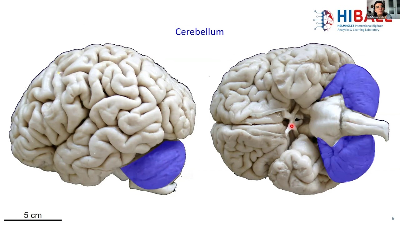
Day 1 - 02. BigBrain anatomy for beginners (Nicola Palomero-Gallagher)
333
Cortex refers to gray matter located on the surface of the brain hemispheres (from Latin 'cortex' meaning bark). Subcortical structures are gray matter located within the forebrain. Different subcortical nuclei can be identified in brain sections, including the caudate nucleus, putamen, nucleus accumbens, claustrum, thalamus, amygdala, globus pallidus, and hippocampus. The caudate nucleus and putamen together form the striatum, while the putamen and globus pallidus together form the lentiform nucleus.
Click to watch →
Central Nervous System | Explained by Dr. Rajeev Ranjan | NEET 2025 | Biology Lectures #neet2025
733.7K
The forebrain consists of the diencephalon (thalamus, hypothalamus, pineal gland) and cerebrum. The diencephalon serves as a sensory relay and homeostatic regulator. The cerebrum is divided into gray matter (cortex) and white matter (internal), with subcortical nuclei (clusters of gray matter) located within white matter. These nuclei form the basal ganglia and limbic system, involved in motor control, emotion, and memory. The corpus callosum connects the two cerebral hemispheres.
Click to watch →

Neuro-anatomy of fore brain 1
292
The forebrain (prosencephalon) is divided into the telencephalon (cerebrum) and diencephalon. The telencephalon consists of the cerebral cortex (gray matter with neuronal cell bodies) and white matter (myelinated nerve fibers), divided into four lobes: frontal lobe (personality, planning, decision-making, motor control), parietal lobe (sensory processing), temporal lobe (auditory processing, memory, language), and occipital lobe (visual processing). The cerebral cortex is folded into gyri and sulci to increase surface area. The diencephalon contains the thalamus (sensory relay), hypothalamus (homeostatic regulation), and limbic system structures (amygdala, hippocampus) involved in emotions and memory. The corpus callosum connects the two cerebral hemispheres, and the basal ganglia (caudate, putamen, globus pallidus) control voluntary movements.
Click to watch →
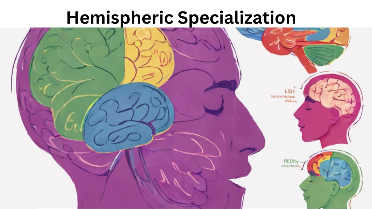
Understanding the Forebrain Your Brain's Control Center
2K
Beneath the cerebral cortex are several important subcortical structures including the thalamus, hypothalamus, amygdala, and basal ganglia. Each of these structures plays distinct roles in various brain functions.
Click to watch →
Deep Dive
Fundamentals of synaptic transmission and the primary neurotransmitters involved in motor and reward pathways (specifically dopamine, GABA, and glutamate).

3.1 Synaptic transmission
11K
Neurons communicate at specialized junctions called synapses. An action potential in a presynaptic neuron travels down the axon to release neurotransmitters that bind to postsynaptic receptors, changing membrane potential. Glutamate is the main excitatory neurotransmitter, increasing firing probability, while GABA is the primary inhibitory neurotransmitter, decreasing firing. Acetylcholine was the first identified neurotransmitter, functioning at neuromuscular junctions. Dopamine is involved in reward-based learning and linked to Parkinson's disease and schizophrenia. Beyond small molecules, neuropeptides like met-enkephalin and oxytocin serve as neurotransmitters, with new ones continually being discovered.
Click to watch →

«Химия» мозга
5.7K
Acetylcholine was the first neurotransmitter discovered, identified in the 1920s through studies of the neuromuscular junction. It plays a crucial role in muscle contraction and in the brain regulates arousal, attention, and learning. Curare blocks acetylcholine receptors at neuromuscular junctions, causing paralysis, while nicotine acts as an agonist at acetylcholine receptors in the brain. Glutamate and GABA are the two most important neurotransmitters in the brain, accounting for approximately 80% of all synaptic transmission. Glutamate is the primary excitatory neurotransmitter, while GABA is the primary inhibitory neurotransmitter. The balance between them is crucial for normal brain function—imbalance can cause seizures (too much glutamate) or excessive sedation (too much GABA). Benzodiazepines enhance GABA effects and are used to treat anxiety, seizures, and insomnia. Dopamine is associated with reward, motivation, and movement, with dysfunction implicated in Parkinson's disease (dopamine loss) and schizophrenia (dopamine excess).
Click to watch →

L 2, Neurotransmitters 1, Biochemistry
526
Neurotransmitters are chemical messengers that transmit signals between neurons or from neurons to other cells. Receptors are protein structures on the cell surface that bind to specific neurotransmitters and trigger cellular responses. There are two main types of receptors: ionotropic receptors directly open ion channels when neurotransmitters bind, resulting in fast synaptic transmission. Metabotropic receptors activate G-proteins and second messenger systems, which then open ion channels, resulting in slower but more complex responses. G-proteins include Gs (stimulatory), Gi (inhibitory), and Gq types. Neurotransmitters can be classified into six main categories: amino acids (glutamate, aspartate, glycine, GABA), amines (dopamine, serotonin, norepinephrine), acetylcholine, purines, peptides, and gases. Glutamate is the major excitatory neurotransmitter, while GABA is the major inhibitory neurotransmitter. During synaptic transmission, an action potential arrives at the presynaptic neuron and opens voltage-gated calcium channels, triggering neurotransmitter release. Excitatory neurotransmitters depolarize the postsynaptic membrane by opening sodium channels, while inhibitory neurotransmitters hyperpolarize the membrane by opening potassium or chloride channels. Glutamate receptors include ionotropic receptors (NMDA, AMPA, and kainate) and metabotropic receptors (mGluRs). NMDA receptors require both glutamate binding and membrane depolarization to open, allowing calcium influx that triggers signaling cascades for synaptic plasticity. After neurotransmitter release, glutamate is rapidly removed from the synaptic cleft by astrocytes through sodium-dependent glutamate transporters. Glutamate is converted to glutamine by glutamine synthetase in astrocytes, and glutamine is transported back to neurons and converted back to glutamate by glutaminase, completing the glutamate-glutamine cycle. GABA is synthesized from glutamate by glutamate decarboxylase, requiring pyridoxal phosphate (vitamin B6) and zinc as cofactors. GABA receptors include ionotropic receptors (GABA-A and GABA-B) and metabotropic receptors. GABA-A receptors are ligand-gated chloride channels that mediate fast inhibitory transmission. GABA-B receptors are G-protein coupled receptors that mediate slower inhibitory effects. Benzodiazepines bind to GABA-A receptors at a site distinct from the GABA binding site, enhancing the effect of GABA by increasing the frequency of chloride channel opening. Barbiturates bind to GABA-A receptors at a different site, enhancing the effect of GABA by increasing the duration of channel opening. Barbiturates can activate GABA-A receptors in the absence of GABA, which can lead to respiratory depression and death.
Click to watch →
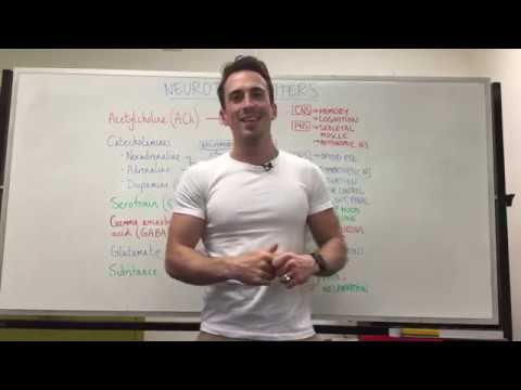
Neurotransmitters | Nervous System
396.4K
Dopamine, a key catecholamine, mediates reward and motivation through D1 (excitatory) and D2 (inhibitory) receptors in the basal ganglia. Dopamine deficiency causes Parkinson's disease, impairing movement initiation and coordination. Serotonin regulates sleep and mood through multiple receptor subtypes (inhibitory 5-HT1/5-HT5 and excitatory 2/3/4/6/7). GABA serves as the primary inhibitory neurotransmitter, preventing excessive neuronal firing through chloride influx causing hyperpolarization; GABA enhancement treats epilepsy. Glutamate, the most abundant excitatory neurotransmitter, activates NMDA and AMPA receptors but excessive release causes neurotoxicity linked to dementia and Alzheimer's disease. These neurotransmitters collectively govern complex CNS functions from cognition to neurodegeneration.
Click to watch →
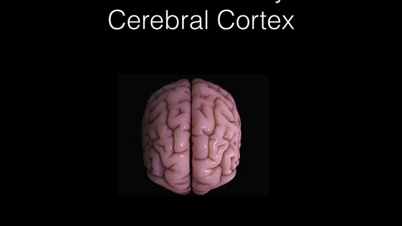
Clinical Neuroscience for Psychiatrists Lecture I: History, Neuroanatomy, Action Potential,Chemistry
1.5K
Neurons communicate through electrical and chemical signals. Action potentials are rapid membrane potential changes generated by sodium and potassium ion movements. Neurotransmitters (dopamine, serotonin, glutamate, GABA) transmit signals across synapses. Dopamine is involved in reward and motor control; serotonin regulates mood; glutamate is the primary excitatory neurotransmitter; GABA is the primary inhibitory neurotransmitter. Synaptic plasticity underlies learning and memory.
Click to watch →
Deep Dive
An introductory understanding of the motor cortex and how the brain plans and initiates voluntary movement.

أحلل حركة إرادية رابعة متوسط علوم طبيعية | الدرس 16
172.7K
Voluntary movement is a conscious, purposeful action performed with awareness and intention to meet body needs, controlled by the motor cortex (specifically the precentral gyrus) which generates motor nerve impulses that travel through motor nerves to effector organs (muscles or glands); the motor cortex exhibits contralateral control where the right hemisphere controls left body movements and vice versa, with different body parts represented in specific regions of the motor homunculus, and motor impulses follow two pathways: direct pathways for head/neck muscles and pathways through the spinal cord for limb muscles.
Click to watch →

La Corteccia Motoria (motoria primaria, motoria supplementare, premotoria)
24.9K
The motor cortex, located in the frontal lobe, consists of three interconnected areas—the primary motor cortex (area 4), premotor cortex, and supplementary motor area (area 6)—that work hierarchically to produce voluntary movement: the parietal and prefrontal areas first decide what action to take, the supplementary motor area and premotor cortex plan the movement strategy, and the primary motor cortex initiates the actual movement by sending signals to motor neurons; these areas exhibit somatotopic organization, meaning different cortical regions correspond to different body parts, and the supplementary motor area is particularly important for coordinating bilateral movements and planning complex movement sequences.
Click to watch →
Ulepszanie ludzkiego mózgu – co już się dzieje a co się (raczej) nie wydarzy? | dr Paweł Boguszewski
97.8K
The motor cortex, located in the frontal lobe, plays a crucial role in planning and initiating voluntary movements. Research using brain-computer interfaces has demonstrated that imagining a movement (without physically executing it) activates the same neural patterns as actually performing the movement. This finding has enabled paralyzed patients to control external devices by simply thinking about desired movements, as the motor cortex generates the neural signals necessary for movement regardless of whether the body can execute them.
Click to watch →

Videoaula 10 Sistema somatomotor (PARTE 2) - COMO MOVEMOS O NOSSO CORPO?
31.6K
Voluntary movements are intentional actions initiated by internal decisions, originating from neurons in the motor cortex. The motor cortex comprises three main areas: primary motor cortex (execution), premotor cortex (planning), and supplementary motor cortex (planning). The prefrontal cortex aids decision-making about which movement to perform. Motor commands follow a pathway: decision-making in prefrontal cortex, planning in premotor and supplementary motor cortices, then execution in primary motor cortex. Motor neurons are organized somatotopically in the primary motor cortex, forming a motor homunculus where larger body part representations correlate with greater movement precision. Commands reach spinal motor neurons via the corticospinal tract, with 90% crossing to the contralateral side in the medulla to innervate distal muscles, while anterior fibers innervate proximal muscles for posture.
Click to watch →

The Motor Cortex and You
11.7K
The motor cortex, located on the top center of the brain, controls voluntary movements through five interconnected regions: the primary motor cortex (generating neural impulses for movement), premotor cortex (preparing and guiding movements), supplementary motor area (planning and coordinating bilateral movements), posterior parietal cortex (transforming sensory information into motor commands), and primary somatosensory cortex (functionally part of motor circuitry). Movement planning is collected in area 6 and delivered to area 4, which then sends commands to specific body areas. The cerebellum regulates the timing and sequence of these movements, ensuring smooth execution.
Click to watch →
Deep Dive
The concept of the brain's reward system and the basic principles of operant conditioning (reinforcement).
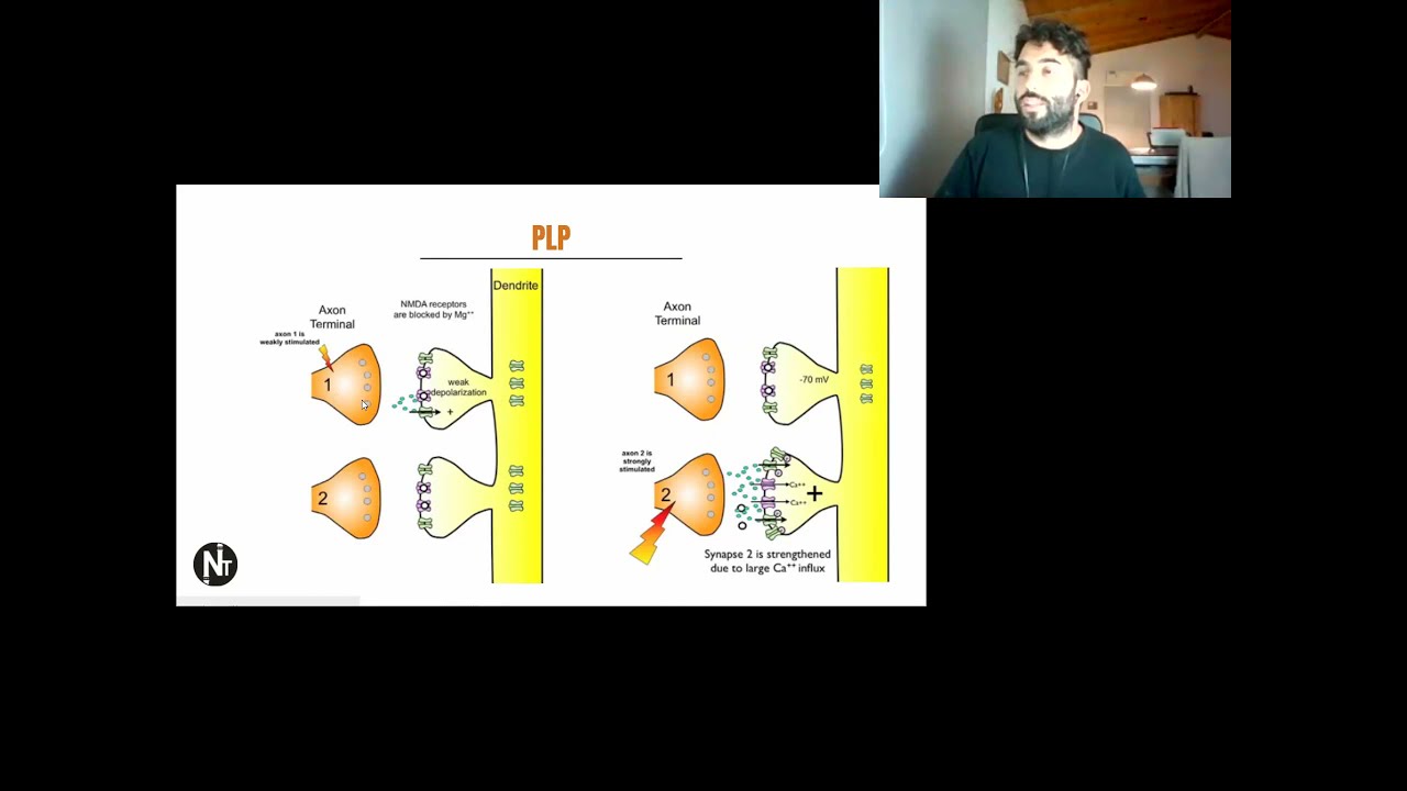
🗣️ CLASES SINCRÓNICAS - SEMANA 10 y 11 - MEMORIA Y APRENDIZAJE
3.2K
Operant conditioning involves learning from consequences: behaviors producing favorable outcomes are strengthened, while those producing unfavorable outcomes are weakened. The brain contains specialized reward circuits that mediate reinforcement. Self-stimulation experiments show organisms will work to produce stimulation in reward circuits, even overriding other drives. The reward system involves two components: 'liking' (pleasure, mediated by opioids) and 'wanting' (motivation, mediated by dopamine). This distinction is important for understanding addiction and motivation.
Click to watch →

REPLAY - Webinar de la section Addictions : "La clinique des addictions revisitée"
411
All animals, including humans, are sensitive to reward, which generates operant conditioning. When the brain encounters an unexpected reward, dopamine is released. Over time, the environment associated with the reward becomes sufficient to trigger reward signals. The brain eventually anticipates the reward and produces reward signals during anticipation, even if the actual reward disappears. This mechanism explains how behaviors can become self-sustaining without the original reward.
Click to watch →
MODÜL 12 BİLİŞSEL DÜŞÜNME BECERİLERİ (Çıkması Muhtemel Sorular 1)
2.5K
Operant conditioning is an organism's adjustment of behavior based on responses to stimuli. Unlike classical conditioning (involuntary, species-specific behaviors), operant conditioning involves conscious responses. B.F. Skinner discovered this through experiments with rats and pigeons. Reinforcement (positive or negative) increases behavior frequency: positive reinforcement adds pleasant stimuli, negative reinforcement removes unpleasant stimuli. Punishment (Type 1 adds unpleasant stimuli, Type 2 removes pleasant stimuli) is less effective. The brain's reward system, particularly the mesolimbic pathway and dopamine, underlies this learning mechanism.
Click to watch →

Comportamentos motivados M4 A5- Papel do sistema dopaminérgico mesolímbico no reforço comportamental
4.8K
Reinforcement strengthens behavior through two mechanisms: positive reinforcement (acquisition of pleasurable responses) and negative reinforcement (removal of aversive conditions). Both increase behavior repetition. Punishment, involving aversive consequences, decreases behavior. Operant conditioning, developed by Thorndike and expanded by Skinner, demonstrated that behaviors with satisfactory outcomes are repeated. Animals learn to associate specific actions (like lever-pressing) with rewarding outcomes, enabling efficient goal-directed behavior.
Click to watch →

Viva Melhor - Dê Atenção ao que Realmente Importa
1.1K
The brain has a powerful reward system that operates through two main pathways: positive reinforcement and negative reinforcement (punishment). When a behavior receives a reward, it is likely to repeat and increase in frequency. Conversely, when a behavior receives punishment or criticism, it tends to decrease in frequency. This fundamental mechanism explains how behaviors are shaped and maintained in our daily lives.
Click to watch →
Prerequisite Knowledge
- Concept 01Basic neuroanatomy, including the location and division of subcortical structures within the forebrain.
- Concept 02Fundamentals of synaptic transmission and the primary neurotransmitters involved in motor and reward pathways (specifically dopamine, GABA, and glutamate).
- Concept 03An introductory understanding of the motor cortex and how the brain plans and initiates voluntary movement.
- Concept 04The concept of the brain's reward system and the basic principles of operant conditioning (reinforcement).
Subsequent Learning
- Step 01The Direct and Indirect Pathways of the Basal Ganglia, illustrating how the striatum modulates movement facilitation and inhibition.
- Step 02Pathophysiology of movement disorders associated with striatal dysfunction, such as Parkinson's disease and Huntington's disease.
- Step 03The neurobiology of addiction, focusing on how substances of abuse hijack the mesolimbic dopamine pathway and the nucleus accumbens.
- Step 04The role of the striatum in habit formation, procedural learning, and goal-directed decision-making.
Defining Striatum
Name Origin
Dorsal/Ventral Split
Basal Ganglia Hub
Movement Inputs
Reward Processing
Reward Circuitry
Defining Striatum
0:05Playing Section
- 1
Striatum refers to a group of structures below the cerebral cortex.
- 2
It includes the caudate, putamen, and nucleus accumbens.
The Continuous Kinematic Control and Vigor Model
While traditional neuroscience views the striatum through the lens of discrete "Go/No-Go" pathways that initiate or inhibit actions, an alternative perspective proposes that it operates as a continuous feedback controller. This model argues that the striatum does not merely select and trigger distinct movements, but continuously regulates kinematic variables such as velocity, force, and vigor during the execution of an action. This challenges the classical binary gating hypothesis by showing that striatal activity dynamically scales with real-time movement parameters, integrating motivation directly into motor performance rather than treating action selection and execution as separate, sequential processes.
Deep Dive
The Direct and Indirect Pathways of the Basal Ganglia, illustrating how the striatum modulates movement facilitation and inhibition.

Basal Ganglia | Code Red 2.0 | Dr. Prachi, MD Psychiatry, (AIIMS Delhi) - "The Final Sprint Mentor"
112
The basal ganglia contain two pathways that modulate movement: the direct pathway facilitates movement, while the indirect pathway inhibits it. Both originate from cortex, go to striatum, then to thalamus, and provide feedback to cortex. The direct pathway involves cortex → striatum → globus pallidus internus → thalamus → cortex. The indirect pathway adds subthalamic nucleus and globus pallidus externus. Dopamine from substantia nigra pars compacta binds to D1 receptors (direct) or D2 receptors (indirect) in striatum. D1 activation disinhibits thalamus, enabling movement. D2 activation activates subthalamic nucleus, which inhibits thalamus, suppressing movement.
Click to watch →
Basal Ganglia | Neuroanatomy | Part 2/3
309.2K
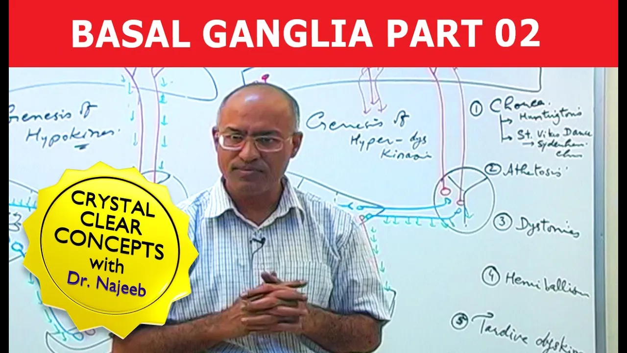
The basal ganglia regulate movement through two parallel pathways: the direct pathway (striatum → globus pallidus internus → thalamus → cortex) which facilitates movement by disinhibiting thalamocortical fibers, and the indirect pathway (striatum → globus pallidus externus → subthalamic nucleus → globus pallidus internus → thalamus → cortex) which inhibits movement by enhancing inhibition. Dopamine from the substantia nigra pars compacta modulates these pathways through D1 receptors (stimulating direct pathway) and D2 receptors (inhibiting indirect pathway), acting as a fine-tuner to initiate and facilitate voluntary movements.
Click to watch →

Basal Ganglia Direct & Indirect Pathways Physiology (7-2025) by Dr Khaled A Abulfadle
216
The direct pathway facilitates movement through disinhibition: cortex sends excitatory (glutamate) projections to striatum, which sends inhibitory (GABA) projections to globus pallidus internus, disinhibiting thalamus to send excitatory projections to motor cortex. The indirect pathway suppresses movement: cortex sends excitatory projections to striatum, which sends inhibitory projections to globus pallidus externus, which sends excitatory projections to substantia nigra pars reticulata, which sends inhibitory projections to thalamus, inhibiting motor cortex. Both pathways work in balance for normal motor control.
Click to watch →

Pathways of the Basal Ganglia
5.2K
The basal ganglia modulate motor control through two complementary pathways. The direct pathway increases excitatory output from the thalamus to the cortex, promoting movement. The indirect pathway reduces this output, inhibiting movement. Key structures include the motor cortex, striatum, Globus pallidus (internal and external), thalamus, subthalamic nucleus, and substantia nigra (pars reticulata and pars compacta). Glutamate provides excitatory signals, GABA provides inhibitory signals, and dopamine from the pars compacta modulates both pathways through D1 receptors (direct pathway, excitatory effect) and D2 receptors (indirect pathway, inhibitory effect).
Click to watch →

DESCOMPLICANDO NÚCLEOS DA BASE E A DOENÇA DE PARKINSON - DESCOMPLICA, NEURO!
3.1K
The basal ganglia have two main pathways: (1) Direct pathway facilitates movement - cortex excites striatum, which inhibits internal globus pallidus, reducing inhibition on thalamus and enabling movement. (2) Indirect pathway inhibits movement - cortex excites striatum, which inhibits external globus pallidus, which excites subthalamic nucleus, which excites internal globus pallidus, increasing inhibition on thalamus and preventing movement. These pathways work together to maintain movement balance.
Click to watch →
Deep Dive
Pathophysiology of movement disorders associated with striatal dysfunction, such as Parkinson's disease and Huntington's disease.

Pathophysiology Week 7 Podcast
104
Parkinson's and Huntington's disease both affect the basal ganglia (brain's movement control center) but in opposite ways. Parkinson's causes overactivity in the 'stop' pathway due to dopamine deficiency, making movement initiation difficult. Huntington's causes underactivity in the 'stop' pathway from genetic mutations, resulting in uncontrollable movements. Both diseases show how delicate the balance between movement pathways must be, and how genetic factors can determine disease outcomes.
Click to watch →
USMLE Neurology 8 Movement Disorders: Parkinson's and Huntington's with Pharmacology
10.2K
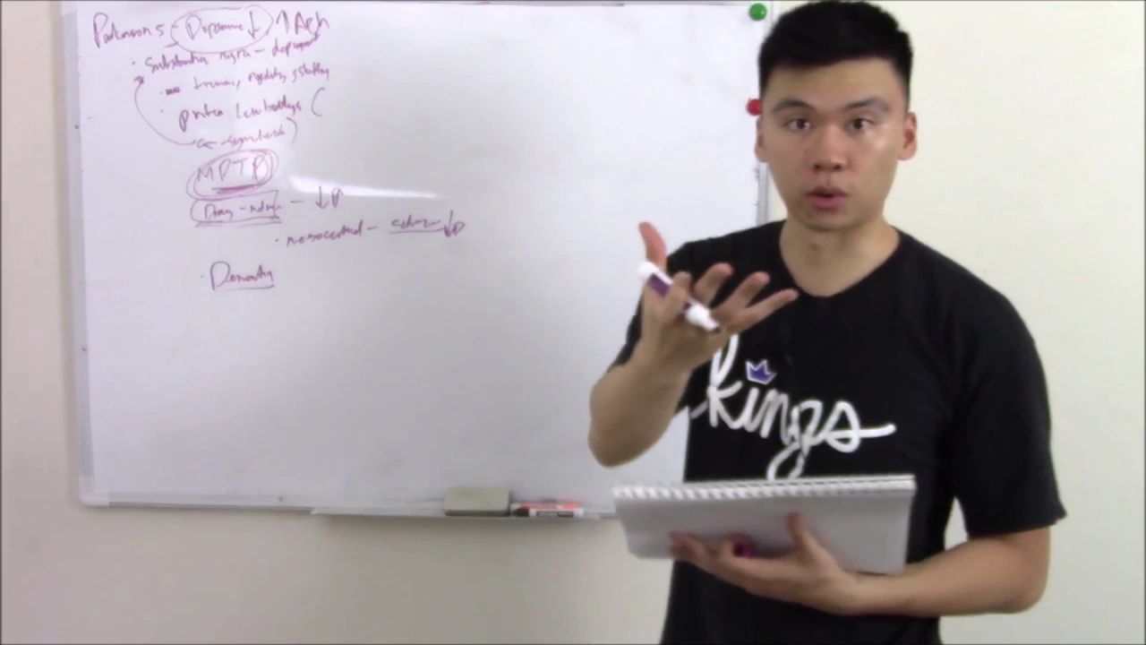
Movement disorders arise from dysfunction in the motor control system involving the cortex, cerebellum, and basal ganglia; Parkinson's disease results from degeneration of dopamine-producing neurons in the substantia nigra, causing resting tremor, rigidity, and bradykinesia, with treatment involving levodopa-carbidopa combinations and dopamine agonists; Huntington's disease is an autosomal dominant trinucleotide repeat disorder (CAG expansion on chromosome 4) that destroys the striatum, leading to chorea and cognitive decline, while Friedreich's ataxia (GAA expansion on chromosome 9) causes mitochondrial dysfunction affecting the cerebellum and spinal cord.
Click to watch →
Parkinson's and Huntington's disease
2.2K
Parkinson's disease is a chronic neurodegenerative disorder characterized by the loss of dopamine-producing neurons in the substantia nigra pars compacta, leading to reduced dopamine levels that cause underactivity of the direct basal ganglia pathway and overactivity of the indirect pathway, resulting in bradykinesia, rigidity, and resting tremor; in contrast, Huntington's disease is an autosomal dominant neurodegenerative disorder caused by CAG trinucleotide expansion on chromosome 4, which destroys striatal neurons and disrupts the indirect pathway, causing chorea (involuntary jerky movements), dementia, and psychiatric disturbances.
Click to watch →
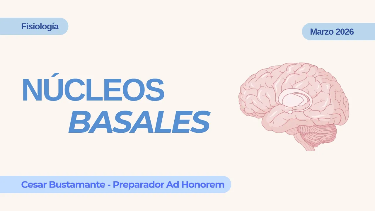
Núcleos Basales // Cesar Bustamante
104
Parkinson's disease results from degeneration of dopaminergic neurons in the nigrostriatal pathway. Reduced dopamine decreases stimulation of the direct pathway and decreases inhibition of the indirect pathway, resulting in net facilitation of the indirect pathway and movement inhibition. Symptoms include resting tremor, rigidity with cogwheel resistance, bradykinesia, micrographia, postural instability, and festinating gait. Huntington's disease is an autosomal dominant disorder from CAG repeat expansion in the huntingtin gene, causing degeneration of GABAergic neurons in caudate and putamen. This destroys the indirect pathway, eliminating its inhibitory effect and causing chorea, tics, dystonia, and psychiatric/cognitive changes. Hemiballismus results from subthalamic nucleus lesions reducing excitation of globus pallidus internus, decreasing thalamic inhibition and causing sudden, unilateral ballistic movements.
Click to watch →

🧠 GANGLIOS BASALES en el control del MOVIMIENTO 🏃🏼♀️
43.5K
Parkinson's disease results from degeneration of dopaminergic neurons in the substantia nigra, blocking dopamine modulation of basal ganglia circuits. Without dopamine's positive modulation of the direct pathway, voluntary movement decreases while the indirect pathway's inhibitory effects dominate, causing bradykinesia, rigidity, and resting tremor. Huntington's disease involves degeneration of striatal neurons, eliminating normal inhibition of the indirect pathway. This causes excessive disinhibition of subthalamic nucleus and GPi, leading to uncontrolled involuntary movements (chorea) while simultaneously impairing voluntary movement control.
Click to watch →
Deep Dive
The neurobiology of addiction, focusing on how substances of abuse hijack the mesolimbic dopamine pathway and the nucleus accumbens.
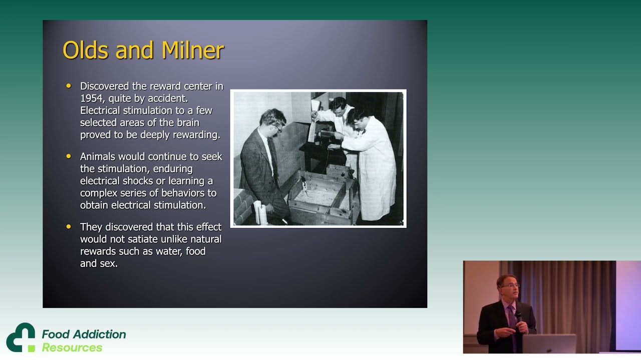
Why do humans develop addiction diseases? by Dr Paul Earley
2.5K
Modern addiction research evolved through three eras: the 1950s-1990s discovery of the brain's pleasure center by Olds and Milner, establishing addiction as a brain disease; subsequent focus on acute brain changes from substances; and current exploration of deeper relapse-promoting circuits. The mesolimbic dopamine system—ventral tegmental area projecting to nucleus accumbens—drives survival behaviors including hunger, nurturing, and safety-seeking. All abusable substances activate this circuit through various neurotransmitters, producing dopamine spikes. Unlike natural rewards which satiate, this circuit can be hijacked by substances to produce persistent, compulsive behavior patterns characteristic of addiction.
Click to watch →

Neurobiology of substance use (A Basic Introductory Lecture for Psychiatry Residents)
5.7K
The neurobiology of substance use primarily involves the mesolimbic dopaminergic pathway (ventral tegmental area to nucleus accumbens) and the cortico-striato-thalamo-cortical (CSTC) loop, with dopamine being the most important neurotransmitter; addiction progresses through a shift from impulsivity (inability to stop initiating actions, driven by the anterior cingulate cortex-ventral striatum-thalamus circuit) to compulsivity (inability to terminate ongoing actions, involving the orbital frontal cortex-thalamus circuit), where the dorsolateral prefrontal cortex normally exerts top-down control over both circuits, while the amygdala mediates reward conditioning and the hippocampus handles memory formation; the reinforcing effects of drugs depend not just on dopamine presence but on the rate at which dopamine increases in the brain, with IV and inhalational routes producing the fastest effects and different drugs having different reward values based on their mechanism of action.
Click to watch →

Clase 2/ 06-02-26 Neuropsicología Clínica y Psicología Terapéutica
1K
This segment explains the neurobiology of addiction through the lens of dopamine pathways. All humans have four dopaminergic pathways: mesolimbic (reward, motivation, pleasure), mesocortical (decision-making, impulse control), nigrostriatal (movement control), and tuberoinfundibular (prolactin regulation). Substance abuse creates unnatural dopamine levels, particularly in the mesolimbic pathway. The mesocortical pathway shows decreased serotonin, impairing decision-making and impulse control. The nigrostriatal pathway affects movement, explaining motor symptoms in addiction. When the brain experiences excessive dopamine from substances, it adapts by requiring higher levels to feel the same pleasure, creating anhedonia where normal activities no longer produce satisfaction.
Click to watch →

“El Secreto para Dominar tus Pensamientos — CÓMO EMPEZAR HOY” – Mario Alonso Puig
3.8K
In the brainstem, the mesolimbic pathway emerges from the substantia nigra (named for its dark pigment) to the limbic system, specifically to the nucleus accumbens. Dopamine is released from the substantia nigra to the nucleus accumbens, producing the pleasure sensation that can lead to addiction. The sustained activation of the nucleus accumbens by constant dopamine release causes the brain to regulate by reducing dopamine receptors, producing more dopamine for the same effect - a vicious cycle. Modern addictions (gambling, pornography, social media) are related to this mesolimbic circuit. The hypothalamus is a humble structure (size of a pea) that controls the body's biochemistry, temperature, and hormones, and without it, one cannot survive.
Click to watch →

The Mesolimbic Pathway: Addiction, Desire & the Hijacked Brain
108
The mesolimbic pathway is a crucial neurocircuit in the brain's reward system, involving dopamineergic neurons projecting from the ventral tegmental area (VTA) to the nucleus accumbens and hippocampus. This pathway is heavily implicated in motivation, reward, learning, and pleasure experiences. It serves as the primary pathway targeted in substance abuse, as individuals with hypoactive mesolimbic systems use substances like cocaine and amphetamines to activate it. The pathway contains multiple receptor types including dopamine receptors (D1 and D2), opioid receptors, muscarinic receptors, and glutamate receptors. Cocaine's addictive properties are rooted in its ability to activate this system, causing compulsive drug use despite adverse consequences and high relapse rates. The mesocortical dopamine system, with projections to limbic and cortical regions, is central to understanding these neuroadaptations.
Click to watch →
Deep Dive
The role of the striatum in habit formation, procedural learning, and goal-directed decision-making.

9 - Reinforcement learning in the brain
468
Different brain regions support habitual versus goal-directed behaviors. In rats, dorsal lateral striatum lesions impair habits but leave goal-directed responding intact, while dorsal medial striatum lesions impair goal-directed behavior but preserve habits. This parallels computational distinctions between model-free (habits) and model-based (goal-directed) learning algorithms. Model-free learners don't immediately adapt to changed reward functions, while model-based learners update their internal models and adjust responses accordingly.
Click to watch →
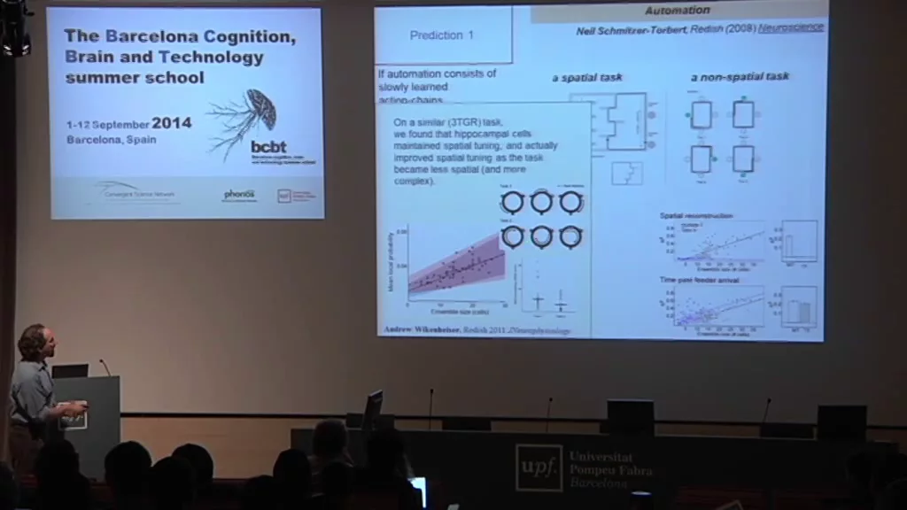
BCBT'14 David Redish
546
Habit formation involves shifting from deliberative to automatic behavior controlled by dorsal striatum. As animals repeat behaviors, hippocampal and ventral striatal representations of deliberation fade, replaced by dorsal striatal representations that encode action-outcome associations. Dorsal striatum develops 'cue-response' cells that fire at specific locations regardless of outcome, reflecting learned stimulus-response associations. This transition explains why hippocampal lesions allow automation of spatial navigation—once habits form, the hippocampus is no longer needed for the behavior.
Click to watch →
Jeffrey Schwartz: You Are More than Your Brain - Science Uprising Extra Content
74.6K
The striatum (caudate nucleus/basal ganglia) serves as a habit center in the brain. When behaviors are repeated, they get wired into this area, allowing the brain to run the outer cortex very quickly and automatically. This creates unconscious, automatic behaviors that make daily life possible—without these mechanisms, even simple actions like getting out of a chair would require thinking through every movement.
Click to watch →
Reward, Reinforcement and the Neural Bases of Decision-making - Bernard Balleine
3.4K
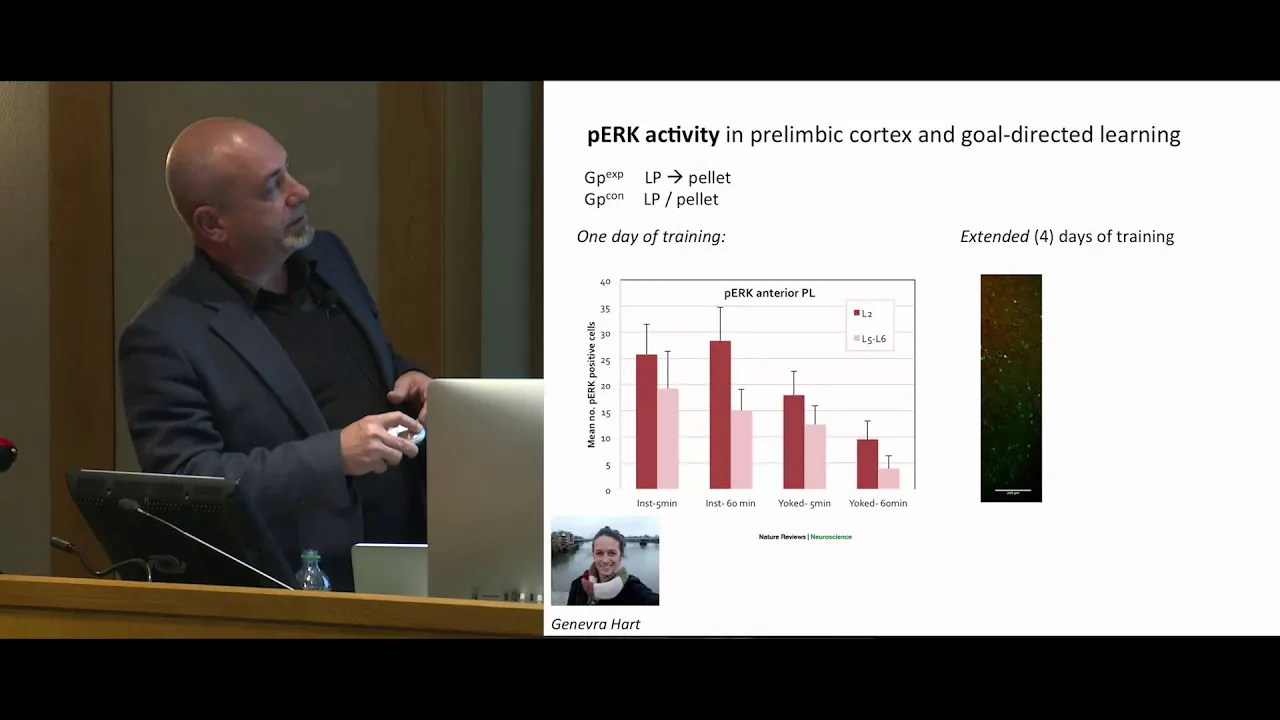
This section presents evidence for parallel neural circuits governing goal-directed and habitual actions. Goal-directed actions involve prefrontal cortical input to dorsomedial striatum, with BLA providing reward signals. Habitual actions involve sensory-motor inputs to dorsolateral striatum, with central amygdala providing reinforcement signals. Lesions to dorsolateral striatum eliminate habitual behavior in overtrained animals, restoring goal-directed control. Electrophysiological recordings reveal depression in D2 receptor neurons of the dorsolateral striatum correlates with habit formation. This post-synaptic depression involves TRPV1 receptors, as capsaicin combined with subthreshold quinpirole produces similar effects and TRPV1 knockout mice fail to develop habits. The basolateral amygdala inhibits the central nucleus, preventing premature habit formation. When this inhibition is removed, habits emerge more rapidly. The infralimbic cortex normally disinhibits the central nucleus, allowing reinforcement signals to drive habit formation.
Click to watch →
A. David Redish: "Cross-species translation through computational analyses - implications for un..."
378
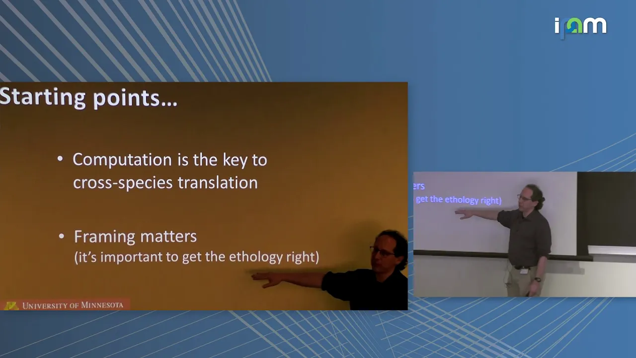
The dorsal striatum supports procedural learning through task-bracketing bursts at action initiations, representing ballistic movements requiring no further decisions. Unlike the hippocampus's prospective coding during deliberation, the striatum represents current location and appropriate actions. As animals learn, they transition from hippocampus-dependent deliberative strategies to striatum-dependent procedural strategies. This flexible switching between computational modes demonstrates how the brain adapts decision-making to task demands, with different neural circuits supporting different aspects of behavior depending on whether the situation requires planning or habit-based execution.
Click to watch →
Defining Striatum
Name Origin
Dorsal/Ventral Split
Basal Ganglia Hub
Movement Inputs
Reward Processing
Reward Circuitry
Defining Striatum
0:05- 1
Striatum refers to a group of structures below the cerebral cortex.
- 2
It includes the caudate, putamen, and nucleus accumbens.
The Continuous Kinematic Control and Vigor Model
While traditional neuroscience views the striatum through the lens of discrete "Go/No-Go" pathways that initiate or inhibit actions, an alternative perspective proposes that it operates as a continuous feedback controller. This model argues that the striatum does not merely select and trigger distinct movements, but continuously regulates kinematic variables such as velocity, force, and vigor during the execution of an action. This challenges the classical binary gating hypothesis by showing that striatal activity dynamically scales with real-time movement parameters, integrating motivation directly into motor performance rather than treating action selection and execution as separate, sequential processes.
Basic neuroanatomy, including the location and division of subcortical structures within the forebrain.
Fundamentals of synaptic transmission and the primary neurotransmitters involved in motor and reward pathways (specifically dopamine, GABA, and glutamate).
An introductory understanding of the motor cortex and how the brain plans and initiates voluntary movement.
The concept of the brain's reward system and the basic principles of operant conditioning (reinforcement).
The Direct and Indirect Pathways of the Basal Ganglia, illustrating how the striatum modulates movement facilitation and inhibition.
Pathophysiology of movement disorders associated with striatal dysfunction, such as Parkinson's disease and Huntington's disease.
The neurobiology of addiction, focusing on how substances of abuse hijack the mesolimbic dopamine pathway and the nucleus accumbens.
The role of the striatum in habit formation, procedural learning, and goal-directed decision-making.
145.2K views1.6Klikes1:55@neurochallengedOriginal Release: 2016-06-25
Welcome to 2 minute neuroscience, where I simplistically explain neuroscience topics in 2 minutes or less.
In this installment I will discuss the striatum.
Striatum is a term used to collectively refer to a small group of structures found below the cerebral cortex.
These structures consist of the caudate, putamen, and nucleus accumbens.
The caudate and putamen are separated from one another by a white matter tract called the internal capsule, but there are many strands of grey matter that cross the internal capsule, giving the structure a striped appearance.
This is why the term striatum, Latin for striped, is used to describe the region.
The striatum is often conceptualized a being divided into dorsal and ventral sections; the dorsal striatum contains the caudate and putamen while the ventral striatum contains the nucleus accumbens.
The striatum is one of the principal components of the basal ganglia, a group of structures best known for their role in facilitating movement.
The dorsal striatum is one of the primary input areas for the basal ganglia, and fibers from the cerebral cortex, substantia nigra, and thalamus all enter the basal ganglia via the dorsal striatum.
The incoming fibers from the substantia nigra, which make up a pathway called the nigrostriatal pathway, are thought to be especially important to movement and are severely affected by neurodegeneration in patients with Parkinson’s disease.
The nucleus accumbens, part of the ventral striatum, has been extensively studied for its role in rewarding experiences.
The nucleus accumbens seems to be involved in reinforcement, reward, and the progression from simply experiencing something pleasurable to seeking it out compulsively as part of an addiction.
The ventral striatum is thus activated when we do something we find pleasurable.
The nucleus accumbens receives fibers from a dopamine-rich structure in the midbrain called the ventral tegmental area.
These fibers are part of a pathway called the mesolimbic dopamine pathway which is a primary component of the reward system.
Up Next

8:26
Reward Pathway in the Brain: Dopamine and Addiction (MCAT)
@khanacademymedicine
527.4K views•2014-06-25

21:55
Deciphering Neural Circuits Controlling Anorexia in Mice | Richard Palmiter
@AllenInstitute
2.3K views•2011-10-10
1:59
How Smell Works: Olfaction Explained in 2 Minutes | Neuroscience
@neurochallenged
303.7K views•2018-01-31

13:03
How Exercise Benefits Your Brain: Science Explained
@TED
11.4M views•2018-03-21
Related Study Plans & Knowledge Roadmaps
Structured learning paths in Neuroscience
neuroscience
Appetite Regulation: Brain Circuits & Hormones
01Foundations of Endocrinology and Brain Anatomy
02Introduction to Energy Homeostasis
03Peripheral Signals: Leptin, Ghrelin, and Insulin
+ 2 more learning modules
14h5 modules
Explore Roadmap
neuroscience
Neuroeconomics: Value, Risk & Brain Mechanisms
01Foundations of Neuroscience and Brain Anatomy
02Introduction to Neuroeconomics and Subjective Value
03Dopaminergic Prediction Errors & Reinforcement Learning
+ 3 more learning modules
18h6 modules
Explore Roadmap
neuroscience
Spatial Navigation: Cells, Grids & Brain Maps
01Foundations of Brain Anatomy and Memory
02Place Cells and Head-Direction Cells
03Grid Cells: The Brain's Coordinate System
+ 2 more learning modules
12h5 modules
Explore Roadmap
neuroscience
BCIs: Neural Decoding & Prosthetic Control
01Neuroscience Foundations: How the Brain Signals Intent
02Signal Acquisition: Recording the Brain's Activity
03Neural Decoding: Translating Brain Waves into Data
+ 2 more learning modules
25h5 modules
Explore Roadmap