Tumors require new blood vessel formation (angiogenesis) to sustain growth beyond a certain size; tumor cells secrete vascular endothelial growth factor (VEGF) and related proteins that stimulate endothelial cell proliferation, basement membrane degradation via matrix metalloproteinases, vessel permeability, and survival of new blood vessels, making VEGF signaling a key therapeutic target in cancer treatment.
Cancer Biology: Angiogenesis, VEGF, and Anti-Angiogenic Therapies
Added:Deep Dive
Basic cellular biology of cancer, including uncontrolled cell division, tumor progression, and the concept of metastasis.

Cáncer biología
4.5K
Cancer is defined in Latin as 'cangrejo' (crab) because it does not release its victim and must be surgically removed. Cancer cells lose control over growth and proliferation, forming tumors. Malignant tumors grow larger, multiply, and can metastasize. Tumors are classified by origin: epithelial (organ surfaces), sarcomas (bone/muscle), lymphomas (lymphatic system), and leukemias (blood). Cancer involves uncontrolled cell division with loss of balance between division and differentiation. Cancer cells become immortal through mechanisms preventing apoptosis, maintaining telomere length via telomerase. Normal cells have controlled growth regulated by P53 and RB proteins, while cancer cells bypass these controls.
Click to watch →

Cancer | benign and malignant tumor | class 12
4.8K
Cancer is characterized by uncontrolled cell division resulting from broken regulatory controls, leading to two basic types of tumors: benign tumors that remain localized and cause minimal harm, and malignant tumors (cancer) that invade surrounding tissues, enter the circulatory system, and spread to form secondary growth areas called metastases; cancer cells are distinguished from normal cells by their reduced differentiation, rapid growth, high nucleus-to-cytoplasm ratio, prominent nucleoli, and increased mitotic activity, and are primarily caused by somatic cell mutations that bring about two fundamental changes: loss of contact inhibition allowing cells to break free from barriers like the basement lamina, and unlimited proliferation without following the body's normal regulatory checkpoints.
Click to watch →
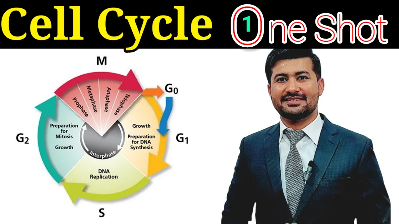
Cell cycle chapter 21 Fsc biology class 12 in one shot
2.4K
Cancer is characterized by uncontrolled cell division resulting from mutations in genes that normally regulate cell division. Cancer cells differ from normal cells by having uncontrolled division, high nucleus-to-cytoplasm ratio, irregular shape, prominent nucleolus, and rapid division. Tumors can be benign (localized, non-invasive) or malignant (invasive, capable of metastasis). Metastasis is the spread of cancer cells from the primary tumor to other body parts through the bloodstream or lymphatic system.
Click to watch →

a) Cancer is one of the most dreaded diseases of humans. Explain \'Contact inhibitions\' and
261
Cancer is a dangerous disease characterized by uncontrolled cell division. Normal cells have contact inhibition, a property where cells stop dividing when they touch neighboring cells. Cancer cells lose this property, allowing them to divide uncontrollably and form tumors. These cancer cells can break away from the primary tumor, enter the bloodstream, travel to distant body parts, and form new tumors at these locations—a process called metastasis. This ability to spread throughout the body makes cancer particularly dangerous and difficult to treat.
Click to watch →

B.Sc.Sem 1 Zoology One Shot Important Questions from Cell Biology, Genetics & Infectious Disease
21K
Cancer is a disease of uncontrolled cell division resulting from cell cycle control failure. When checkpoints fail, damaged cells continue dividing and accumulate, forming tumors. Benign tumors remain localized while malignant tumors can metastasize to other body parts. This occurs because normal regulatory mechanisms that limit cell proliferation are compromised.
Click to watch →
Deep Dive
The anatomy and physiology of the circulatory system, specifically how blood vessels (endothelial cells and capillaries) deliver oxygen and nutrients to tissues.

Cardiovascular System | Summary
222.2K
Blood vessels are lined with endothelial cells, which are simple squamous epithelial cells. Their flat structure facilitates rapid diffusion of substances across the vessel wall. Oxygen diffuses from red blood cells through endothelial cells into tissue cells, while carbon dioxide diffuses in the opposite direction. This single-cell barrier enables efficient exchange between blood and tissues.
Click to watch →
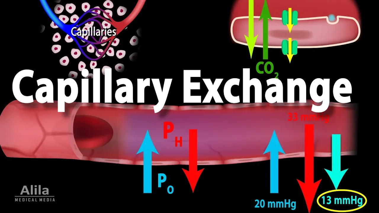
Capillary Exchange and Edema, Animation
736.6K
The circulatory system delivers oxygen and nutrients to tissues while removing wastes through capillaries—smallest blood vessels with single-layer endothelial walls. Substances cross capillary walls via diffusion (lipid-soluble molecules), facilitated diffusion (glucose via transporters), osmosis (water), and transcytosis (vesicles). Bulk flow occurs through intercellular clefts and fenestrated pores. Filtration happens at the arterial end where higher capillary blood pressure pushes plasma outward, while reabsorption occurs at the venous end. Hydrostatic pressure (higher inside capillaries) drives outward flow, opposed by oncotic pressure from blood proteins (mainly albumin). Since arterial ends are closer to the heart, pressure decreases along the capillary bed. Net filtration exceeds reabsorption, leaving ~15% fluid in tissues, which the lymphatic system returns to circulation.
Click to watch →

CARDIOVASCULAR SYSTEM, CVS, PART-1, BLOOD CIRCULATORY SYSTEM, ANATOMY, PHYSIOLOGY, BASIC FUNCTION,
12.9K
The cardiovascular system (CVS), also known as the circulatory system, is a major organ system consisting of the heart, blood, and blood vessels that delivers nutrients, oxygen, hormones, and immune cells to all body cells while removing metabolic wastes like carbon dioxide; the heart acts as a natural pump that generates blood pressure to propel blood through arteries (which carry blood away from the heart to organs), veins (which return blood back to the heart), and capillaries (which surround body cells to deliver and absorb oxygen and nutrients).
Click to watch →

THESE Foods Can STARVE Tumors (Scientific Proof) | Dr. William Li
15.7K
Capillaries are the finest blood vessels, thinner than a human hair, responsible for delivering oxygen and removing metabolic waste from tissues. At the tips of these blood vessels are endothelial cells, which line the interior walls of blood vessels like tiles in a kitchen. These endothelial cells secrete protein factors called survival factors that keep adjacent tissues alive and healthy.
Click to watch →

VAPE, CIGARRO e MACONHA destroem os vasos sanguíneos
370.6K
Endothelial cells are the cells that form the inner lining of capillaries, which are the smallest blood vessels in the body. These capillaries are responsible for transporting oxygen and other nutrients to the brain and other organs throughout the body.
Click to watch →
Deep Dive
Fundamental concepts of cell signaling, including how extracellular ligands bind to cell-surface receptors to trigger intracellular cascades.

Cell signaling| GPCR| types of receptors| type of ligands|cell surface receptors|cell communication|
167
Cell signaling is the communication process between cells, essential for organism development and function. Cells must receive and respond to signals from their surroundings to perform modifications and division. The three main stages are: (1) Receiving the signal when a signaling molecule binds to a receptor, (2) Signal transduction transmitting the signal from exterior to interior, and (3) Response where the cell reacts. Signaling molecules (ligands) are classified by polarity: hydrophilic molecules (peptides, amino acids, neurotransmitters like dopamine, histamine, serotonin, cytokines) cannot cross the plasma membrane and require cell surface receptors, while hydrophobic molecules (steroid hormones, retinoids, nitric oxide, vitamin D) can cross the membrane and bind intracellular receptors. There are four main types of cell signaling: (1) Autocrine signaling - cells respond to signals they themselves secrete, (2) Paracrine signaling - cells respond to signals from nearby cells, (3) Juxtacrine signaling - cells communicate through direct physical contact via transmembrane proteins, (4) Endocrine signaling - signaling molecules travel through the bloodstream to distant target organs. Receptors are classified into cell surface receptors (integral membrane proteins on the plasma membrane) and intracellular receptors (in cytoplasm or nucleus). Cell surface receptors bind hydrophilic signaling molecules and have three domains: extracellular (ligand binding), transmembrane (hydrophobic), and intracellular (signal transduction). Cell surface receptors are further classified into: (1) Ion channel-linked receptors - directly open ion channels when activated, (2) G protein-coupled receptors (GPCRs) - activate trimeric G proteins (alpha, beta, gamma subunits) to transmit signals, (3) Enzyme-linked receptors - have enzymatic activity (like tyrosine kinase) activated by ligand binding.
Click to watch →

CELL SIGNALING MPSET 2026 | COMPLETE BIOLOGY CLASSES | MPSET, NET 2026 BY SHIRIN MA’AM
4.6K
Cell signaling is the process by which cells communicate through chemical signals. A signaling cell creates molecules (ligands) that carry signals to target cells. The target cell has receptors that catch these signals and generate a response. Receptors are proteins located in the plasma membrane with three domains: extracellular domain (outside the cell, hydrophilic), transmembrane domain (embedded in the membrane), and cytoplasmic domain (inside the cell, hydrophilic). A ligand is a signal molecule that binds to receptors on target cells, also called primary messengers. Primary messengers carry signals from outside the cell to the cell membrane but do not enter the cell. When a primary messenger binds to a receptor, it triggers the activation of secondary messengers inside the cell. Extracellular signaling includes three main types: (1) Endocrine signaling - hormones released into bloodstream for long-distance communication; (2) Paracrine signaling - signals transmitted between nearby cells, such as neurotransmitters between neurons; (3) Autocrine signaling - cells signal to themselves, seen in cancer cells and T-cells. Juxtacrine signaling involves direct physical contact between cells through plasmodesmata in plants or cell junctions in animals. Cell receptors are classified into two main types: (1) Extracellular receptors - located on the cell surface and interact with signals outside the cell, including GPCRs, RTKs, and TGF receptors; (2) Intracellular receptors - located inside the cell (cytoplasm or nucleus) and interact with signals that can cross the cell membrane.
Click to watch →
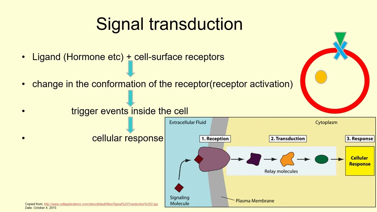
Cell Membrane & Its Proteins, Signal Transduction, Receptor & Messenger | Lecture 13c
108
Cellular signaling begins when extracellular ligands bind to cell surface receptors, causing conformational changes that trigger intracellular events. Receptors are classified as extracellular (embedded in plasma membrane, converting extracellular signals to intracellular signals) or intracellular (regulating gene expression). Nuclear receptors directly bind DNA to control gene transcription. Examples include G protein-coupled receptors (seven transmembrane domains, associated with heterotrimeric G proteins), receptor tyrosine kinases (with kinase domains), integrins (connecting cells to extracellular matrix), and ligand-gated ion channels (opening upon ligand binding).
Click to watch →
LIGAND AND RECEPTOR (PART-1) || CELL SIGNALING || CSIR NET || GATE LIFESCIENCE
54K
Ligands are signaling molecules secreted by signaling cells that bind specifically to receptors on target cells, inducing conformational changes to transmit signals; ligands are categorized into membrane-bound and secretory types, with secretory ligands further divided by distance traveled (endocrine, paracrine, autocrine) and by chemical nature (lipophilic molecules crossing the plasma membrane to bind intracellular receptors, versus hydrophilic molecules binding cell surface receptors).
Click to watch →
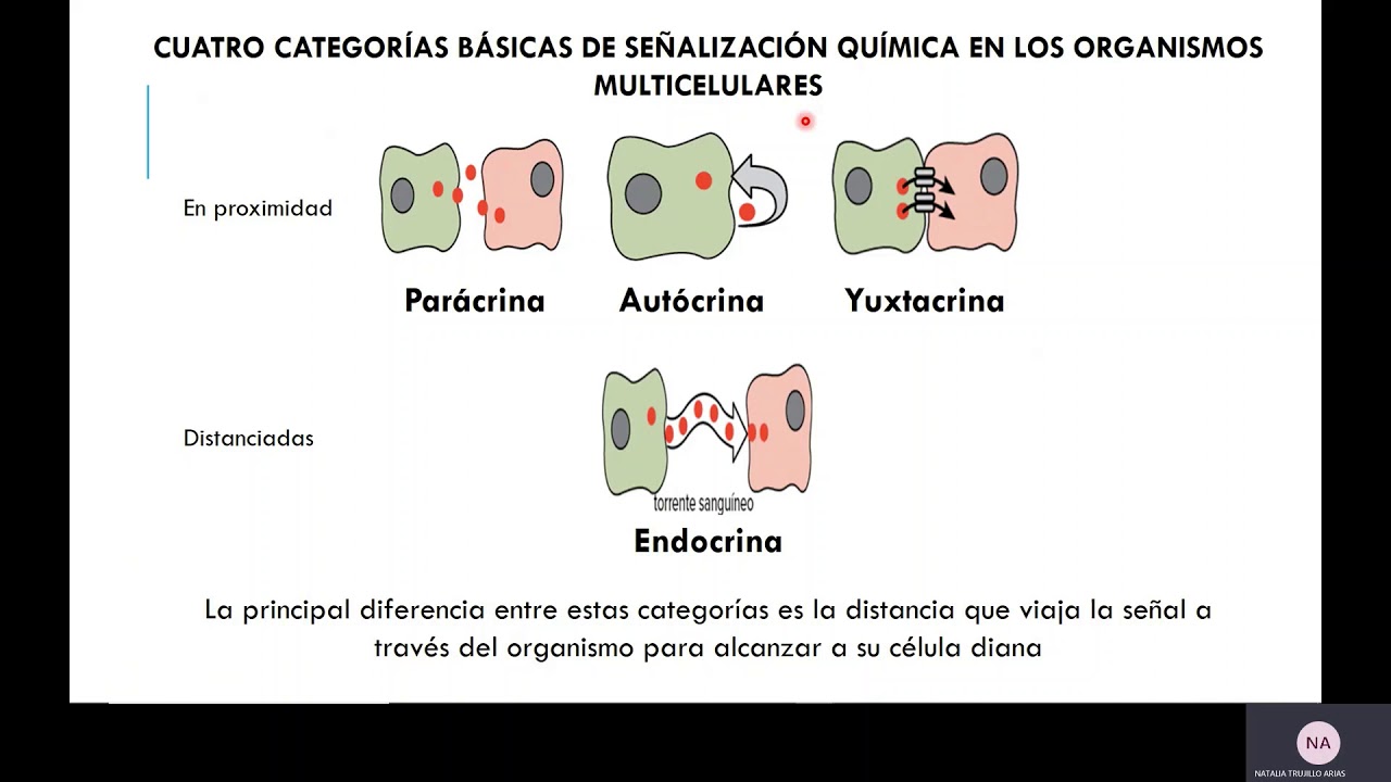
Señalización celular -Generalidades_I
4.6K
Cellular signaling (cellular communication) is the capacity of cells to exchange information through signals that generate specific responses in other cells. It involves an emitter (signal-sending cell) and a receptor (signal-receiving cell). This process affects all aspects of cellular structure and function, including gene activation/deactivation for protein production, tissue formation, and physiological processes like muscle movements. The process is highly specific—only cells with matching receptors can process signals. Signals are primarily transmitted through exocytosis, though some ligands can diffuse through membranes. Communication only occurs when ligands bind to specific receptors, triggering intracellular cascades. Biological responses include gene modification, cell division initiation, and apoptosis. Cellular signaling is categorized into three types based on distance: paracrine (short-distance, cells coordinate with neighbors), autocrine (cells signal themselves), and endocrine (long-distance through circulatory system). Juxtacrine signaling (contact-dependent) involves ligands remaining bound to the signaling cell and binding to receptors on adjacent cells. Ligands are categorized as intracellular (small, hydrophobic molecules like steroid hormones that cross membranes to bind cytoplasmic or nuclear receptors) or extracellular (water-soluble molecules like neurotransmitters that bind membrane-bound receptors). Membrane-bound receptors include ligand-gated ion channel receptors, enzyme-linked receptors (like receptor tyrosine kinases), and G-protein coupled receptors (GPCRs) with seven transmembrane domains that transmit signals through G-proteins.
Click to watch →
Deep Dive
The cellular response to hypoxia, particularly how low oxygen levels trigger transcription factors like Hypoxia-Inducible Factor (HIF).
LIVE: Cercetare de Nobel în biologia moleculară
7K
Hypoxia-inducible factor (HIF) acts as a master transcriptional regulator that coordinates the cellular response to low oxygen conditions. When activated by hypoxia, HIF regulates the expression of hundreds of genes involved in oxygen sensing, energy metabolism, and vascular development. This central role makes HIF a critical target for understanding and treating diseases characterized by abnormal oxygen metabolism. The HIF pathway represents a fundamental mechanism that cells use to adapt to their oxygen environment, and its dysregulation contributes to various pathological conditions including cancer and chronic kidney disease.
Click to watch →
Pearson Edexcel level 3 GCE biology B. 9BIO/01. June 2022. Part 3 of 3
299
Hypoxia is defined as inadequate oxygen supply to tissues. The body responds by releasing hypoxia-inducible transcription factors (HIF), which act as transcription factors binding to gene promoters to increase glycolysis enzyme production. During hypoxia, the electron transport chain cannot function without oxygen, so cells switch to anaerobic respiration via substrate-level phosphorylation. HIF1 and HIF2 show different expression patterns: HIF1 rises quickly then declines, while HIF2 remains elevated, reflecting their distinct roles in early versus sustained hypoxic responses.
Click to watch →

Special situations in cells 2026
343
Hypoxia-inducible factor (HIF) is a transcription factor that senses oxygen levels in cells. HIF consists of alpha and beta subunits. Under normal oxygen conditions, HIF-alpha is continuously degraded by prolyl hydroxylases, which require oxygen, iron, and alpha-ketoglutarate. When oxygen is scarce, HIF-alpha is stabilized, translocates to the nucleus, and forms a dimer with HIF-beta to activate genes for hypoxic adaptation. HIF-1 primarily regulates metabolic adaptations, while HIF-2 stimulates erythropoietin and VEGF production. HIF signaling is slower than AMPK because it requires gene transcription. Mutations in the VHL protein cause von Hippel-Lindau syndrome due to inability to degrade HIF-alpha. In hypoxia, cells upregulate glycolysis, lactate dehydrogenase, and glucose transporters while downregulating the electron transport chain, Krebs cycle, and mitochondrial biogenesis.
Click to watch →
الكيمياء الحيوية 2 - المحاضرة السابعة ( الجزء الثالث ) د. أحمد المحمد
219
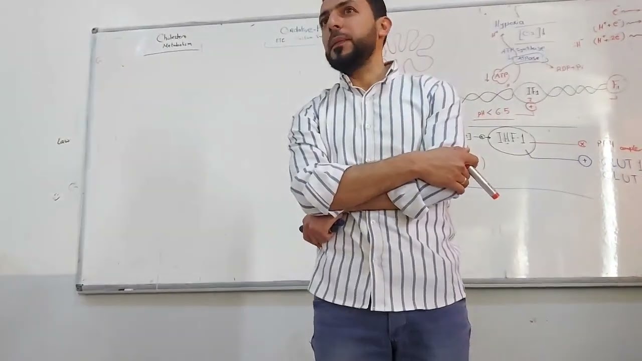
When oxygen levels are reduced, cells activate hypoxia-inducible factor 1 (HIF-1), a transcription factor that regulates gene expression. HIF-1 increases glucose transporter expression (GLUT1, GLUT3) to enhance glucose uptake, and induces changes in Complex IV of the electron transport chain by promoting subunit replacement through proteasome-mediated degradation. This allows cells to adapt to low oxygen conditions by modifying their respiratory machinery and maximizing energy production from available glucose.
Click to watch →

Extreme situations in cells
1.3K
HIF (hypoxia-inducible factor) is a transcription factor that senses oxygen levels in cells. Under normal oxygen conditions, HIF-alpha is rapidly hydroxylated by prolyl hydroxylases and destroyed by the proteasome. Under hypoxic conditions, prolyl hydroxylases cannot function, so HIF-alpha accumulates in the cytoplasm. The accumulated HIF-alpha then enters the nucleus, forms a dimer with HIF-beta, and activates transcription of hypoxia-responsive genes. HIF activates anaerobic glycolysis through hexokinases and lactate dehydrogenase, and increases glucose transporter expression (GLUT1 and GLUT3). HIF also activates erythropoietin (stimulating red blood cell production) and VEGF (promoting angiogenesis). HIF downregulates the electron transport chain and Krebs cycle because these processes require oxygen. In hypoxia, multiple regulatory pathways are activated simultaneously: HIF-1 alpha is activated by low oxygen, AMPK is activated by reduced ATP production, and mTOR activity is decreased. These signals can sometimes conflict, but the cell integrates all signals to determine the appropriate response. The outcome depends on the context and other cellular signals.
Click to watch →
Prerequisite Knowledge
- Concept 01Basic cellular biology of cancer, including uncontrolled cell division, tumor progression, and the concept of metastasis.
- Concept 02The anatomy and physiology of the circulatory system, specifically how blood vessels (endothelial cells and capillaries) deliver oxygen and nutrients to tissues.
- Concept 03Fundamental concepts of cell signaling, including how extracellular ligands bind to cell-surface receptors to trigger intracellular cascades.
- Concept 04The cellular response to hypoxia, particularly how low oxygen levels trigger transcription factors like Hypoxia-Inducible Factor (HIF).
Subsequent Learning
- Step 01Mechanisms of drug resistance to anti-angiogenic therapies, such as compensatory upregulation of alternative angiogenic factors like FGF and PDGF.
- Step 02The clinical concept of 'vessel normalization' and how anti-angiogenic drugs are strategically combined with chemotherapy and immunotherapy.
- Step 03The systemic side effects of VEGF inhibitors, including hypertension, arterial thromboembolism, and impaired wound healing.
- Step 04Next-generation angiogenesis inhibitors, including endogenous inhibitors like angiostatin and endostatin, as well as novel gene-therapy delivery methods.
Angiogenesis
Targeting Vessels
Angiogenesis
0:03Playing Section
- 1
Tumors trigger VEGF pathways to grow new blood vessels.
- 2
Multiple proteins aid vessel invasion and survival.
Vessel Co-option and Compensatory Resistance to Anti-Angiogenic Therapy
While anti-angiogenic therapies like bevacizumab (Avastin) were designed to starve tumors by blocking VEGF-mediated blood vessel growth, clinical outcomes have revealed significant limitations. A major counterpoint to the 'starve-a-tumor' model is that cancers quickly adapt to VEGF inhibition. Tumors can bypass these drugs through 'vessel co-option,' where cancer cells hijack existing healthy blood vessels instead of growing new ones, or 'vasculogenic mimicry,' where tumor cells themselves form fluid-conduction channels. Furthermore, the extreme oxygen deprivation (hypoxia) induced by anti-angiogenic drugs can backfire. Hypoxia activates hypoxia-inducible factors (HIFs), which trigger the release of alternative growth factors (such as FGF and angiopoietins) and promote epithelial-mesenchymal transition (EMT). This transition often makes the tumor more aggressive, invasive, and prone to metastasis. Consequently, anti-angiogenic therapies often extend progression-free survival only temporarily, without significantly improving overall survival, challenging the paradigm of VEGF inhibition as a standalone magic bullet.
Deep Dive
Mechanisms of drug resistance to anti-angiogenic therapies, such as compensatory upregulation of alternative angiogenic factors like FGF and PDGF.
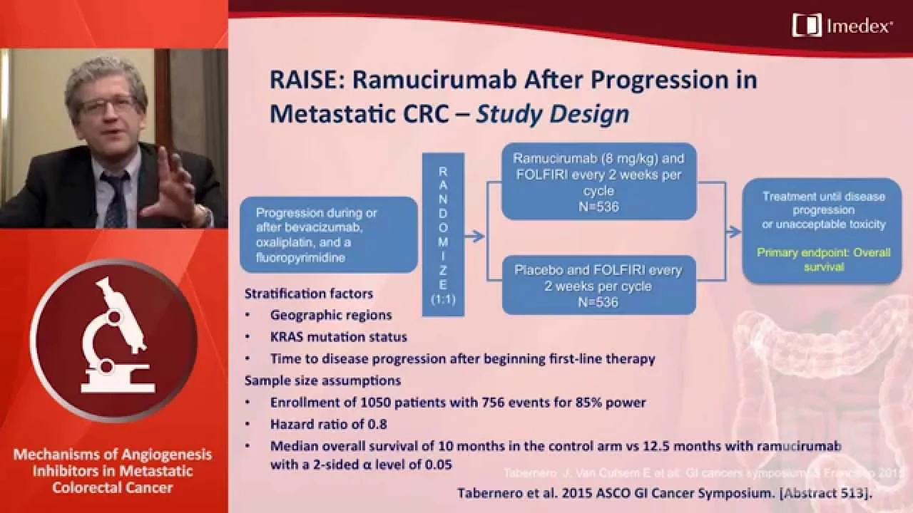
Mechanisms of Angiogenesis Inhibitors in Metastatic Colorectal Cancer
4.2K
Angiogenesis is essential for tumor growth beyond minimal size, involving vasculogenesis (de novo formation) and angiogenesis (new vessels from existing ones). Hypoxia triggers VEGF production, initiating chaotic tumor vasculature through the angiogenic switch. Tumor angiogenesis differs from normal angiogenesis through disorganized vasculature, abnormal pericyte support, and increased permeability. Four therapeutic strategies target VEGF pathways: monoclonal antibodies (bevacizumab, ramucirumab), fusion protein traps (aflibercept), and tyrosine kinase inhibitors (regorafenib). VEGF overexpression correlates with poor prognosis across cancer types. Resistance mechanisms involve compensatory increases in alternative growth factors like FGF and PDGF.
Click to watch →

М.Ю. Федянин, «Современные подходы к проведению 2-ой и последующих линий терапии».
112
Resistance to anti-VEGF therapy develops through compensatory mechanisms. When VEGF blockade is implemented, the tumor upregulates other angiogenic factors, particularly placental growth factor (PlGF). This compensatory response can lead to treatment resistance. Understanding these mechanisms is important for developing combination therapies that target multiple angiogenic pathways simultaneously.
Click to watch →
Why has everything beyond bevacizumab failed in angiogenesis inhibition?
854
Cancer cells develop sophisticated resistance mechanisms to anti-angiogenic therapy through compensatory pathway activation. Following VEGF blockade, tumors upregulate alternative growth factors including FGF, PDGF, and EGF, enabling continued angiogenesis. EGFL7, an extracellular matrix protein enriched in tumors, promotes vascular regrowth by facilitating perivascular tract formation after anti-angiogenic drugs impair angiogenesis. Despite these findings, several critical questions remain unanswered: optimal dosing and duration of bevacizumab treatment, potential benefit of continuing therapy beyond disease progression, and identification of best combinations with anti-angiogenics. Better-designed trials with prospective translational endpoints and broader patient access to clinical research opportunities are needed to advance this field.
Click to watch →

Expert talk by Dr Arup Das on anti-VEGF @drishtieyeinstitute
113
Treatment resistance to anti-VEGF therapy occurs through multiple mechanisms: (1) compensatory upregulation of alternative VEGF ligands (VEGF-C and VEGF-D) that are not neutralized by standard anti-VEGF agents; (2) continuous VEGF secretion that overwhelms neutralization capacity; (3) absence of reperfusion of non-perfused areas, meaning lost vessels cannot be restored; and (4) presence of other inflammatory mediators that drive disease independent of VEGF.
Click to watch →

Aula 1, parte 2: Angiogênese
135
Os mecanismos de escape da terapia anti-angiogênica incluem: produção de fatores angiogênicos alternativos, mudança para outros mecanismos de vascularização (vasculogênese, mimetismo vascular), e formação de novos vasos sem depender do VEGF. O mimetismo vascular ocorre quando células tumorais passam a exibir fenótipo endotelial e formam vasos sanguíneos sem depender de células endoteliais normais. Além disso, a terapia anti-angiogênica pode selecionar células tumorais mais agressivas e com maior potencial invasivo e metastático. A hipóxia seleciona células com maior capacidade metastática e resistência a terapias convencionais, representando um efeito maléfico da própria terapia.
Click to watch →
Deep Dive
The clinical concept of 'vessel normalization' and how anti-angiogenic drugs are strategically combined with chemotherapy and immunotherapy.
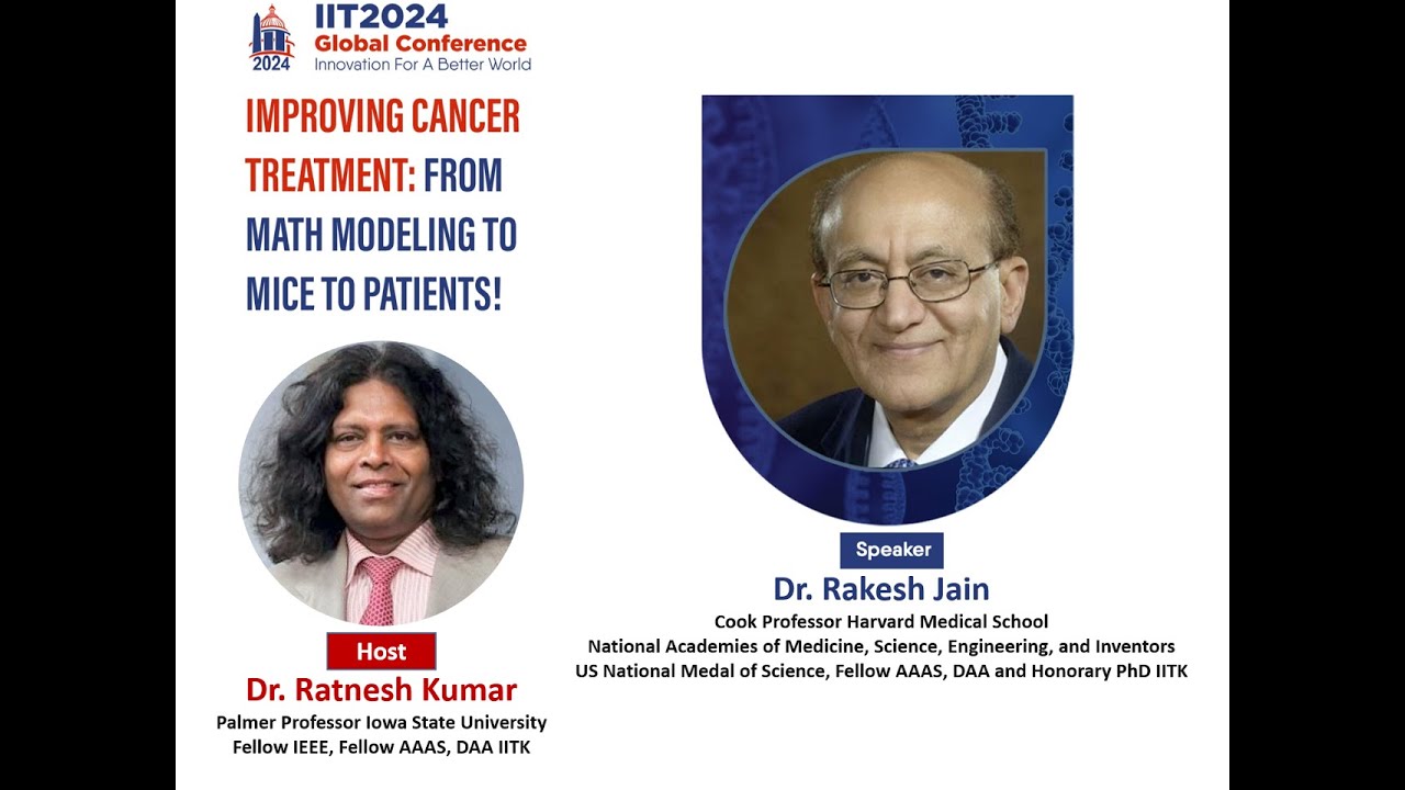
IIT2024 Talk - Improving Cancer Treatment with Rakesh Jain, Jan 14, 2024
309
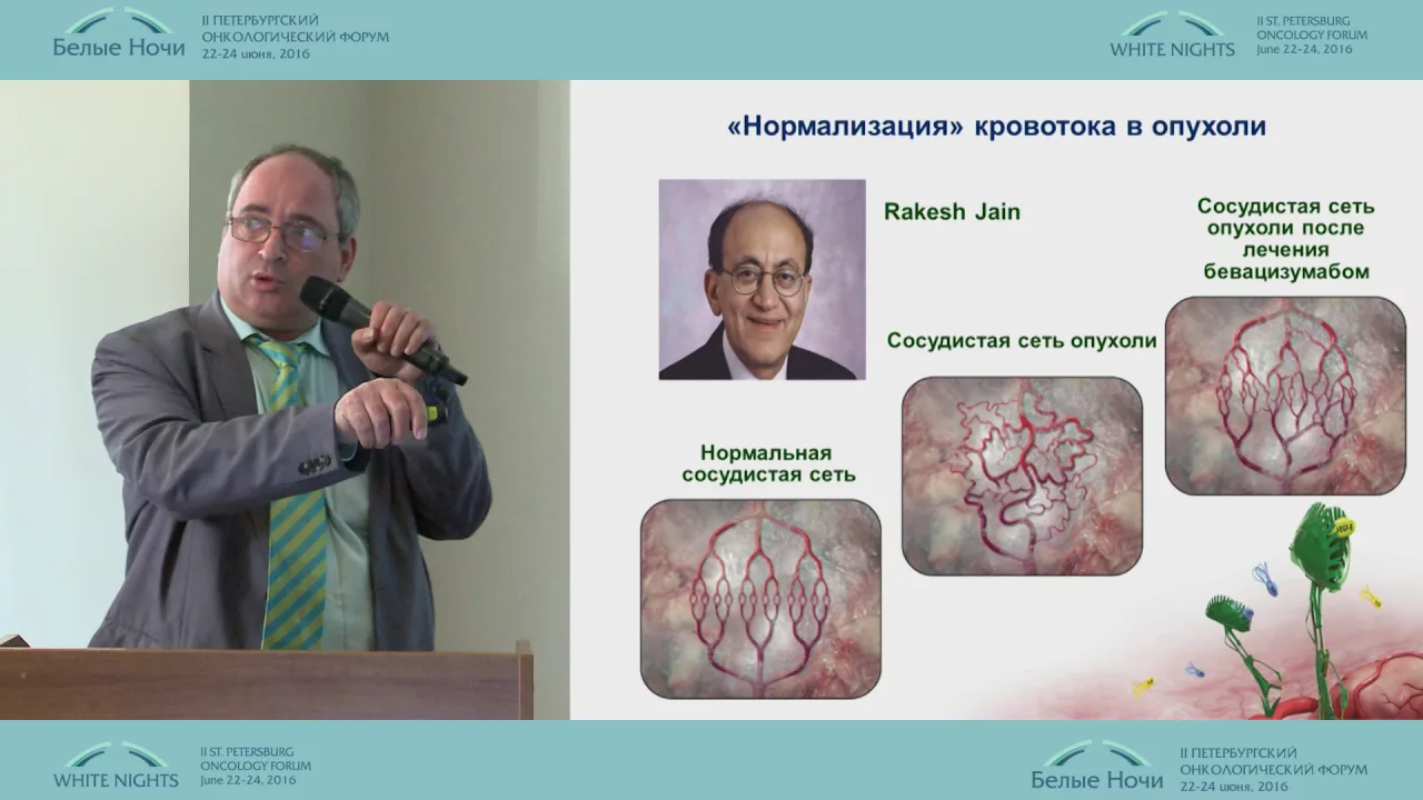
Abnormal tumor blood vessels create physical barriers that prevent effective drug delivery to cancer cells; by using low-dose anti-angiogenic therapy to normalize rather than destroy these vessels, researchers can improve blood flow and oxygenation within tumors, thereby enhancing the effectiveness of chemotherapy and immunotherapy treatments.
Click to watch →
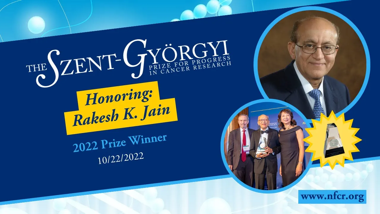
Rakesh K. Jain, Ph.D. Award Speech - 2022 Szent-Gyorgyi Prize for Progress Winner - NFCR.org
406
Multiple clinical trials confirmed that increased tumor perfusion correlates with better survival, contradicting decades of belief that reducing blood supply would kill tumors. Vascular normalization benefits immunotherapy by improving perfusion and polarizing macrophages toward anti-tumor phenotypes. FDA approved seven combinations in four years, with most trials now testing anti-VEGF plus immune checkpoint blockers. This principle also applies to non-malignant diseases like macular degeneration and neurofibromatosis.
Click to watch →

MolBiol: Angiogénesis
21.6K
Anti-angiogenic therapy targets the VEGF pathway to inhibit tumor vascularization. Three main approaches exist: (1) Anti-VEGF monoclonal antibodies like bevacizumab that bind VEGF and prevent receptor activation; (2) VEGF traps that sequester VEGF molecules; (3) Tyrosine kinase inhibitors (sunitinib, sorafenib, pazopanib, regorafenib, axitinib) that block VEGFR signaling. These drugs inhibit endothelial proliferation and normalize chaotic tumor vasculature, improving chemotherapy delivery. Metronomic chemotherapy exploits anti-angiogenic properties of cytotoxic agents. Approved indications include metastatic colorectal cancer, cervical cancer, non-small cell lung cancer, ovarian cancer, clear cell renal carcinoma, hepatocellular carcinoma, and gastrointestinal stromal tumors. Combination with chemotherapy produces synergistic anti-tumor effects. While anti-angiogenic therapy has transformed cancer treatment, challenges remain including resistance mechanisms and optimal combination strategies for different tumor types.
Click to watch →
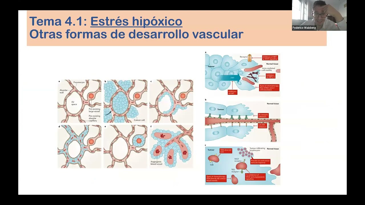
31 10 2025 - BIOLOGÍA - Estres celular: Hipóxico - Metabólico - Oxidativo - Proteostático
111
Anti-angiogenic drugs inhibit VEGF signaling, causing tumor vessels to transition from abnormal, dysfunctional states to more normal, quiescent states. This normalization improves vascular function by reducing permeability and improving blood flow, enhancing drug delivery to tumors. The effect is particularly important because tumors generate aberrant vasculature that is inefficient at drug delivery. Anti-angiogenic therapy is most effective in tumors dependent on angiogenesis, such as renal cell carcinoma.
Click to watch →
Биология метастатического колоректального рака. Новые мишени для антиангиогенной терпаии
2.4K
Anti-angiogenic therapy has undergone significant conceptual evolution over two decades. Initially proposed by Judah Folkman, the theory that tumors require angiogenesis for growth was dismissed as implausible. Early monotherapy trials showed disappointing results, revealing that simple angiogenesis inhibition was insufficient. The modern paradigm now emphasizes vascular normalization—destroying immature, leaky tumor vessels to restore blood flow and improve drug delivery rather than directly killing tumor cells. This shift explains why anti-angiogenic agents enhance conventional chemotherapy efficacy by normalizing the chaotic tumor vasculature, fundamentally changing how these agents are understood and applied in clinical oncology.
Click to watch →
Deep Dive
The systemic side effects of VEGF inhibitors, including hypertension, arterial thromboembolism, and impaired wound healing.

Episode 375: Pharmacology 101: VEGF Inhibitors
246
VEGF (Vascular Endothelial Growth Factor) inhibitors are a class of anti-cancer drugs that block tumor angiogenesis by interfering with VEGF signaling pathways, either by binding to VEGF itself or its receptors on endothelial cells; this prevents new blood vessel formation and reduces vascular permeability, thereby limiting tumor growth and improving chemotherapy delivery. The class includes monoclonal antibodies (bevacizumab, ramucirumab, aflibercept) and oral tyrosine kinase inhibitors (sunitinib, cabozantinib, lenvatinib, pazopanib, etc.), used across multiple solid tumor types including colorectal, ovarian, renal cell carcinoma, hepatocellular, and non-small cell lung cancers. Common side effects include hypertension (up to 67%), thrombotic events, bleeding risks, and wound healing complications, requiring careful monitoring and patient education on recognizing symptoms like headaches, blurred vision, or abdominal pain. Healthcare professionals should collaborate between nursing and pharmacy teams to manage these adverse events effectively.
Click to watch →

Cardio-oncology simplified-Part 1
4.3K
Targeted therapies cause cardiovascular side effects because the receptors and kinases they target are overexpressed in both cancer cells and normal cells including myocardial cells, skin cells, and gastrointestinal cells. The five major cardiovascular side effects are: hypertension (very common with VEGF-targeted therapies), heart failure and cardiomyopathy (1-10% incidence with VEGF and HER2-targeted therapies), arterial thrombosis including MI (1-10%), venous thrombosis and thromboembolism, atrial fibrillation risk increase, and QT prolongation. Skin problems and impaired wound healing also occur due to effects on skin cell growth.
Click to watch →

Management of toxicities related to systemic therapy for metastatic renal cell carcinoma
1.7K
Systemic therapy for metastatic renal cell carcinoma, including VEGF inhibitors (Avastin, Sutent, Votrient, Nexavar, Inlyta) and mTOR inhibitors (Torisel, Afinitor), causes predictable toxicities that can be managed through early recognition, supportive care, and dose modifications; VEGF inhibitors commonly cause fatigue, hand-foot syndrome, diarrhea, hypertension, and hemorrhagic side effects, while mTOR inhibitors uniquely cause hypoglycemia, hyperlipidemia, and interstitial pneumonitis, with most toxicities being reversible upon dose adjustment or discontinuation.
Click to watch →

胃がん/抗がん剤副作用 2023年版
30.1K
This section explains the mechanisms and side effects of molecular targeted therapy drugs. TRASTUZUMAB blocks HER2 receptors and can cause heart failure. RAMUCIRUMAB blocks VEGF receptors to prevent angiogenesis, causing hypertension, bleeding tendencies, and impaired wound healing (requiring 1-2 month waiting period before surgery). The section details how these drugs work by blocking specific protein pathways that cancer cells use to grow and survive, and explains the unique side effect profiles that differ from cytotoxic drugs.
Click to watch →
A Case-Based Introduction to Onconephrology | NephroPlus
142
VEGF inhibitors (bevacizumab, aflibercept) cause endothelial injury leading to thrombotic microangiopathy (TMA). The mechanism involves VEGF balance disruption - too little VEGF causes endothelial injury/TMA, while too much can cause collapsing FSGS. Hypertension is often the first symptom of endothelial injury. Risk factors include GU cancers, older age, hypertension, and hypomagnesemia. The VEGF balance is critical - too little causes TMA, too much causes collapsing FSGS.
Click to watch →
Deep Dive
Next-generation angiogenesis inhibitors, including endogenous inhibitors like angiostatin and endostatin, as well as novel gene-therapy delivery methods.

Concepts of Angiogenesis in Oncology-Dr.(Maj)Mirza Qaiser Baig
972
Angiogenesis is regulated by a balance between pro-angiogenic stimulators (VEGF, bFGF, TNF-α) and endogenous inhibitors (thrombospondin-1, angiostatin). VEGF promotes angiogenesis through five key mechanisms: sensitizing endothelial cells, inducing migration, enhancing survival, mobilizing progenitor cells, and relaxing vascular smooth muscle. Anti-angiogenic drugs neutralize these factors or induce endogenous inhibitors. Metronomic chemotherapy and doxycycline increase thrombospondin-1 levels. These therapies normalize tumor vasculature, improve drug delivery, and enhance chemotherapy/radiotherapy efficacy.
Click to watch →

Oxford BioMedica
559
Oxford BioMedica, a UK-based gene therapy company, utilizes lentiviral vectors for gene delivery, which can carry larger genetic payloads and infect dividing cells, enabling permanent gene expression. Their pipeline includes Retinostat, a subretinal injection producing angiostatin and endostatin for wet AMD, and Oncostatin, a topical version for preventing neovascularization in corneal transplants. The company has established partnerships with Novartis, GlaxoSmithKline, and Sanofi, demonstrating the commercial viability of their lentiviral platform for treating diseases with no current treatments.
Click to watch →

Your Feet & Legs Are Warning You! - Signs Of Poor Health & A Decreased Lifespan | Dr. William Li
963.6K
Gene therapy research involves delivering DNA coding for growth factors directly to leg muscles. Clinical trials showed that this approach can stimulate angiogenesis (new blood vessel growth) in oxygen-starved legs. While still experimental, successful cases demonstrate the potential of this futuristic treatment approach.
Click to watch →

CBS Evening News - 1999-11-19
526
Angiogenesis inhibitors represent a new class of experimental drugs for cancer treatment. The theory behind these drugs is that tumors need to create new blood vessels to grow, a process called angiogenesis. By inhibiting or stopping new blood vessel formation, these drugs can starve the cancer, stop it from growing, and potentially shrink tumors. Several drugs are in development, including SU5416 (tested at UCLA), endostatin (in clinical trials at Dana-Farber), and combretastatin (tested at Cleveland). Early results show promise, with some patients experiencing complete remission or tumor stabilization. Doctors suggest these drugs may help make cancer a chronic illness that can be medically managed, representing a significant advancement in cancer treatment for the 21st century.
Click to watch →

Neovascularización corneal
257
The cornea possesses multiple endogenous anti-angiogenic mechanisms. PEDF interferes with endothelial signaling and induces apoptosis through caspase-3. Angiostatin, derived from plasminogen fragments, inhibits endothelial proliferation. Endostatin, from type XVIII collagen, interferes with endothelial proliferation. Thrombospondins (TSP-1, TSP-2) bind to CD36 and inhibit MMPs. Tissue inhibitors of metalloproteinases (TIMPs) prevent unregulated matrix degradation. The balance between MMP activity and TIMPs determines matrix degradation and neovascularization progression.
Click to watch →
Angiogenesis
Targeting Vessels
Angiogenesis
0:03- 1
Tumors trigger VEGF pathways to grow new blood vessels.
- 2
Multiple proteins aid vessel invasion and survival.
Vessel Co-option and Compensatory Resistance to Anti-Angiogenic Therapy
While anti-angiogenic therapies like bevacizumab (Avastin) were designed to starve tumors by blocking VEGF-mediated blood vessel growth, clinical outcomes have revealed significant limitations. A major counterpoint to the 'starve-a-tumor' model is that cancers quickly adapt to VEGF inhibition. Tumors can bypass these drugs through 'vessel co-option,' where cancer cells hijack existing healthy blood vessels instead of growing new ones, or 'vasculogenic mimicry,' where tumor cells themselves form fluid-conduction channels. Furthermore, the extreme oxygen deprivation (hypoxia) induced by anti-angiogenic drugs can backfire. Hypoxia activates hypoxia-inducible factors (HIFs), which trigger the release of alternative growth factors (such as FGF and angiopoietins) and promote epithelial-mesenchymal transition (EMT). This transition often makes the tumor more aggressive, invasive, and prone to metastasis. Consequently, anti-angiogenic therapies often extend progression-free survival only temporarily, without significantly improving overall survival, challenging the paradigm of VEGF inhibition as a standalone magic bullet.
Basic cellular biology of cancer, including uncontrolled cell division, tumor progression, and the concept of metastasis.
The anatomy and physiology of the circulatory system, specifically how blood vessels (endothelial cells and capillaries) deliver oxygen and nutrients to tissues.
Fundamental concepts of cell signaling, including how extracellular ligands bind to cell-surface receptors to trigger intracellular cascades.
The cellular response to hypoxia, particularly how low oxygen levels trigger transcription factors like Hypoxia-Inducible Factor (HIF).
Mechanisms of drug resistance to anti-angiogenic therapies, such as compensatory upregulation of alternative angiogenic factors like FGF and PDGF.
The clinical concept of 'vessel normalization' and how anti-angiogenic drugs are strategically combined with chemotherapy and immunotherapy.
The systemic side effects of VEGF inhibitors, including hypertension, arterial thromboembolism, and impaired wound healing.
Next-generation angiogenesis inhibitors, including endogenous inhibitors like angiostatin and endostatin, as well as novel gene-therapy delivery methods.
424.2K views3.8Klikes3:59@MechanismsInMedicineOriginal Release: 2012-10-29
as the tumor grows it will eventually reach a size where it will need to have additional vasculature to sustain continued growth to achieve this the tumor cells excrete certain proteins to stimulate blood vessel growth into and around the tumor a process called angiogenesis one of the major Pathways involved in angiogenesis involves vascular endothelial growth factor or vegf and its family of receptors there are seven subtypes of vegf and three receptors that bind differently vegf affects the endothelial cells that line the blood vessels in a number of ways it can cause them to proliferate by activating the extracellular Kinesis and map kyes signal transduction Pathways it can induce proteins that break down the basement membrane to allow endothelial cells to migrate and invade these proteins include Matrix metalloproteinases or mmps eurocin type plasminogen activator or UPA and its receptor up as well as the tissue type plasminogen activator it makes vessels more permeable allowing molecules and fluids to leak out when MMP is secreted into the extracellular space it degrades The extracellular Matrix to to allow pro-angiogenic factors to reach the vasculature with the extracellular Matrix degraded pro-angiogenic factors including vegf can reach receptors on the endothelial cells of blood vessels surrounding the tumor thus stimulating the angiogenic signal in the vessel VF also helps the new endothelial cells survive by upregulating Inhibitors of apoptosis VF also activates the endo IAL cells to express the proteins necessary to allow the new blood vessels to form the end result is the growth of new blood vessels into the tumor with the growth of new vessels into the tumor additional nourishment can be delivered to the tumor new blood vessels in the tumor facilitate further tumor growth strategies targeting VF and its receptors have been used successfully in clinical practice bism or avastin is an antibody that binds vegf and prevents its binding to its receptor another therapy is sunitinib or suant which is a small molecule inhibitor with high binding affinity for VF and pdgf receptors The receptors on the surface of normal and tumor cells can be inhibited directly camab or herbit is an antibody that binds directly to the epidermal growth factor receptor on the normal and tumor cells and competitively inhibits The Binding of EGF and other ligans such as TGF Alpha as we have seen in this presentation tumorogenesis is a complex multi-step process that results from genetic changes and causes malignant transformation of normal cells a summary of the characteristics common to many cancers are abnormal signal transduction resulting in uncontrolled cell proliferation loss of apoptosis or programmed cell death tissue Invasion and metastasis permitting spread of the cancer and angiogenesis leading to enhanced blood supply of tumors many of the components of these mechanisms are potential targets of anti-cancer Therapies
Up Next
5:41
p53 Tumor Suppressor Protein: DNA Damage Response Explained
@WEHImovies
100.3K views•2020-06-26

50:12
Bacterial Communication: Quorum Sensing and Biofilm Formation Explained
@JHUAAP
12.6K views•2011-06-28

5:35
Enteric Nervous System Explained: The Gut's Brain | Neurobiology Lecture
@alumniu6029
438 views•2018-09-12

7:08
Bacteriophages: Earth's Deadliest Killers and Future Antibiotics
@kurzgesagt
34.6M views•2018-05-13
Related Study Plans & Knowledge Roadmaps
Structured learning paths in Biology
biology
RNAi: Biogenesis, RISC & Gene Silencing
01Molecular Biology Foundations: DNA to Protein
02Introduction to RNA Interference (RNAi)
03Molecular Biogenesis of miRNA and siRNA
+ 2 more learning modules
10h5 modules
Explore Roadmap
biology
Mycorrhizae: Signaling, Biophysics & Ecology
01Foundations of Mycorrhizal Symbiosis
02Molecular Signaling and Root Colonization
03Biophysics and Bioenergetics of Nutrient Exchange
+ 1 more learning modules
11h4 modules
Explore Roadmap
biology
Circadian Clocks: TTFL, SCN & Periphery
01Foundations of Molecular Biology & Gene Expression
02Introduction to Chronobiology and Actograms
03The Core TTFL: Molecular Architecture of the Clock
+ 3 more learning modules
14h6 modules
Explore Roadmap
biology
Population Genetics: Math, Drift & Selection
01Genetic & Probability Foundations
02The Hardy-Weinberg Principle and Baseline Models
03Mathematical Modeling of Natural Selection
+ 3 more learning modules
18h6 modules
Explore Roadmap