


























![Les sciences du sommeil et du rêve [TenL117]](https://i.ytimg.com/vi/7CIvfkOdmHo/maxresdefault.jpg)
Sleep serves as a critical 'offline' period for synaptic renormalization, counteracting the net synaptic potentiation that occurs during wakefulness; during wakefulness, learning and adaptation cause synapses to strengthen, consuming more energy, occupying more space, and eventually saturating signal-to-noise ratios, while sleep provides the necessary mechanism to restore synaptic strength to sustainable baseline levels, thereby enabling continued plasticity and preventing cognitive saturation.
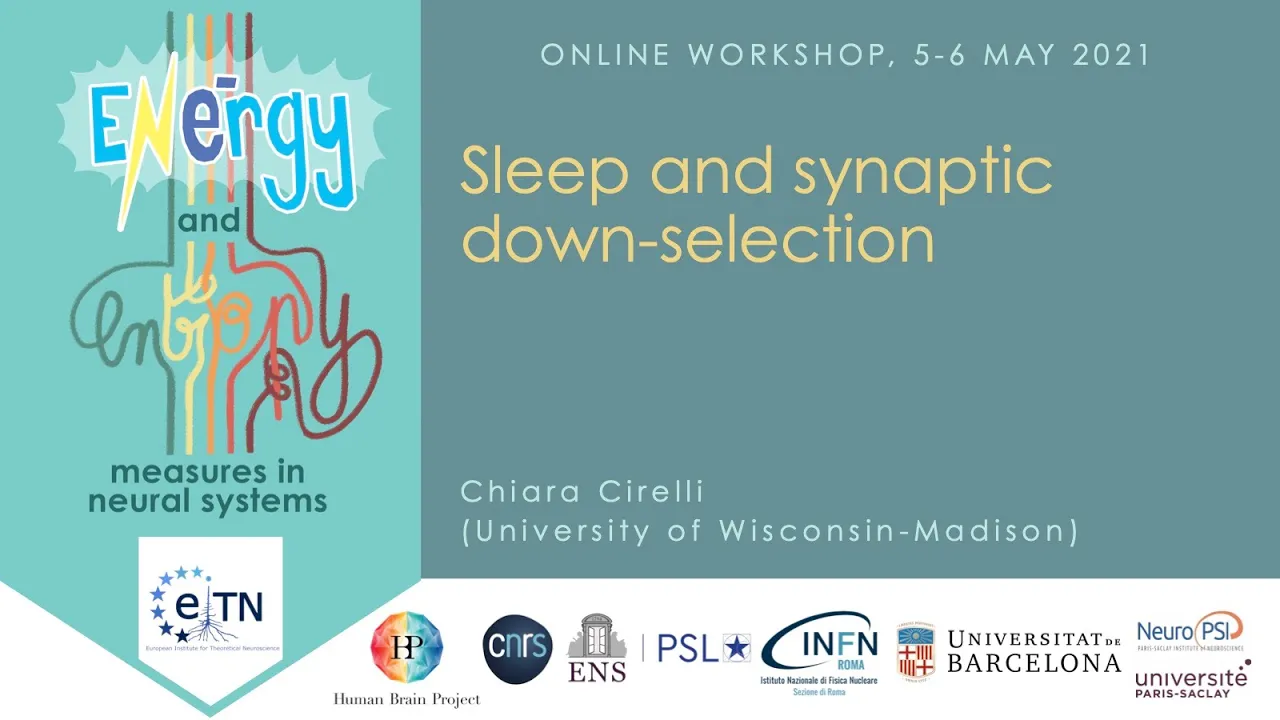
Sleep Function & Synaptic Homeostasis | Giulio Tononi | Allen Institute
Added:Basic neuroanatomy and synaptic plasticity, specifically the mechanisms of Long-Term Potentiation (LTP) and Long-Term Depression (LTD).
The standard architecture of sleep cycles, including the distinction between Non-Rapid Eye Movement (NREM) and Rapid Eye Movement (REM) sleep.
Fundamental principles of homeostatic regulation in biological systems.
The basic concept of memory consolidation and how neural networks encode information during wakefulness.
The specific molecular and cellular mechanisms of synaptic downscaling, such as AMPA receptor internalization during sleep.
The Integrated Information Theory (IIT) of consciousness, also developed by Giulio Tononi, which explores how conscious experience relates to neural integration.
Clinical implications of synaptic homeostasis disruption in neurodegenerative diseases (like Alzheimer's) and chronic sleep disorders.
The comparative neurobiology of sleep across different animal species to understand the evolutionary necessity of synaptic downscaling.
28.2K views209likes19:45Original Release: 2011-10-07
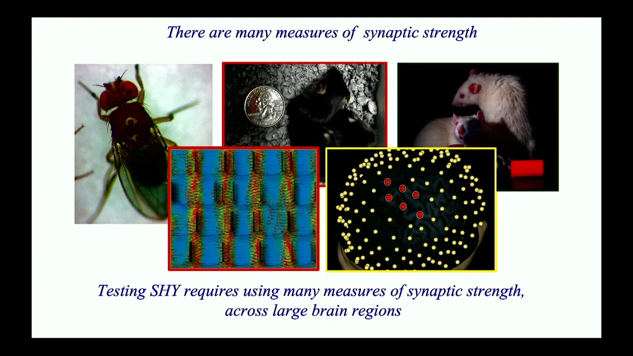
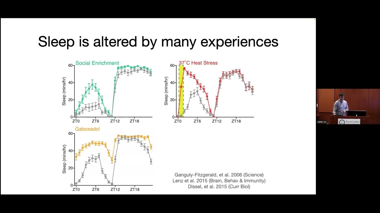
thank you um among the open questions in Neuroscience I believe the functional sleep remains perhaps one of the most embarrassing we don't know why every night our brain needs to put us and presumably itself to sleep for several hours and uh there is every indication that sleep performs an essential function if we only knew which one the indications are that it is first of all dangerous obviously you are you know available to Predators if you fall asleep and you don't respond to stimuli it is pervasive we do this from the Cradle to the Grave every animal that has been studied careful do does so you know going from fruit flies and now several other species have been shown to sleep to ourselves there's basically no single exception it is also irresistible as we all know but uh if you try and do experiments to keep animals awake or humans awake for that matter there is no way that uh you can overcome the need for sleep you can even use pain shocks at some point the animal will fall asleep it's also tightly regulated with a big portion of the brain and certainly the brain stem and the hypothalamus all kind of complicated connectivity is set up to put us to sleep and then wake ourselves up and finally if you don't sleep or you sleep too little it's clear there are serious consequences According to some people death would be one consequence in fatal familiar insomnia after a few months of essentially lack of sleep you die rats die after two weeks but the most obvious consequences are cognitive consequences there are all kind of problems we become extremely bad cognitive make mistakes of all sorts and we become extremely irritable so now let me before I go and discuss the issue of the functional sleep remind you with some perhaps novel work what's happening in the brain when we sleep this is uh an indication of the fact that when you go from wake to non-rm sleep which is 80% of sleep in humans most mammals and birds what you see are those big slow waves in theeg that are abundant early in the night and then less so later in the night and corresponding to those slow waves you actually see units neurons this now been shown in humans and and cats rodents monkeys they fire roughly at the similar firing grate as in Wake they just have these brief periods of Silence lasting 100 to 100 millisecond something like that then they resume firing and intracellular as shown by mad and his collaborators what happens is neurons are firing tonically and then they stop briefly they hyperpolarize 15 20 mols and then they go back this is what's happening thousand times per night in our brain and this is triggered and associated with changes in neuromodulators like the ones that Michael just described acidin or epinephrine serotonin histamine oraxin and so on they all change dramatically between wake and sleep and obviously produce that very different way of functioning of the brain now here I want to use an all large scale simulation with many many tens of thousands of neurons and millions of connections of two cortical areas like a primary and a super higher order visual area and a piece of Thalamus the lgn and the reticular thalamic nucleus divided into three layers and just to give you a feeling of the tonic firing during wakefulness with the corresponding EG or local field potential so the typical low voltage fast activity of wakefulness and neurons fire in this random way as if we were Imaging them with Optical recordings and then we turn the neurom modulators down and we send the model to sleep and when we send the model to sleep it continues firing but there are these very brief interruptions that happens roughly once every second and that is most of what sleep is made of there are additional things like sleep spindles Etc but the slow waves that correspond to neurons doing that is the major feature of sleep again if we knew why the brain is doing that because when it is doing that it is also obliterating to a large extent our own Consciousness so now we know a few more things about this one of the things we have learned is that each of these slow waves especially the big ones start in a different place in the brain and then travels it's literally a traveling wave that invades other regions of the brain and when doing Source modeling we actually saw that many of these uh slow waves the big one start around the left insula also on the the right side so it's distributed but there are some hot spots and then they travel and they propagate throughout the brain along the main super highways of the uh connectum if you wish especially along the medial surface of the hemispheres and then they spread out laterally invading much of the brain now a recent finding that was done in collaboration with itak freed by yaler is that if you record in humans in let say 10 different regions in the brain when the human is entering nonre sleep what you find is that while there are indeed Global waves that invade most regions the ones we typically see with the high density EG on the scalp there are plenty in fact the majority of waves that are local they may happen in one brain region and not in others or in two brain regions and not in the others so a lot of what you see with the scalp EG is actually a reflection only of the biggest phenomena there are local ways happening all the time throughout the human brain and as we now know also the rat and the mouse brain this is a recording again from yval near of uh uh Rat a is auditory cortex and F is frontal cortex you see the EG you see the local field potential you see the multi unit activity and you see individual spikes and you can see these periods of Silence in the array recordings those are the down state of the slow solation underlying the slow wave and very often those downstate happen in one region or cortex for instance and not in another region prefrontal cortex so this is happening a thousand times a night in different ways and a sort of a synchronous manner sometimes throughout the brain why so the Enigma of sleep function has been there for a long time all kind of ideas have been proposed what I'm going to discuss today is the synaptic hypothesis which has been an attempt to understand what may be the fundamental the core function of sleep in every animal and the idea here in short is that sleep is the price we pay for plasticity of course plasticity should happen primarily during wake when we adapt to the external world now I will not discuss the theoretical motivation for this except for this very brief sketch which suggests the following that you know neurons either Spike or they don't that's how they communicate spikes are more expensive than nons spikes in terms of their post synaptic consequence in terms of energy and everything else so spikes should be reserved and as far as we can tell they largely are to Signal important events events that convey a lot of information now when you need to adapt to a changing World which is basically all the time especially during development what you need to do is to shift strength of synapsis or even add synapsis to make sure that you're are firing for important events for importance changes dynamically in the environment and you do that and you know every neuron tries to do that to make sure it signals important stuff Downstream in this complicated brain where every neuron is in a sea immersed in a sea of other neurons it doesn't know what it's getting from where it doesn't know what it's signaling and doesn't know where it's sending it so in all of this uncertainty for any learning system which is as complicated as a brain there is a problem in the end which is you tend to strengthen to make sure your signal but that becomes biologically untenable because stronger synapses consume more energy occupy more space require more supplies and finally they saturate signal to noise basically then neurons start firing for everything and that can't be good so there is a need for renormalization to make sure that total synaptic strength is constant and we think that that renormalization is not only essential but it better happen offline when you actually can sample in an unbiased way the environment of a neuron and every neuron by itself does it in the course of the night that's what we think sleep is fundamentally for and without again elaborating on the theory let me just then illustrate in practice what this hypothesis suggests it suggests that at the minimum during wake there is a net increase in synaptic strength potentiation and during sleep this is renormalized to go back to a baseline value which is sustainable biologically and from the point of view of information transferring the brain by synaptic strength I refer both to the uh molecular aspects like the number of Amper receptors and their phosphorization state and to the electrophysiologic and aspect of force how effective is a preoptic spike in producing post synaptic depolarizations and here I want to very quickly go through evidence about this molecular evidence actually started a long time ago you see there sections through a rat brain and we see the expression of fce in Wake when it's white and in sleep when it's not white at all many other genes behave like that and here I put a very nice recent study from the Allen Institute in which in a much more thorough way many many different Amar have been mapped and there to Arc for instance is much more expressed in an animal who has been awake than in an animal who has been asleep the general rule is while many many genes change the one that seem to change most dramatically between wake and sleep are genes involved in synaptic potentiation and that kind of plasticity here you see for instance the phosphorated crab in the cortex is high in wake and is very low in sleep and similarly bdnf and bdnf Mark fibers are much higher in Wake than in sleep here you see a more detailed study using synaptoneurosomes of the D it of Amper receptors uh gl1 and they are much increased like 50% or 80% in the working cortex or OC campus for that matter compared to a sleeping cortex and a more recent study by the group of ulich has also shown something like this with a completely different technique I want to move briefly to the electrophysiological evidence and you know I can tell you already goes in the same direction here is a recent study in which we looked at meis that is the spontaneous release of transmitter you can see both the amp ude and the frequency of the spontaneous release is higher in slices this is both mouse and rat of animals who had been awake in the previous 2 three hours than animals who had been asleep and here is the slope of devoke potential a classic measure of synaptic efficacy so you given anal stimulation you see how big and how steep their response is and after an animal has been awake from w0 to W1 the response get bigger and steeper after an animal has been asleep from s0 to S1 for 3 4 hours to response goes down and in humans you can see something very similar use instead of electrical stimulation use TMs and then you use high density G to record the response and again the more you stay awake the more excitability goes up the response gets bigger and steeper and then you sleep and it goes back to a normal level the most recent evidence that we have obtained about this is actually structural evidence we reason that if these things change at the functional molecular level maybe also they change at the structural level some indication this burp which is a very strong preoptic marker in fruit flies some of the indications are that bur protein is now highly expressed for instance in all over the brain of a fly who has been awake and much much less in the brain of a fly who has been asleep we showed quite some time ago that FES sleep very much like we do so this is an indication that even at the level of synapsis things change in the same direction and Dan bushe and KY recently were able to look at this using conf focal microscopy and here focus on one particular area of the fly brain Loba plate visual system number one but this true in two different structure that we analyze you can see that dritic branching and the punter in an animal who had been asleep then an animal has been exposed to a typical and Rich waking environment and you can see that the number of synapsis goes up as well as the richness of the dudic branching when the animal goes to sleep afterwards the branching and the synapsis go down that does not happen if the animal is kept awake meaning sleep is actually necessary to produce the synaptic pruning and branching simplification you see something similar now this is a other recent study that was done by Stephanie maray and Ugo faraguna using two Photon Imaging on the mouse up there sort of like we saw before we did several sessions in the same animal that's pretty hard to do and counted synapses in adolescent mice and after they've been awake number of synapses tends to go up slightly and after I've been asleep for say 7 to 8 hours it tends to go down so there too there is a change in the number of the synapsis as a function of having been awake and having been asleep and perhaps one of the most unexpected to me findings just came out by a Japanese group of yokoyama and coauthors have shown that the in the number of neurons in the olfactory bulb which is as you know always undergoing very very strong ter over is heavily regulated so lots of neurons die well it turns out that they die in the postprandial sleep period so they die they disappear they are selected away when the animals sleep so this pruning so to speak seems to be rather Universal in the fruit fly in the rat in the mouse and it is also pretty extreme at times including the number of neurons so now I want to move briefly to uh as second aspect a corollary of this hypothesis if what changes between wake and sleep is the net I strength and perhaps even structural markers of that can we see that expressed in the usual parameters that have been used for many many years to record sleep that is the slower activity in deeg well the hypothesis says that if you strengthen during wake then this increased net synaptic uh strength is going to produce stronger coupling among neurons which should at the minimum result in a higher synchronization and therefore in bigger and steeper slow waves when you go to sleep then the renormalization of synapsis during sleep so they tend to go down will reduce this coupling and reduce the amplitude of connections down to a reasonable level and that is what we first tested with large scale models if we increase the strength of connections to plasticity during wake when the model goes to sleep as you saw before the slow waves are bigger and more synchronous and then at the end of sleep they become smaller and less synchronous and this fits with what we actually measure in the slow of activity in both the model rats and humans now if you try and manipulate this process you can actually see some local component to it you can for instance ask an animal to locomote a lot which fits very well with what Michael was saying a moment ago that would produce several marks of plasticity andrich in the cortex like bdnf and as a result of that when the animal goes to sleep the slow activity will be larger and if instead you keep an animal awake for the exactly the same duration but uh it's doing very little it's moving very little then the be will go up less and so will other markers of plasticity and the slower activity in denight will be reduced we can actually interestingly don't have the slide here but leion the nergic system one of these key neurom modulators and that will produce similar results even in an animal who is locomoting so it will decrease the amplitude of this in a local sense you can do a learning task here is a rotation learning task done by humans in front of a computer where they sort of adapt unconsciously to a rotating display and the area of the brain that's involved in learning that task which we sort of new is Right parial cortex after you learn and you go to bed shows bigger slow waves conversely if you immobilize an arm uh the corresponding controlateral Cal sensory cortex will show less slow waves when you go to bed so this indicates that the kind of plasticity and where the plasticity occurred during waking is simply read out by the amplitude and steepness of the slow waves during sleep you can see this here for instance we apply this in Behavior therapy for Aphasia so aphasic patients underwent a very intense behavioral therapy in which they exercise and exercise then they go to bed and we can see that the relevant areas show more slower activity so presumably this could be a way clinically to test whether plasticity occurred how much of it and by the way this predicts recovery of function uh I want to finish by telling you only in a few words what are therefore the consequences of the net increase in synaptic strength that I believe happens in every animal when we learn for the theoretical reasons I only briefly sketched you end up in the evening with more and or stronger synapses those consume more energy even doing nothing they occupy more space and we saw from sinowski presentation this morning how little space there is there they will require a lot of supplies going from mitochondria to membranes to proteins that need to deliver to the synapsis and by cost be increasing they will decrease the signal to noise ratio so when sleep comes in it renormalizes we think in a very smart way the overall synaptic strength putting all of those functions back to where they need to be I want to finish instead of discussing so much the evidence for how sleep fulfills its functional role there by asking what might happen if you don't sleep long enough and what is a typical reaction of neurons so this is a recent study by Vlad vasovski in which you know that you see that a rat who is not as awake as he looks in other words he looks completely awake he runs around he does all the normal things rats do and his EG is a perfect waking EG but if we keep him awake for three four hours more than he normally would what you can see is that after sleep deprivation you start seeing off periods very much similar to those that normally happen only during sleep in a completely awake animal and this can be Global but more often they are local may happen in one brain region and not in another so you have this rat running around in whom the behavior is a wake Behavior the EG is a wake e you couldn't really tell that anything is wrong and one particular area just goes offline briefly for a fraction of a second when when that happens the rat makes mistakes if it happens in the area that's controlling Behavior at that point so it is not a long short to imagine that something similar happens to us and we now have indications uh that at least at the EG level with high density EG if for instance you stay awake too much and in one case you do lots of talking and listening in dim light in the other case instead you do a lot of driving simulation with no talking and listening you actually tire off as expressed there by waking thetap power progressively more certain regions than others everything gets tired but some regions more than others and those sudden theta waves in the Wake EG probably corresponds to this of periods at the neuronal level if we could ever show that also in humans and when that happens we make mistakes of all kind thank you very much [Applause]
Up Next
28:12
Memory Consolidation During Sleep: Hippocampal Replay Mechanisms | MIT
@mitocw
6.3K views•2018-04-03

21:55
Deciphering Neural Circuits Controlling Anorexia in Mice | Richard Palmiter
@AllenInstitute
2.3K views•2011-10-10

4:10
Vagus Nerve (CN X): Anatomy, Nuclei & Functions Explained
@Alilamedicalmedia
305.2K views•2022-10-31

13:03
How Exercise Benefits Your Brain: Science Explained
@TED
11.4M views•2018-03-21
Related Study Plans & Knowledge Roadmaps
Structured learning paths in Neuroscience
neuroscience
Appetite Regulation: Brain Circuits & Hormones
01Foundations of Endocrinology and Brain Anatomy
02Introduction to Energy Homeostasis
03Peripheral Signals: Leptin, Ghrelin, and Insulin
+ 2 more learning modules
14h5 modules
Explore Roadmap
neuroscience
Neuroeconomics: Value, Risk & Brain Mechanisms
01Foundations of Neuroscience and Brain Anatomy
02Introduction to Neuroeconomics and Subjective Value
03Dopaminergic Prediction Errors & Reinforcement Learning
+ 3 more learning modules
18h6 modules
Explore Roadmap
neuroscience
Spatial Navigation: Cells, Grids & Brain Maps
01Foundations of Brain Anatomy and Memory
02Place Cells and Head-Direction Cells
03Grid Cells: The Brain's Coordinate System
+ 2 more learning modules
12h5 modules
Explore Roadmap
neuroscience
BCIs: Neural Decoding & Prosthetic Control
01Neuroscience Foundations: How the Brain Signals Intent
02Signal Acquisition: Recording the Brain's Activity
03Neural Decoding: Translating Brain Waves into Data
+ 2 more learning modules
25h5 modules
Explore Roadmap