
























X-ray attenuation is the exponential decrease in X-ray intensity as it passes through a medium, described by the equation I = I₀ × e^(-μx), where I₀ is the incident intensity, I is the transmitted intensity, μ is the linear absorption/attenuation coefficient, and x is the distance traveled. Different materials have different attenuation coefficients—bone has the highest μ (most attenuation), while air has the lowest. The half-value thickness (x₁/₂) is the distance required for intensity to drop to 50% of its original value, calculated as x₁/₂ = ln(2)/μ. This exponential behavior explains why different tissues appear with varying contrast on X-ray images.
X-Ray Attenuation and Absorption | A2 Physics | Cambridge Syllabus
Added:Basic wave properties and the electromagnetic spectrum, specifically focusing on the high-frequency, high-energy nature of X-rays.
The mathematical concept of exponential decay and the application of natural logarithms to solve equations.
The physical definition of wave intensity and how energy is distributed per unit area.
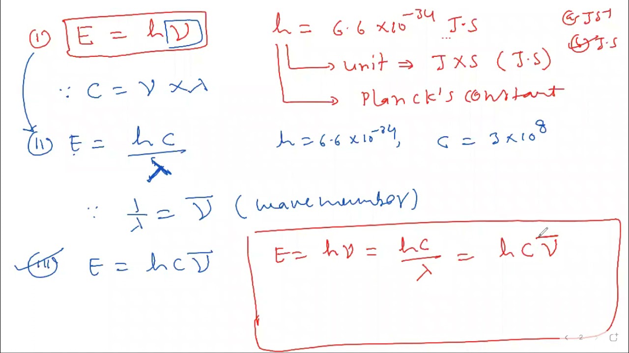
Fundamental atomic structure and quantum ideas, such as photon energy (E=hf) and basic photon-matter interactions.
The physics of Computed Tomography (CT) scanning, which utilizes multiple X-ray attenuation measurements to construct 3D images.
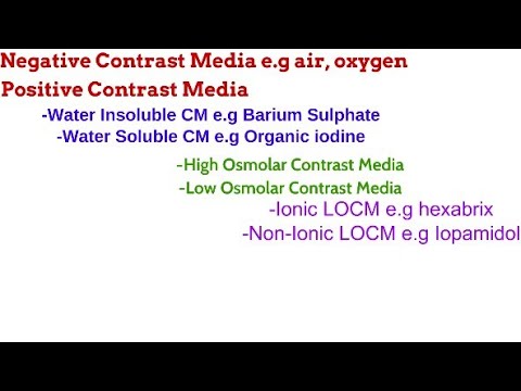
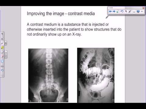
The clinical use of contrast media (such as barium or iodine) to artificially increase attenuation in soft tissues.
Principles of radiation protection, dosimetry, and the calculation of absorbed dose in medical physics.
Comparative analysis of other medical imaging techniques, such as Ultrasound (acoustic impedance) and Magnetic Resonance Imaging (MRI).
18.5K views354likes20:22Original Release: 2021-03-03
previously we learned about how to adjust the x-ray image for the best sharpness and a good contrast we can see good pictures but now we get these pictures on actually why is bone muscle tissue blood water all different different color on the film what affects that to know that we need to know about attenuation of x-rays so here we go firstly what is attenuation that's such a pretty we can say that attenuation is the exponential decrease in what in power energy and intensity of a wave i mean electromagnetic wave is x-ray right so these are still talking about waves so exponential decrease in power energy intensity amplitude you wanna throw it in there sure of a wave as it travels true a medium so travels through a medium so it's a decrease in energy though in other words it's kind of like miss isn't this like friction well yeah kind of but this one is due to absorption so i'm going to make a note here wire due to absorption energy or due to absorption processes let's just call it that x-ray get absorbed by the body or something happen okay absorption processes okay so in other words this is similar to your idea of damping if you remember from the oscillation chapter the idea of a decrease or energy loss you're something oscillating but over time the amputee becomes smaller and smaller you lose energy same idea here except that now it's in x-ray and you call it a fancy name attenuation so let's set up a scenario to understand this better what is attenuation let's say i draw a box this box represents the medium that i am going to shoot my x-ray through okay so like a bona tissue whatever is a box so now you have your incident intensity rays are coming in at some kind of intensity i'm going to call this a incident intensity x-ray coming with a certain energy intensity and this is going to be called i naught the zero is called not n a u g h t then something happened happened inside there and then when the x-ray come out oh the transmitted intensity is not so big already so this one after the x-ray pass through a certain medium this is what we call the transmitted intensity whatever that is intensity come up at the end so what happened in between the wave get attenuated so what happened to the wave okay remember electromagnetic waves come in right that's your wave coming in so i'm gonna can i draw that in here sure let me try drawing but then what happens inside this thing called attenuation happens so if your middle is let's say a wave middle is like this all then your exponential decrease or damping looks something like this so then your wave maybe start off with a pretty big amplitude but then it gets smaller and smaller and smaller and smaller and smaller and smaller lose energy lose power then come out transmitted so this is what we call damping but now we call it attenuation this shape of this graph you notice that this damping curve well that makes this pattern decreasing amplitude this is what we call an exponential decrease exponential decrease so if we want to plot a graph of intensity against distance you have traveled in the block okay maybe you start from here you travel a certain distance until here and we call this x how would the graph look like so here's our graph we're going to label the axis on the vertical axis we're going to call this intensity a we use eila again let's call this distance x it's maybe a certain meter or millimeter whatever that is i call millimeter since body mass cm or millimeter also can so your wave coming with what an initial intensity we're going to call this i naught and then exponential decrease right it's like z p go down okay so you draw the the thing that goes down like this go down this is the exponential curve what can we know about this thing well a few things we can tell is that once you go further away your intensity will drop that's all we can say and this curve have a very special equation so the equation of this curve let's go down a bit has this equation called uh let's see where shall i write this right here is exponential decrease equation and this is the intensity at any point maybe you come until let's say you come on do you hear la this point x is the original intensity times this e what is this e this is exponential it's exponent e to the power of negative mu x this is the equation of this graph and yeah you might have to use it a little bit but you have to do ratios for this thing so to do ratios with exponents so what is all these oh my okay so we're gonna break down this equation and look more carefully of what existing uh quick notes this equation applies for parallel beams or almost parallel beam if came collimated parallel beams so the ray come in like that though okay if the ray can spread out ah well then that one your power is changing your area is changing too many changing so this equation is for parallel b okay collimated nice horizontal ray coming in okay so what what does this equation mean it means that let's say the distance you travel a certain distance let's say this x i'm going to call this x half y x half ah because once i have traveled this distance away inside once the wave goes inside the material already then the intensity could drop down to 50 so 50 here means i can say this is half of the original which is half i naught which is 50 of the original then maybe you travel another same distance the same amount okay you travel under here i'm going to call this 2 times of the x half what would that be so now your your your 50 percent divide by 2 again become what 25 percent so you drop again here's what we call exponential law here drop their drop okay where is my line are they so this one now become one over four i naught this is what we call an exponential decrease so now drop to 25 percent okay and you can keep going like half and half and half and half and half until everything is gone so let's draw some dotted lines to show that this is corresponding to this diagram up here like that okay so what is the mu the mu mu this mu greek letter has a very important thing that we need to know this mu is what we call the linear absorption linear because parallel beams so linear absorption coefficient this is why we go such pains to make sure the beam is parallel if not it would be very complicated linear absorption coefficient or you can say the attenuation coefficient that's the name for it so whenever you see these words are absorption coefficient attenuation coefficient you are talking about the [Applause] symbol this one me would the intensity drop down okay so different parts of the body you can imagine do you think they all have the same meal as the x-ray pasture no lay that's how you tell from different different parts you measure the intensity coming through and you can tell ah this is the picture i'm looking for you can tell what is the thing that you just passed through so these are all your meal values each part of the body have you which one got the highest you look and see highest meal whoa do you see the biggest value bone so this one has the highest absorption or attenuation coefficient which one is the lowest er higher air you don't even see on the x-ray film okay like air is the lowest lowest so on on a film very few x-rays can go through the bone and reach the other side but in air everybody just fly through neela okay so that's the meal that you can draw here so if you want to draw a so if you are wondering like how the graph will change i have got different different mu well you think about it this mu is what uh in the equation mu is this e to the power of negative mu how does that affect the graph that we see here let's say this graph here is a bone i mean a bone you just send a electromagnetic wave to a bone so you have that kind of shape going down what if you change it to something with a less mew like maybe what's the other thing fat let's change to to fat or water water okay we change the water now i sand the beam through water how will this graft change first you can think of it through water you don't lose energy so quickly like this so your decreased intensity shouldn't be so steep so your mean will look like this ayah it should it will eventually go down towards the x-axis la but my drawing is a bit charged so let's label this if this is water or anything that has a lesser or smaller mu let's call it smaller mu new value smaller mu will affect this graph if you if you study the graph of exponential in maths please apply your knowledge how does this negative mu this constant mu affect the shape of the graph go check out your graphs anyway bone a very large mu so we have this thing here go down okay so remember o now will cause a lot of energy loss when the x-ray passed through dos energy go out water most of the x-ray can still pass through with two not too much problem okay so the first thing you need to know very importantly is this that will keep on appearing in all your past years and usually they will give you the value i'm not sure let's write it down too so the value they'll throw give you in the table and the units by the way is per cm or in millimeters per mm it's just a coefficient okay a constant for this mu okay so larger absorption like bone here larger mu lesser absorption smaller mu because after all this is called the absorption coefficient so if you have very large meal you have very large absorption over here now the other thing i want to highlight before i wrap up this section is this x half what is x half ah look at the graph again how do we come up with x half x half is how far you can travel okay a certain distance x before your intensity will drop from original to 50 percent so i'm gonna put a highlight here hey x half is how far you can travel before you drop to 50 there's a very fancy name for it so i'm going to name it down here the x half which is half value half of the value thickness i thickness there we go so this one is the other thing that they will also ask you once in a while to do in parcels how do you find the thickness how far it go is there equation for it kind of but we need to know the derivative so you see all if you have this chunk of whatever that is here bonla metalla and then you send x-ray in how far will this x-ray go how many cm or mm before the amplitude or intensity or power will drop down to half of its original well looking at intensity okay how far the intensity how far before intensity dropped to 50 percent so we have to do some little bit of the equation here so to find the half value thickness i'm gonna put here a sometimes we call it the hvt i don't know why they call it that i guess it's just the acronym of half value thickness so how do we find that question mark there's an equation for it let's derive it together so firstly we know that we have this equation up here this i intensity equation i goes so i naught the original times e negative mu x so we're going to rewrite that but in terms of a fraction i over i naught equals to e negative mu x now at half value thickness what is the fraction of i over i naught means the current intensity over the original [Music] okay so this means you have already dropped by 50 so this ratio of i over the original is half okay so this one is half uh we can write that down there so this will be equal to e negative mu x how do we get the x this is x half by the way that is the half value thickness with we want the x half but how do we get it out it's stuck inside the exponent so we have to do this thing called log and lawn so we need to get the x out so this is similar to what you do in practical paper five we need to take out the thing so first let i'm going to take the lawn the natural log of both sides so long half and lon of e to the power of negative x now this one got very nice magic because of when you take a log inside the bracket if there are any powers or you can take it out like this so x half times the natural log of e and this is lon of half the nice thing about lon of e is that this thing you press calculator what's the natural log of log of e become one so it's gone bye bye okay very nice so all we have now is just uh x i'm going to rewrite this x which is the half value thickness equals to negative lon 1 over 2 divided by which is the loss how how quickly you lose energy in passing through a medium negative sign here okay so another way to write this is you want to give a negative sign negative of a long number you can reverse the fraction inside so instead of 1 over 2 it becomes 2. what does that work oh there's some long long magic there that we can do so this is how we can calculate the half value thickness if we know the mean of a certain object how far will your x-ray travel before it drops down to 50 so you can adjust your setting law contrast intensity everything to make sure you have this in mind when you are trying to scan somebody you want to scan the backbone but your ray also cannot really reach the backbone then how okay so remember all these things okay so that is the idea of attenuation basically like damping energy loss or the proper terms in x-ray is absorption the orange curve low absorption smaller mu means low absorption or attenuation whereas the bone a lot of extreme lose energy when you go through the bone so this one is large absorption large mu so the computer is very clever one it will measure all these computer actually the film you can see whatever x-rays make it true it will cause a different shades of color so maybe like this one here this one is hu but you can replace this with mula it's a different unit but you can replace this column with you a mu then you set a certain value okay i want uh this mu to be gray color so you adjust the machine wow 256 shades of grey interesting okay so you said water is great then everything else that is a different mule can be either white or either black above or below water so it's up to you to what you want to set as grey color to adjust accordingly to have the best contrast to see what you want to see but the first place where you have different mew is because you have different parts and different linear absorption coefficient or attenuation coefficient okay so a very quick recap of this equation i'm going to label this now in case some of us are don't remember what is this already so i here is the intensity of the x-ray at any point any position this i naught this one is the initial intensity when it enters the medium e is an exponent it's a certain number miu represents this decrease in energy and this is our attenuation coefficient or absorption coefficient and lastly the x here it's just how far you travel inside the object law distance cm units whatever you want to call that all right so that is the idea of this thing and remember these only can work for parallel beam the condition is only parallel beam because for parallel beam uh the area of the beam does not change like this one so we have parallel beam coming in this is very collimated and nice okay we try to adjust the endnote everything the setting we talked about before to make sure it's nice and parallel because if you have something that is spreading out too much your equation will not be quite right because here ah your area is changing so your intensity is also changing because intensity is power per unit area so in the first one your area is one area is same throughout the beam or almost the same but if your beam is spreading out too much this area keeps changing so we don't want this we only parallel beam we use this so we try our best to get parallel beams all right so that's all for this attenuation idea hopefully that was helpful in helping you understand how x-ray travel how x-ray beams travel through objects human body metal wall plastic bone anything you want to scan a luggage metal and remember it's an exponential decrease [Applause] as you go through an object pretty much like damping of an oscillation kind of thick but now electromagnetic wave radius all right so that's all for this video i will see you in the next one we'll look at some examples on how to do calculation related problems for attenuation in the body right that's all for this video i will see you in the next one
Up Next

10:44
A-Level Medical Physics: X-Ray Attenuation Mechanisms Explained
@PlymCollSci
36.5K views•2013-03-05

12:53
21cm Hyperfine Transition in Neutral Hydrogen: Radio Astronomy Basics
@AaronRobertParsons
12.4K views•2011-10-13
114:06
A2 Medical Physics Review: MRI, Ultrasound, X-Ray & CT | 9702
@ETphysics
13.6K views•2021-04-30

27:14
Entropy and the Second Law of Thermodynamics Explained
@veritasium
27.5M views•2023-07-01
Related Study Plans & Knowledge Roadmaps
Structured learning paths in Physics
physics
Nuclear Physics (Decay, Fission & Reactors)
01Atomic Structure and the Binding Force
02Radioactive Decay and Half-Life
03Nuclear Fission Mechanics and Chain Reactions
+ 2 more learning modules
12h5 modules
Explore Roadmap
physics
Soft Matter (Polymers, Colloids & Rheology)
01Foundations of Soft Matter & Thermal Forces
02Polymer Physics and Macromolecules
03Colloids, Surfactants, and Self-Assembly
+ 2 more learning modules
38h5 modules
Explore Roadmap
physics
Analytical Mechanics: Lagrangian & Hamiltonian
01Mathematical Foundations: Calculus of Variations
02From Newtonian Mechanics to Constraint Dynamics
03The Principle of Least Action & Lagrangian Mechanics
+ 3 more learning modules
28h6 modules
Explore Roadmap
physics
General Relativity (Spacetime & Cosmology)
01Foundations of Relativity: Special Relativity
02The Geometry of Spacetime: Equivalence Principle & Curvature
03Einstein's Field Equations: The Mathematics of Gravity
+ 2 more learning modules
55h5 modules
Explore Roadmap