














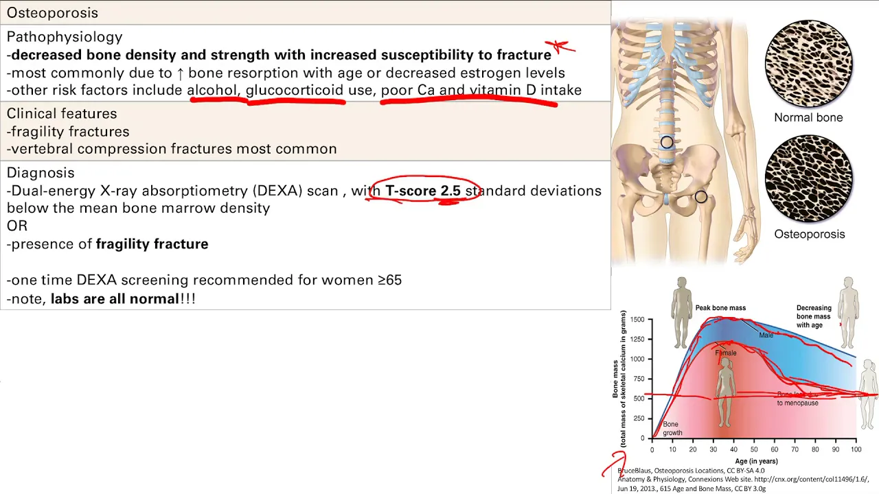
Metabolic bone disorders affect different components of bone: rickets and osteomalacia cause defective mineralization (osteoid formation is normal but mineralization is impaired), with rickets occurring in immature skeletons and osteomalacia in mature skeletons, both due to vitamin D deficiency leading to calcium and phosphate deficiency; scurvy results from vitamin C deficiency causing defective collagen synthesis, leading to brittle bones, pseudo-paralysis, and characteristic radiological findings including ground glass appearance, pencil-thin cortex, metaphyseal white line of Frankel, Pelcon spur, Trummerfeld zone, and Wimberger sign.
Orthopedics: Metabolic Bone Disorders & Bone Tumors | MBBS Final Year Crash Course
Added:Normal bone histology and physiology, including the roles of osteoblasts, osteoclasts, and the bone remodeling cycle.
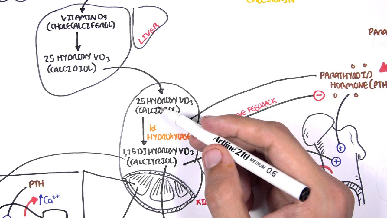
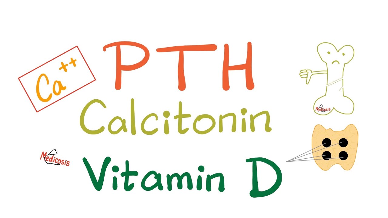
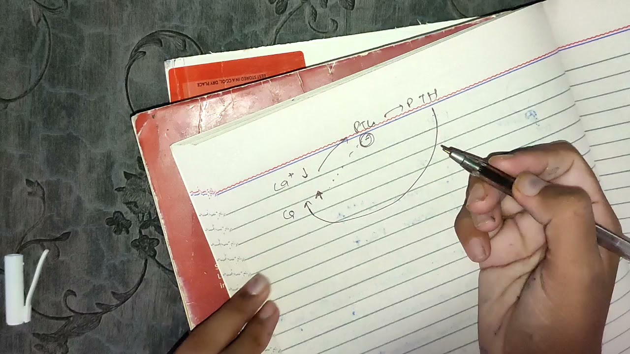
Endocrine regulation of calcium, phosphate, and magnesium homeostasis, specifically the functions of parathyroid hormone (PTH), Vitamin D, and calcitonin.
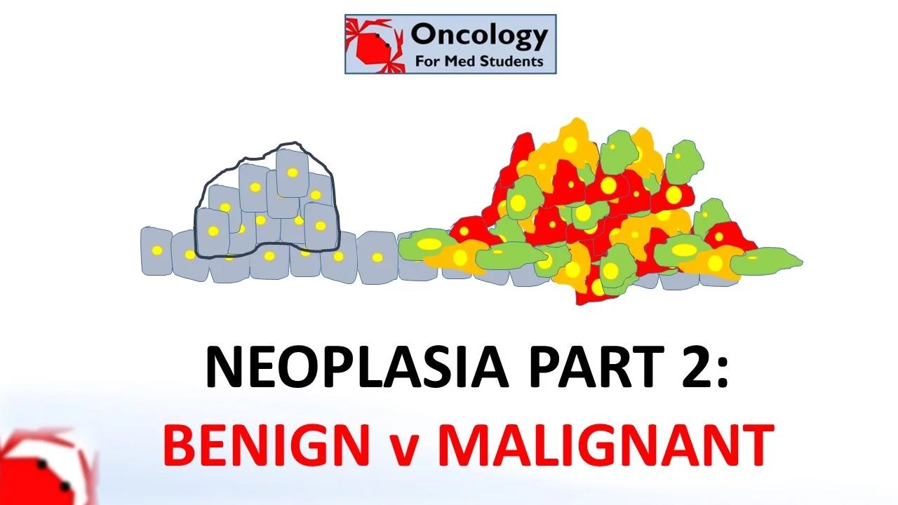
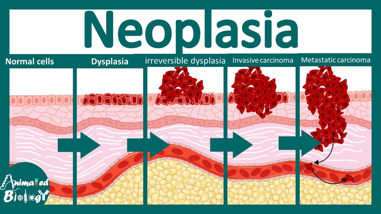
Basic pathology of neoplasia, including the biological differences between benign and malignant tumors, staging systems, and pathways of metastasis.
Fundamental musculoskeletal radiology, focusing on identifying normal anatomy and basic cortical or medullary lesions on plain radiographs.
Advanced oncological management of bone tumors, including limb-salvage surgical techniques, tumor resection, and reconstruction options.
Clinical pharmacotherapy guidelines for metabolic bone diseases, focusing on the mechanism and administration of bisphosphonates, SERMs, and monoclonal antibodies.
The clinical workup and emergency management of pathological fractures secondary to osteoporosis or metastatic bone disease.
Pediatric metabolic bone disorders and genetic skeletal dysplasias, such as Rickets, Osteogenesis Imperfecta, and Achondroplasia.
42.2K views890likes1:35:55Original Release: 2020-12-13
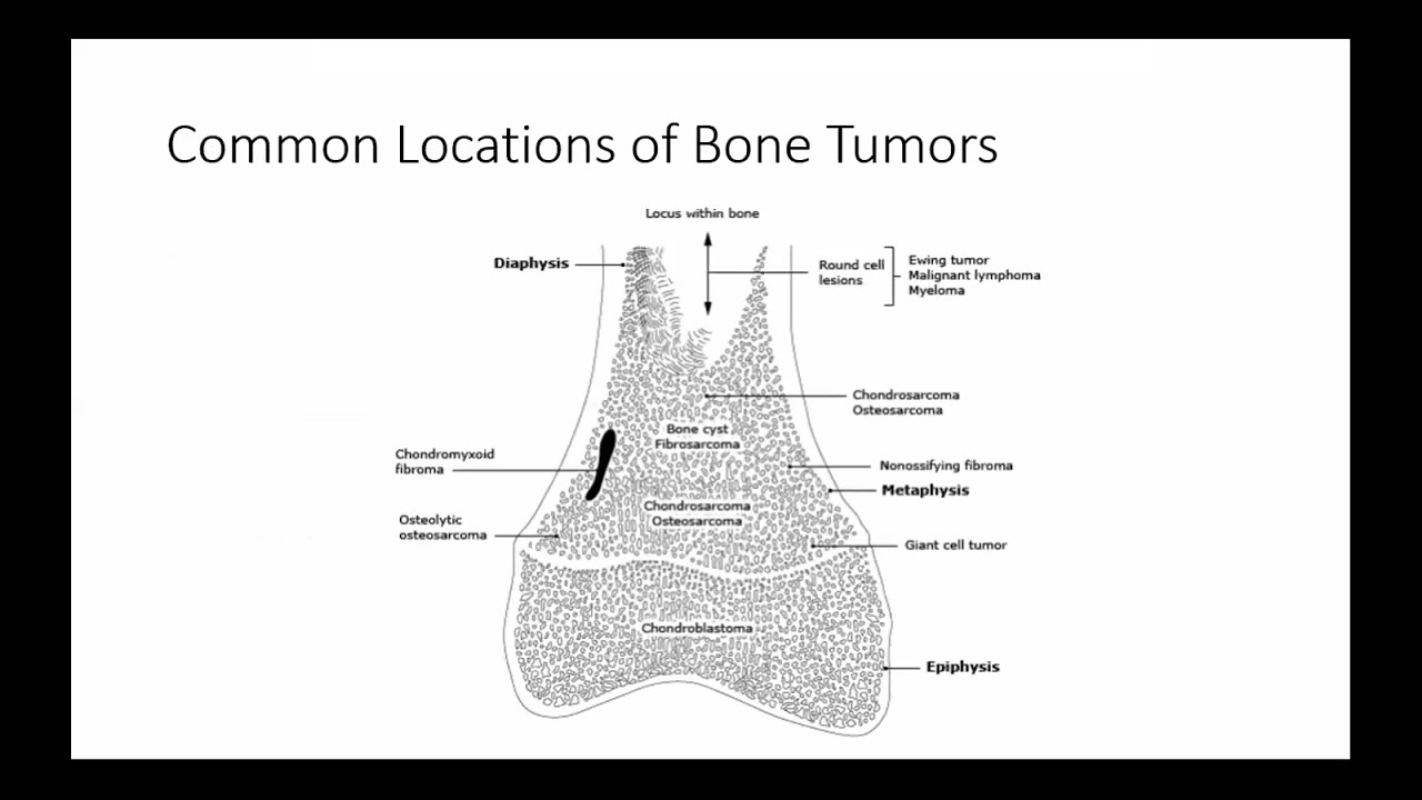
good morning everyone can you see me I'm on live please tell me in comment box then he'll start okay parag beta so let's start so I'm Dr arind I am faculty in Department of Orthopedics s GS Medical College and km Hospital Mumbai in s GS Medical College and km Hospital Mumbai we have very structured teaching courses but due to this situation of covid-19 we were not able to take our physical classes for students so it was idea of my head of the department that do of Dr Sunil Kumar ASA to arrange some online classes for the students and at that time he advise me to invite all students besides km Hospital you should invite students from other medical colleges also so that is why today I'm taking lecture so it was the idea of Dr SK Shasta head of the Department of Orthopedics in km hospital so here I am going to discuss only important topics which are very very very important for your University examinations okay so in a day where I am going to take 6 to 8 hours lecture I cannot cover full Orthopedics but believe me take my words whatever I'm teaching today if you remember those things only you will be able to clear your University exam with very good marks okay whatever will be remaining say 1 hour or two hours that I can take next Sunday or whenever you will say so let's start with the lecture first I will give you the just brief idea of bone Anatomy whenever we are going to discuss any of the disorders we will be mentioning the parts of the bone the components of the bone so it is very very important to know the basics in bone Anatomy so first we will discuss bone components bone is mainly composed of two major components one is inorganic part which makes 65% of bone mass the second will be organic Matrix which makes 35% of bone mass now this inorganic component which is the major component of bone is nothing else but the mineral in the bone and the main mineral in the bone is hydroxy appetite C A10 P4 6 O2 so this mineral makes 65% of the bone mass now the second component is organic Matrix which is also known as osteoid if we remove this mineral from the bone the remaining part will be osteoid only that is why this unmineralized bone is also known as osteoid and this have been asked in neat exam unmineralized bone is known as osteoid one time they asked if we remove mineral from the bone the remaining part will be this osteoid or organic Matrix now this organic Matrix or osteoid consist of two components one is cells osteoblast osteoclast and osteocytes that we are going to discuss in next Slide the second component is protein which is the major structural component of this organic Matrix or osteoid and this protein part which makes major component of organic matrix consist of type one collagen predominantly so major component of this organic Matrix and Osteo major structural component of this organic Matrix or osteoid is type one collagen that is why the proteinous part we divide into collagenous and non-collagenous because collagen is the main important structural component of osteoid the non-collagenous protein may be proteoglycans osteocalcin osteonectin and fibro nectin so these are the major component of the bone now in cells we have three types of cells osteoblast osteoclast and osteocytes these osteoblast are the major bone forming cells they form protein in the bone that is osteoid or organic Matrix part and they do mineralization of the bone both of the both the processes are done by osteoblast so these osteoblast are the cells responsible for bone form formation and during bone formation there is mineralization of bone so these are the cells for bone formation and Bone formation requires alkaline medium remember whenever there is formation of bone that will require alkaline medium and that alkaline medium is provided by alkaline phosphatase in the osteoblast that is why the marker of osteoblast as well as a mark for bone formation is alkaline phosphatase bone specific alkaline phosphatase which is the marker for bone formation the second type of cells are the Osteo class these are the cells responsible for bone destruction or bone resorption these are the cells which are derived by Fusion of mononuclear macras system cells that is why these are multinucleated G cells okay they are derived from fosic cells but they don't have phagocytic function their major function is bone destruction now bone resorption or bone destruction is bone destruction or bone resorption requires acidic medium because all hydrolytic enzymes which destroy things they act in acidic medium to provide that local acidic environment for bone destruction these Osteo class consists of two enzymes one is acid phosphatase this acid phosphates is tartrate resistance so this trap tartrate resistant acid phosphates and Carbonic anhydrates these enzymes will provide local acidic environment which is necessary for bone destruction so marker for bone resorption major marker for bone resorption or bone destruction is this trap tartrate resistant acid phosphatase which is the marker of Osteo class now the third type of cells are osteocytes these cells have mechanos sensory function and ion exchange what is mechanos sensory function Whenever there are normal stresses along the line of there are mechanical stresses along the lines of weightbearing or something there will be formation of bone so those stresses those mechanical stresses will be sensed by these Osteo sites and they will send the signal then these osteoblast will form the bone so mechanosensory function is done by osteocytes the major the second major function of these osteocytes is Ion exchange any type of ion Exchange in the bone is done by these osteocytes calcium ion sodium ion anything so ion exchange is done by Osteo sites now these osteocytes are derived from osteoblast these osteoblast during bone formation what happens when there is development of organic Matrix formation of organic matrix by these osteoblast some of the osteoblast are enclosed in that developing Matrix only they become osteocytes so during bone formation around 20% of osteoblast are converted into osteocytes only 20% of osteoblast are converted into osteocytes still these osteocytes are the most abundant cells in the bone because they are longest living cell in the bone so this was about cells in the bone osteoblast major bone forming cell containing alkaline phosphates osteoclast bone resorption cell containing trap and Carbonic anhydrates and osteocytes having mechanos sensory function and ion exchange function now we will see parts of the long bone whenever you are you will be listening to some of the disorder like bone tumors so we will be saying epipal tumor metaph fasial tumor diapal tumor so which are these part in a long bone so in a long bone if we are starting from one end say for from this end towards the center of bone we are going in this direction the terminal most part of the bone is episis okay so this terminal part of the bone is epifisis this part of the bone B is growing end of the bone growing end the bone will grow in this direction whenever growth will be occurring bone will be growing in this direction epipal direction okay so this is the growing end of the bone just inside the epifisis is fisis also known as growth plate or epipal plate okay so remember this epifisis is different epipal plate is different in my Yugi days I always used to get confused between these two these are two different epifisis is different this epipal plate is growth plate or fisis now this growth plate or fisis is the area in a growing bone where actual growth is occuring so this is actively growing part of the bone and this fisis is not a bone it is Highline cartilage okay this Highline cartilage during the growth is always mineralizing there is mineralization of this Highline cartilage that leads to Bone formation so what happens this is Highline cartilage at this area from metap fasial side this is metapas just below the fisal plate from metap fisal side there will be always mineralization when there is mineralization this area will be converted into bone and further cartilage proliferation occur in this direction so epip episis will go away further cartilage formation in this area and further mineralization from this side so this is how this growth will take place now this is metaphysis just below the growth plate which is metabolically the most active part of bone because it is the most vascular part and it consists of Maximum kilus bone now the center shaft of the bone it is known as diasis which is the strongest part of bone and the major side for having saroma so from one end towards the center we have epiphysis then just inside the episis f is or growth plate then metaphysis and diaphysis so these are the parts of long bone in a growing skeleton immature skeleton in mature skeleton what will happen this mineraliza this growth plate will fuse with the bone there is complete completion of growth so complete fisal plate will be mineralized so in a developed skeleton mature skeleton we will not have episis or growth plate we we have epifisis metaphysis and diaphysis so these are the parts of long bone now we will see microscopically types of bone microscopically there are two types of bone Lamar bone which is the mature bone and woven bone immature bone it is known as woven bone because here there is arrangement of this Collision in wavy pattern random pattern wavy pattern that is why this is weak bone and this immature bone you can remember immature so it will be seen in fetal skeleton all fetal bone intra utrine life they will be woven bone and in postnatal life they will be converted into laminar or mature bone in postnatal life there are only few conditions where you will find this woven bone in the body one is fracture healing that hard Kus is nothing else but the woven bone initially second some of the pathological condition like fibrous dysplasia and ped disease where you can get these bone otherwise woven bone immature bone in fetal skeleton and in postnatal life all bones are mature bone or Lamar bone where there is arrangement of these osteocytes and collagen in structured manner in the form of lam the bone is arranged in the form of lam that is why it is known as Lamar bone and all bones after postnatal life are Lamar bone or mature bone now this Lamar bone is further divided into two types one is cortical or compact bone which is hard bone and here Lam are densely packed concentrically in the form of haversian system or oston which is the structural unit of cortical bone and this part of bone forms the outer cortex of bone the second type of bone is cancelous bone or tabular bone it is soft sponge like bone where Lam are Loosely packed they are Loosely packed that is it is soft sponge like bone this bone is seen at the end of long bone in the metap fasial area and in the center of vertebral body so it will be at the end of long bones and inside the vertebral body now very interesting fact that this cortical bone makes around 20 80% of bone mass and this Con cancelous bone makes only 20% of bone mass still this cortical bones has 70 to 80% metabolic activity because this cancelous bone as you can see has very large surface area and it is the most vascular part of the bone okay so though the cancellus bone makes only 20% of bone mass it has maximum metabolic activity and it is the most vascular part of the bone so types of bone immature that is woven mature your Lamar now this Lamar bone is two type cancellus bone and cortical bone now this the structural unit of cortical bone known as oston or herian system where there is concentric arrangement of collagen and Osteo side around a longitudinal Canal this longitudinal Canal known as aversan canal which consist of neurovascular bundles so there will be these are the longitudinal canals around which there is concentric arrangement of lam now this longitudinal or herian canals are connected by horizontal or transverse canals known as workman's Canal so this is the structural unit of a bone now the covering of the bone the outer covering of bone here you can see this is outer covering of bone it is periostium and the inner covering of bone from inside it is endium now this periosteum is a very very important covering layer it provides blood supply to Outer 2/3 of the cortex as well as it is osteogenic this periostium has two layers inner cambium layer outer fibrous layer the inner C ium layer which is attached to Cortex that is osteogenic and in bone tumors whenever you are reading those terms codman's triangle Sunray appearance onion peel appearance those are nothing else but these bone formation by these this periostium cambium layer of periostium inner layer of kostium that is why those things are known as periostal reaction Sunray appearance codman's triangle onion peel reaction these are nothing else but the periostal reaction whenever this periostium inner layer of periostium that is cambium layer of periostium is irritated or stimulated either by infection or by growth of tumor it will form bone that is known as periostal reaction okay so these are the part of a long bone which we have already discussed this is the episis terminal part this is growth plate this is metaphysis this is diaphysis now this is the cortical bone here you can see this is cortical bone cortex of a bone so cortical bone will be Maxim seen in maximum thickness in the shaft area diaphysial area here inside this met cortical bone in the metaphases where cortex is very thin there will be cancelous bone so maximum cancelous bones will be seen in this metap fasial area here so cancellus bone cortical bone medular cavity episis fisis or growth plade metaphysis diaphysis outer covering periosteum inner covering osum okay so these are the parts of long bone and this was about basics in bone Anatomy if anyone of you beta have any query you can ask me then we will proceed to next chapter yes anyone clear to everyone better so we will move ahead with next chapter Metabolic Bone disorders as the name is suggesting Metabolic Bone disorders disorders which are mainly affecting the metabolism in the bone so disorders which are affecting components of the bone will be Metabolic Bone Disorder so just we have discussed components so bone is mainly composed of two components one is mineral that is inorganic part the second is organic metric or osteoid this osteoids consist of two part protein part and the major protein was type one collagen and the second parts are cells now these Metabolic Bone disorders will be affecting any of these so mineral part this mineral part is affected by rates and Osteo Malaya so rates and osteomalacia will affect this mineral part this organic Matrix or osteoid part in these disorder will be normal now type one collagen which is the major protein in the bone major structural part of organic Matrix will be affected in scurvy and osteogenesis imperfecta so scurvy and osteogenesis imperfecta will affect type one Collision that is why they will affect this osteoid part in these this mineralization part will be normal now in cells osteoclast the bone will be affected through osteoclast by Paget's disease osto petris and Hyper primary hyper par thyroidism okay so these disorder will affect the bone through Osteo class now the most common Metabolic Bone disorder that is Osteo porosis which affects both mineral and organic Matrix both will be decreased there is generalized decrease in bone density both will decrease mineral will decrease and as as well as organic metrix or osteoid will decrease so these are the important metabolic Bond disorders this is the most common metabolic Bond disorder overall most common metabolic Bond disorder in children is rates most common Metabolic Bone disorder in adults is osteomalacia overall most common Metabolic Bone disorder is osteoporosis now in this chapter we are going to discuss these three rates Osteo Malaya and scurvy because the if they are going to ask any Metabolic Bone disorders these are the three Metabolic Bone disorders which have maximum probability to come in the exam and beta if anyone of you have any suggestion to take lecture on any of the disorders after the lecture you give the suggestion I will keep one lecture either next Sunday or whenever you say next week okay so here we are going to discuss retes osteomalacia and scurvy so first we will discuss rates and Osteo Malaya so here definition is very important in our UGI days whenever we are reading these retes osto malasia always we think of deficiency of Calcium deficiency of phosphate isri deficiency of vitamin D is rates these are the causes of rates actually what is rates rates is defective mineralization of bone is rates in immature skeleton in homalia in mature skeleton so rates and osteomalacia these are Metabolic Bone disorders which are characterized by defective mineralization of bone so mineral ation process is defective if it is in immature skeleton or growing skeleton it is rates if it is in mature skeleton it is Osteo malaia so basically to understand the pathophysiology I will give you a quick recap how bone is formed during bone formation first there is formation of osteoid or organic Matrix part and we have discussed the major structural component of organ Matrix or osteoid is type one Collision so what will happen first osteoblast will lay down this framework of type 1 Collision there is framework of type one Collision so during bone formation first there is osteoid in which there is formation of framework of type one Collision the next step in bone formation will be maturation of this osteoid by mineralization so what will happen there will be deposition of these min mineral crystals hydroxyapetite crystal in this area in this framework of collision so there will be formation of mature bone okay so first formation of osteoid then maturation of that osteoid by mineralization what happens in retes and osteomalacia this mineralization part is defective okay so osteoid formation is normal osteoid formation is normal but maturation of that osteoid by mineralization that step is defective okay this step is defective mineralization is defective so what will happen there will be defective osteoid maturation osteoid formation is normal organic Matrix formation is normal but maturation of that osteoid is abnormal defect in maturation of that osteoid by mineralization so osteoid maturation time will increase in rates and Osteo Malaya so this is the Bic basic pathophysiology in rates and Osteo Malaya now we'll see each one by one rates Metabolic Bone disorders of immature skeleton developing skeleton due to defective mineralization of bone and the basic deficiency is calcium or and phosphate in the bone and this deficiency in turn is due to vitamin D deficiency okay so the most common cause of retes is vitamin D deficiency which leads to insufficient availability of calcium and phosphate okay now as you all know in vitamin D activation or metabolism of vitamin D three organs are involved skin liver and kidney in skin 7 dehydro cholesterol it is converted into Vitamin D3 kly calciferol in skin by UV ray exposure now this kic Calif or vitamin D3 under goes 25 hydroxy by 25 hydroxy in lever to form 25 hydroxy kic califo or 25 hydroxy vitamin D3 pardon me MAA my handwriting is very bad but I will try to make you understand clearly now this 25 hydroxy vitamin D3 in kidney undergoes one hydroxy by one alpha hydroxy so there is formation of 125 dihydroxy colic calciferol or 125 dihydroxy vitamin D3 which is also known as caly triol which is the active form of vitamin D so this caly triol is the main acting vitamin D in the body now you can see if there is any skin disorder or lack of UV exposure there is any liver disorder or there is any kidney disorder all these will lead to defective metabolism of vitamin D they can cause rates or Osteo malasia now classification of rates the rates is simply classified into two types one the most common type where there is some problem in vitamin D so type one is vitamin D related there is some problem in vitamin D either there is deficiency or there is defective metabolis or there is some problem genetically so in nutritional deficiency either there is deficient intake in diet or there is some malale absorption syndrome defective metabolism which we have just discussed in last slide either liver disorder kidney disorder or lack of sunlight or skin disorder so in that there will be sorry problem in defect problem in active formation of vitamin D active form of vitamin D calc Tri will be not forming calc trial will not be forming that is why it will cause rates now genetic vitamin D dependent rates where there is dependence on vitamin D so it may be either type one vitamin D dependent rates where there is deficiency of one alphahydroxy in kidney which is the rate limiting step in Activation so there will be formation up to 25 hydroxy vitamin D3 in liver but one hydroxylation in kidney is not there so there is no formation of active vitamin D the type two vitamin D dependent R is where there is normal formation of active vitamin D or calcy Tri but the receptors are insensitive they are not responding to active vitamin D okay now don't get confused with this type one vitamin D dependent rates type two into these type one and type two this classification is for vitamin D dependent rates which is genetic so type one vitamin D dependent type two vitamin D dependent both are subtypes of type one rates overall where there is rates due to vitamin D deficiency or metabolic now type two is due to type two rates which is due to phosphate deficiency it may be either due to dietary deficiency or malale absorption so in these condition you will find combination of these two type one and type two because both vitamin D and phosphate will be deficient or it may be due to defective reabsorption in kidney like in xlink hypophosphatemic rate a genetic condition fenon syndrome or proximal renal tubular acidosis so these are the type one and type two rates now in pediatric they have classified into three type type three this genetic vitamin D dependent rates they have classified into type three so here mentioning so you don't get confused okay so this is the simplest one we type one vitamin D related type two phosphate related now what is the pathophysiology in rates in rates I'm sorry as we have discussed in our first few slides that during normal growth of the bone there is mineralization from metap fasial side in this Highline cartilage of growth plate and bone will grow in this direction but in rates this mineralization is defective so what will happen there is continuation continuous proliferation of cartilage but no mineralization so what will happen there will be thickening and broadening of growth plate and this metaph fasial area as there is no mineralization will be soft so what will happen Whenever there is weight bearing exal loading of bone either by crawling of child or walking so this epifisis it will be pushed into soft metaphysis like this piston in cylinder effect so what will happen there will be cupping there will be sping of metaphysis so exal pressure will cause cupping and SP saying of metaphysis further as there is defective mineralization there will be fraying of metaphases metaphases will be irregular here you can see this is normal bone clearly defined metaphases here in rates cing playing and fraying irregular fraying of metaphases you can see here it is clearly defined and there is thickening and widening of growth plate which we have discussed and due to defective mineralizations bone are weak which will lead to deformity of bone boing of legs now clinical manifestations of rates clinical manifestations of retes can be divided into Regional which are more important to remember and systemic so Regional manifestations are more important and it will be easier to remember if you go systematically from head down in skull there is cranio tapes that is softening of skull bone which is the earliest clinical manifestation there will be widening of skull sutures there will be delayed closure of fonton nails and there is Caper quadrum hot cross bun skull due to widening of skull suture there will be cruciate pattern in the skull and skull will look like something this hot cross bun so that is known as cross bun skull there will be prominence of frontal and perial Bones so there will be frontal and parital bosing so these are the finding in skull now from Skull we are going down in the mouth there will be delayed eruption of the teeth and there will be defective enamel part of the teeth in chest we will have prominent costoc condal Junction due to enlargement of cartilage at costoc condal function which is known as retic Rosary there we practice carinatum or pigan test where sternum will be protruding anteriorly because these soft ribs they cannot hold the sternum inside they lose their elasticity because of defective mineralization so sternum will protrude outside like this pigeon so this is known as pigon chest or Pectus carinatum and at the lower margin of rib C there will be sulcus known as Harrison sulcus or Harrison Groove as ribs are soft with every effort of respiration diaphragm will pull this soft rib inside which will create this Harrison sulcus now on abdomen we have protruded abdomen Port belly after finishing anterior part of trunk posteriorly in spine we will have kyotic deformity and finally in limbs there will be widening of wrist and ankle because we have seen there is widening of growth plate that is why whenever you are palpating in a child of retes there will be widening of ankle and rest and these are the areas where we first initially palpate to see clinically the findings of retes there is kavara that is decrease neck shaft angle in femur there can be ggam or noi there can be genu varas and there can be wind deformity what is wind s deformity one side galgas and one side garus like wind is sweeping something away wind swep deformity wind is blowing something so that is known as wind swep deformity in children it is due to rates in adults it is due to rheumatoid arthritis there will be boing of legs once child starts working due to weakening of bone there is Boing of flx in systemic manifestations we will have growth retardation and hypotonia and muscle weakness so quick recap from up down in head cranio tapes widening of suture delayed closure of fonton nail hot cross bun skull and frontal and parital bosing then come to mouth delayed eruption of teeth defective enamel then come to chest suas retic Rosary pigan chest or Pectus kinum then abdomen Port belly back kyotic deformity then limbs wiing of wrist and Ankle cavara ggas or garus wind Swip deformity and boing of legs so these are the important clinical manifestation of retes very very very important topic for your University exam now radiologically this features we have already discussed in our pathophysiology part the earliest finding will be around wrist because child first starts crawling so first the exal pressure will be around rest so there will be cupping playing and fraying of metaphases there will be cupping of metaphases sping of metaphases and fraying of metaphases these are the important radiological findings of rates now the second disorder Metabolic Bone disorder to discuss here is Osteo malaia it is the adult or mature counterpart of rates where there is defective mineralization in mature bone so due to defective mineralization this osteoid osteoid is forming normally but mineralization of that osteoid is defective so what will happen there will be relative increase in osteoid volume and delayed maturation of that osteoid so in increase maturation time of osteoid that is the basic pathophysiology both in retes and Osteo Malaya now Osteo means bone Malaysia means soft so actually Osteo Malaysia means softening of Bones clinically most of the patients are asymptomatic and whenever they are presenting with some symptoms pain is the most common presenting symptom so bone pain it may be either in back or lymphs and there may be muscle weakness Osteo malesia can cause proximal myopathy sometimes there may be pathological fractures as bones are soft or weak so these are the clinical manifestation now radiological features are very important radiologically the one of the characteristic finding is loser Zone also known as milkman's fracture or pseudo fractures these are the incomplete fractures along the lines of me meical stress here you can see in pubic Remi in proximal part of femur on medial side so this is the area which has maximum mechanical stress because weight is transmitted in this direction so this is the area where you will get these loser zone or milkman's fractures that is superior and inferior pubic Remi and medial side of proximal femur either in the neck or in the shaft around lesser troas so this is the area where you get these pseudo fractures okay so these are the pseudos fracture in complete fractures here you can see this second is TR radiate pelvis in females especially triangular shaped pelvis Proto acetabuli so along with acetabulum fural head is protruding inside the pelvis Proto acetabuli if it is bilateral it is known as autop pelvis cod fish vertebra Bon cave vertebra with bonx intervertebral disc so what happens normally this is vertebra this is vertebra this is intervertebral disc in osteomalacia as well as in osteoporosis where there is weakening of bone this intervertebral disc on weight bearing bulges inside the soft vertebrae so this intervertebral disc will become biconvex and it will make these vertebrae by concave so by concave vertebra by convex intervertebral dis codfish or fish mouth vertebra so these are the radiological features of rates and uh sorry Osteo Malaysia triradiate pelvis pseudos fracture or milkman's fracture cfish vertebra and Proto acetabuli now in treatment sorry first we will discuss serum markers of rates and Osteo malesia so these serum markers are very easy to remember if you have concept clear in your mind so both are due to deficiency of vitamin D due to deficiency of vitamin D what will happen blood calcium level will decrease and this serum calcium is the most important stimulus for paraone so there is increased secretion of paraone that is secondary hyper parathyroidism now this paraon as you all know has major function it tries to increase calcium level in the blood so it will try it tries to normalize the blood calcium so ultimately calcium will be either slightly decreased or normal the second function of par hormon is to decrease phosphate reabsorption from kidney so in blood phosphate level will be decreased and compensatory activity of osteoblast is increased in rates which will lead to increased alkaline phosphates so now you can see these are in fullblown rates what will be happening normal or slightly decrease calcium decrease phosphate increase alkaline phosphates and increase paraone secondary hyper parathyroidism so calcium will be either low or normal phosphate will be low alkaline phosphate will be increased and Hyper parathyroid will be there now one of the condition where in rates phosphate is not decreased is renal osteodystrophy chronic renal failure because kidney is not acting properly it is unable to excrete phosphate from the blood though there is secondary hyper parathyroidism phosphate is not excreted by kidney because it is not functioning so in that condition phosphate is also increased okay so this is the only exception renal Osteo distrophy or chronic renal failure so these are the important markers for bone formation oh sorry for rates now in treatment of rates and osteomalacia the major treatment most of the time it is conservative treatment unless there is fracture pathological fracture so calcium and vitamin D supplementation we will be giving and in children if there is severe deity in rates we will go ahead with osteotomy for correction of the deformity and remember one very important fact if you are going ahead with surgery or correction of deformity first you will control the disease Pro es otherwise even after correction there will be recurrence of deformity so how will you come to know whether the rates is under control or not serological markers that is alkaline phosphates which is coming to the normal second radiological findings in healing rates you will see this cup shaped white line of metaphysis in the metaph fisal area this is the finding of healing rates so radiological and serological finding of healing rates after that that you will going for surgical correction okay now the next metabolic Bond disorder which we are going to discuss is scurvy scurvy is due to deficiency of vitamin C if you just remember the important functions of vitamin C inside the body scurvy is over for you so the most important function of vitamin C in the body is synthes of collagen it helps in post translation modification during Collision formation so this is the most important function it is one of the most important antioxidant or reducing agent in the body it also helps in generation of norepinephrine and serotonin you must be reading in your biochemistry methylation steps then it also helps in utilization of iron and folic acid in the body now you can see four important function you remember scur is over for you now due to defective collagen synthesis in scurvy what will happen collagen is the major structural component in vessel wall so there will be bleeding from the vessels gum and sub periostal bleeding collan is a major structural component of organic Matrix or osteoid part of the bone which we have discussed in our first slide so in Bones there will be defective osteoid formation so these bones will be brittle and tender and due to tenderness there will be pseudo paralysis tenderness is due to defective osteosynthesis and due to recurrent sub periostal bleeding so what the child will cause the child will lie in Flex posture like this so it seems that the child is having paralysis but actually there is no motor or motor deficit or there is no muscle paralysis still child is looking to having uh to have paralysis that is why it is known as pseudo paralysis and there will be scorbutic Rosary in rates there is enlargement of cartilage at costochondral Junction which causes scor it causes Rosary retic Rosary in scurvy what happens at costr condal Junction and there is defective ligaments collagen is defective there is separation of costoc condal Junction that lead to scorbutic rosary and in teeth the dentine part of the teeth will be affected in rates it is enal part which is affected in scurvy it is the dentine part which is going to be affected now due to defective synthesis of norepinephrine and serotonin there will be irritability and other psychatric symptoms there will be defective utilization of iron and folic acid which leads to anemia and as it is one of the most important antioxidant reducing agent there will be free radical injury and decreased immunity so these are the findings of rates radiological features are very very important the initial finding will be around the knee joint lower end of humor and upper end of tibia and we have discussed in pathophysiology as there is defective osteosynthesis it will lead to ground glass appearance of bone so this a is indicated ground glass appearance of bone and cortex will be thin Pencil Thin cortex the other very important F important finding is white line in the metaphysis Frankl line metap fasial white line so this will be white line of Frankle here this is C now this white line of Frankle there is prolongation of this white line laterally Beyond cortex this lateral spur it is known as pelcon spur as there is defective pooid formation this area just below the metap fisal white line there will be destruction of bone radiolucent area this is known as Tremor fail zone so there will be Tremor fail [Music] Zone and finally the most important finding this epipal part of bone this epifisis is ring shaped known as wimberger sign wimberger ring sign or simply ring sign okay so Pencil Thin cortex ground glass appearance of bone metap fisal white line of Frankle pelcon spur Tremor Fel Zone and wimberger sign or ring shaped epip so these are the important radiological features of scurvy now we'll go with the next chapter if anyone of you have any query beta regarding this Metabolic Bone disorder you can ask anyone so we should proceed should we proceed okay so now we'll start the next chapter bone tumor which is very very important chapter for your your University exam as well as for PG entrance examination still it is the most underere subject under chapter in the ug days so now we'll start with bone tumors and remember I am giving you initial seven slides that is generalized overview of bone tumor if you remember those seven slides in your PG entrance exam you will be able to solve more than 80% of question on bone tumors and in your University exam also if you do per permutation combination permutation combination of those seven slides you will be able to do writing of your University exam question also that I will be showing and I will explain how you can do that so first bone tumor we are going to discuss generalized overview bone tumors then we will discuss individual tumors first benign bone tumor malignant bone tumor in generalized overview I will give you whole important information regarding bone tumors so we are not going to go into the details of all bone tumors here we will discuss two important benign tumors that is osteochondroma and Gant cell tumor two important malignant bone tumor that is aing saroma and osteogenic saroma but in this generalized overview only I will give you full information of that particular chapter I have compiled that information in very concise way okay beta soan I have already explained the uh radiological changes in rates is due to aial pressure episis is pushed into metaphases due to aial pressure or weightbearing and you know initially child starts crawling not walking first child starts crawling so there is exal pressure along these wrist that is RIS xray are taken first okay betas soan okay so I I will first explain this generalized overview which is a very very important if you cannot remember anything in bone tumor remember these seven slides I will give you example these are the seven slides and in these seven slides I will give you a age distribution of various tumors location of bone tumors whether it is metaph fasial diaphysial or epipal and the most common s for tumor radiological features important most common types of bone tumors and workup and treatment so let's start with age distribution in your exam in your PG entrance exam if you are getting clinical question and image based questions first always look for this information age distribution of the tumor what is the age of patient patient and all important Bor tumors which are as in your PG entrance exam I'm saying which are as in your PG entrance exam or your University exam they can ask anything any age tumor but usually common tumors which they ask and they ask in your PG entrance exam most of them arise in first two decade of Life less than 20 to 25 years so if you cannot remember that specific age group for tumors remember this line most are in first two decade of Life less than 20 to 25 years you will have to just remember the exceptions so this fibrous dysplasia the usual age group is 20 to 30 years gen cell tumor usually seen in 20 to 40 years condr saroma usually seen between the age of 40 to 60 years metastasis and multiple Myoma these are the tumors of old age more than 50 to 60 years so you remember the exceptions only other tumor will be falling in this age group which are important for University exam and PG entrance examinations now the second important information which you should always look in your McQ is location of the tumor whether the tumor is epiphysial metaph fasial or diaphysial for epipal tumors the pneumonic is General condition or general category General condition gen cell tumor also known as osteoclastoma condr blastoma so these are the two important epipal tumors for metap fasial tumors the pneumonic is C simple bone cyst anonda aneurismal bone cyst condr saroma osteochondroma osteogenic saroma and nonossifying fibroma so these are the important metap fasial tumors diaphysial tumors are fade o fibrous displasia adamantinoma aing saroma most important diapal tumor and osteoid osteoma so these are the important tumors epipal tumor General condition Gant cell tumor condr blastoma metap fisal tumor cun simple bone cyst androma aneurismal bone cyst condr saroma osteochondroma osteogenic saroma and non oying fibroma diapal tumor fed o fibrous displasia adenoma aing saroma and osteoid osteoma so these are the over most common location for the tumors now remember some of the tumors are uh some of the tumors can arise in other location also like this remember this fibrous displasia and osteoid osteoma overall most common location is diaphysial but they can be metad diaphysial tumor that is in metaph fasial region and diapal reason and most common location for both these two is metad diaphysial reason of proximal femur but in other bones like in tibia there in diaphysis only that is why overall most common location is diaphysis okay so remember this this is the overall most common location for these tumors first remember age then location of tumor now most common site for various important tumors so how you can remember easily you go systematically always go systematically so that you can remember first for aial skeleton then for appendicular skeleton and for exal skeleton we will go from up down most common location for Ivory osteoma is Bones of frontal sinus most common location Ivory osteoma bones of frontal sinus most common location for Amo blastoma it is mandil most common location for multiple Myoma metastasis that is secondary St bone osteoblastoma and coroma these four tumors most common location is Verte vertebral column this Corda is particularly column in sacrum in vertebral column it is a sacrum where the Corda arises most commonly and most common location for condr saroma is pelvis so this was about exal skeleton now in appendicular skeleton first we will see Upper Limb and we will go proximal to distal unial bone Sy or simple bones is condr blastoma most common location is proximal humorus most common location for anonda small bones of hand fingies and metacarpal followed by small bones of feet now these seven tumors how you remember these seven tumors pneumonic is f quadriple o fibrous dysplasia aneurismal bone cyst aing saroma osteoid osteoma osteochondroma osteoclastoma and osteogenic saroma these seven tumors the most common location is fur now in femur the first letter of this Fe and first letter of this quadriple o these two fibrous dysplasia and osteoid osteoma these arise most commonly in proximal femur so first of Fe first of quadral O and having saroma as you all know it is a diaphysial tumor so aing saroma will be seen in mid shaft remaining four tumors aneurismal bone cyst osteochondroma Osteo ASA or gen cell tumor and osteogenic saroma they will be seen most commonly in distal femur and adenoma most common location is tibial diaphysis so this is very important slides showing most common location for various tumor now in some of your coaching classes or the guides they will give some of the tumor most common location in proximal tibia like gen cell tumor osteogenic common no it is distal femur which is the most common location followed by proximal tibia okay so this is the most common sites for various important tumors now we will see important characteristic radiological features so here you can see ground glass appearance of bone ground glass appearance of bone that is fibrous dysplasia now bending of upper end of femur like a band at upper end of stick of a shepher so this is shepher croak deformity it is also seen in fibrous displasia so in fibrous displasia you will find ground glass appearance and Sheffer crook deformity now this this is soap bubble appearance and here you can see it is in the epipal area this is Joint line and just adjacent to Joint line will be epipal so soap bubble appearance in epipal area it is Gen cell tumor if soap bubble appearance is seen in diaphysis especially diaphysis of tibia it will be adamantinoma so soap bubble appearance in diaphysis adenoma in episis J cell tumor now you can see this cystic leion cystic leion where one of the cortical fragment is falling in the cavity this is known as Fallen fragment sign or Fallen Leaf sign here this cortical fracture fragment is not falling it is still attached like a hinge so this is hinge fragment sign or trap door sign both are seen in unial bone SE cyst or simple bone cyst now you can see this area metaph fasial area this is episis this is fisis or go plate this metap fisis expansile leion in metap fisal area expansile leion in metaph fasial area which gives these septated appearance loculated appearance this is aneurismal bone cyst now the difference between simple bone cyst and aneurismal Bone cyst in aneurismal Bon simple bones there will be single cavity whereas in aneurismal Bones there will be multiple cavities here you can see these are multiple capacity so it can also give rise to soap bubble appearance of giant cell tumor I will show you the X-ray of differences between two ABC and GCT but you can confuse it with jant cell tumor also the second important fact the width of the tumor simple bony is either nonexpensive or mildly expansile so width of the tumor will never be more than the width of the growth plate whereas this aneurismal bone cyst is an expansile Leon where width of the tumor is more than the width of the growth plate so width of the tumor is more than the width of the growth plate so aneurismal bone cyst expansile simple bone cyst either nonexpansile or mildly expansile aneurismal bones is multiple C cystic spaces simple bone cyst or unial bone Cy single cavity that is why it is known as unial bone cyst now the next x-ray nonhomogeneous calcification okay so non homogeneous will be like this any type of and homogeneous calcification will be dense homogeneous like this so nonhomogeneous calcification which can takee any shape cporn punctate coma shaped that is characteristic feature of cartilagenous tumus it is characteristic feature of calcification or rific in cartilagenous Matrix here you can see here you can see this is anonda this is condr saroma so enoma condr saroma condr blastoma all these will be having nonhomogeneous type of calcification now this you can see this is homogeneous calcification you can see in the center dense homogeneous calcification this is SunRay or sun bust appearance and here you can see this codman's triangle this is osteogenic saroma now here you can see this lated L of periostal reaction so this is onion peel periostal reaction this is seen in having saroma so here is a quick recap slide showing various important radiological features ground glass appearance of bone and Sheffer crook deformity fibrous dysplasia soap bubble appearance gen cell tumor adenoma Fallen Leaf sign trap door sign unical bone cyst non homogeneous calcification either punctate popcorn coma shaped stippled cartilagenous tumors homogeneous calcification osteogenic saroma Sunburst appearance or cben triangle osteogenic saroma onion peel perio reaction having saroma so these are the important radiological features of bone tumors now commonest in bone tumors most common benign tumor of bone is osteochondroma but it is usually not considered as true tumor it is considered as developmental defect so most common true benign tumor of bone is osteoid osteoma most common malignant tumor of bone it is metastasis or secondaries to Bone most common primary malignant tumor of bone is multiple Myoma most common primary malignant bone tumor of long bones and most common primary maligant tumor in children it is osteogenic saroma because these metastasis and multiple Myoma though they can involve long bones mostly they are seen in flat bones like skull vertebrae pelvis okay so most common primary malignant tumor of long bones is osteogenic saroma now we'll see work up in a patient with bone suspected bone tumor first investigation always is X-ray and after x-ray most of the time it is m MRI because MRI will help us to plan biopsy as well as it will exactly shows the extent of tumor and after MRI we will confirm the diagnosis by biopsy that is histopathological examination and first always you will have to do histopathological examination confirmation of tumor only after that you can give this treatment Okay so first x-ray MRI then diagnosis by tissue biopsy now here is the slide showing treatment of choices for various tumor so these are the single best treatment of choices if they have not specified any condition so four tumors aneurismal bone cyst Jan cell tumor androma and condr blastoma the T treatment of choic is extended curage along with bone grafting for unial bone cyst in Upper Limb the treatment of choic is aspiration of cystic fluid along with injection of either scel roing agent bone marrow aspirate or cortico steroid so this is in Upper Limb in lower limb treatment of choice is simple curage lower limb now what is the difference between simple curage and extended curage in simple curage we just cure it out we take out the cystic mass with the help of an instrument known as curate we just curate out the cavity we remove manually the tumor Mass cystic mass whereas in extended curach what we do we try to kill microscopic fukai in the wall of that cystic cavity after doing manual curage we put some chemical like phenol or liquid nitrogen and we keep that chemical for some time in the cystic cavity so that it can kill some of the microscopic folai in the wall of that c which can give rise to recurrence okay so in extended curage we put some chemical to kill microscopic folai in the wall so extended catas is done by phenol or liquid nitrogen liquid nitrogen is preferred treatment of choice for osteochondroma is exision of the T tumor treatment of choice for osteogenic saroma and aing saroma is combination of surgery and chemotherapy first we will give pre-operative or NE Adent chemotherapy followed by surgery followed by postoperative or uent chemotherapy treatment of choice for multiple Myoma is chemotherapy treatment of choice for osteoid osteoma is radi frequency aolion of tumor under City guided so City guided radio frequency ablation of tumor nidus treatment of choice for Hemangioma of vertebra is radiotherapy so this is what this was about the generalized overview of bone tumor so now you can see there are only seven slides which you will have to remember first slide age distribution second location of the tumor third most common tumors fifth characteristic radiological tumor Futures six commonness tumor then work up and treat seven slide so you will have to remember seven slides and all important information is there in these seven slide for bone tumors now in any bone individual bone tumors you will have to remember these headings so that will be easier General introduction of the bone tumor for example if this is androma you will be saying it is cartilagenous tumor kroma cartilagenous benign tumor of bone so that will be General introduction of tumor then age group location common sight including most common sight pathology or histology it is important for at your level only for two tumors Gant cell tumor and having saroma that we are going to discuss with individual tumors now clinical features all bone tumors either will be presenting pain growing mass or pathological fractures radiological features which have discussed and treatment so among these in those seven slides we we discussed age group we discussed location epipal metapas diapal we discussed most common site we discussed radiological features we discuss work up and treatment so for each and every tumor I have given you important information if you cannot remember anything remember those seven slides and whatever they are asking in your exam you can take your information from those seven slides now we will discuss two important benign tumors then two important malignant tumors which are are very very important for your University exam also one is osteochondroma and Gan cell tumor among benign tumors in malignant tumor we will discuss having saroma and osteogenic saroma so first osteochondroma also known as exostosis because growth is exophytic outside the bone like this the growth will be like this so exophytic growth that is why it is known as exostosis it is a cartilagenous tumor arising from folai of aberant cartilage in metap fasial area in our initial slides we have discussed how the growth of bone takes place this is fisis or growth plate this is epifisis this is metaphysis Now growth takes place by mineralization of this phisal cartilage from metap fisal side and growth will take place in this direction so this Highline cartilage or growth plate is respons responsible for bone growth what happened in osteochondroma some of aberant mass of this cartilage it is also present in metap fasal area along with phisal area it is also present in metap fisal area so as the bone is growing in this direction this aberant mass will be going in this direction this will form the tumor that is why as soon as the growth stops at skeletal maturity growth of this tumor will also stop because there will be no further mineralization of Highline cartilage this is characteristic feature growth of the tumor stops after attaining skeletal maturity so this is the cartilaginous tumor due to aent mass of fisal cartilage at metaphysis it is most common benign growth of the bone because it is not considered as true tumor so this will be most common benign growth of bone age group we have discussed in first slide more most of the tumor which are asked they are in first two decades of life and here the specific age group is 5 to 15 years tumor grows till skeletal maturity after that there is topage in growth that we have discussed here males are affected more than females it is a metap fasial tumor which we have discussed in those several slides and the most common site is distal femur which we have also discussed followed by proximal tibia and proximal humorus now in pathology there is a pedunculated mass most of the time growing outside the bone exophytic growth sometimes it may be cile without any stock and this mass of the tumor is covered by cartilaginous cap Highline cartilage that is why this tumor is bigger to Pulpit rather than on x because this Highline cartilage cartilaginous cap is a soft tissue structure you cannot see on x-ray so on x-ray the mass size of the mass will be up to this area only but on palpation you will be palpating this cartilagenous cap also so this is the tumor where clinical size of tumor is more than the radiological or x-ray size okay so swelling is covered by Highline cartilage and this tumor grows from metaphysis towards diis so growth will be occurring in this direction growth of the bone will be in this direction and the growth of the tumor will be in this Direction one particular syndrome is known as diapal easis where there is multiple osteochondromas multiple osteochondromas in diaphysial area normally it is in metap fasial area so in this syndrome what happens this apparent apparent mass of cartilage instead of in metaph fasial area it is located in diaphysial area where it give rise to these osteochondromas so remember this tumor diaphysial Eis multiple osteochondromas clinically most of the time the most common presentation is painless slowly growing Mass sometimes it may cause pain and the most common cause of pain is btis there is development of advantageous baa over the bone growth over the growth of this tumor there will be development of baa in this area due to continuous friction with soft tissue there is development of advantous Bara which can get irritated inflamed or infected causing barstis which is the most common cause of pain there may be pathological fracture in stock the growth can compress adjacent neurovascular bundle or the most dangerous complication malignant transformation which can give rise to pain in this tumor and most of the time malignant transformation will occur into condr saroma because osteochondroma is a cartilagenous tumor kroma so malignancy will be cartilagenous condr saroma sometimes however there may be secondary osteogenic saroma also but mostly it will be condr saroma the other presenting symptom may be limitation of joint movement of adjacent joint as joint will be there in this area if tumor is very large it can hemper the growth bending of the joint further so these are the important clinical features in radiological findings or x-ray finding you will see well defined exophytic growth in metap fasial area the medular cavity of bone will be continuous with medular cavity of tumor cortex of bone will be continuous with the cortex of tumor this is known as cortico medular differentiation in asymptomatic patients usually no treatment is required if patient is symptomatic that is there is continuous symptoms of pain or limitation of joint movement in that cas case extra periostal excision of the tumor is a treatment of choice now the second important benign tumor to discuss Jan cell tumor or osteoclastoma and it is overall the most common individual tumor which you should read okay if someone will ask you read one tumor only it should be J cell tumor it is an osteolytic tumor arising in epipal area of bone it is benign but locally aggressive tumor 5% may be malignant and in 3 to 5% there may be metastasis to lung also so remember it is benign bone tumor but which can have metastasis also age distribution we have discussed 20 to 40 years usually females are affected more than males location wise it is epipal tumor and the most common location is distal femoral epiphysis followed by proximal tibial epiphysis and one other important typical location for Gant cell tumor is distal radius it can also arise in spine okay so here in this slide also you can see maximum information is coming from those initial seven slides age group distribution location most common site all those are coming from those seven slides histology of giant cell tumor is very very important whenever you are doing histological examination there are two important types of cells in the tumor one is multinucleated Gen cell here you can see this multinucleated giant cell and the second is mononuclear spindal cells here these are mononuclear spindle cells so many mon nucleus spindle cells now this is very interesting fact these mononuclear spindle cells these are the tumor cells these are the proliferative component of the tumor not these genant cells so this Gant cell tumor is a misnomer because tumor cells are these mononuclear spindle cells not the giant cell okay so remember this histological finding is very important tumor cells are the spindle mononuclear cell not the giant cells clinically patient will be presenting pain and swelling at the tumor site one of the very important clinical finding but for theoretical purposes not we are not for doing practically that is ex shell crackling of tumor on palpation whenever you are palpating the tumor there will be breaking of overline Cortex like you are breaking an breaking a shell of an boiled egg because there is thinning of the cortex that is why this finding is there but usually this is not allowed to do because you will break the overlying cortex and the tumor will spread to soft tissue so this is just for theoretical purposes exell crackling is characteristic of G cell tumor and there may be pathological fractures radiologically eccentrically located litic area typically giving so soap bubble appearance okay so soap bubble appearance in epipal area remember soap bubble appearance in epipal area is characteristic of J cell tumor but soap bubble appearance in metaphysis it can be due to aneurismal bones in diaphysis it is due to adenoma now you see aneurismal bones versus Jan cell tumor both are looking same here you can see this is looking same this is looking same so how will you differentiate first two slides always remember age and location age and location if they are not giving age in the question look at the bone here you cannot see open fisis so fisis is closed that means the bone is mature so mature skeleton and growth is in episis just below the joint line mature skeleton epifisis ground glass appearance it will be gen cell tumor if you can see open fces that is immature skeleton so this is epifisis this is growth plate or fisis and this is metap fisis so immature skeleton because growth plate is open and tumor in metaphysis so this will be aneurismal bone sis this is how you will differentiate between gen cell tumor and aneurismal Bone cyst now treatment for Gen cell tumor is always Surgical and as a single best option treatment of choice is extended curage and bone grafting which is the most commonly used treatment sometimes instead of bone graft we can also use bone cement and if overline cortex is breached then excision of the tumor along with reconstruction of that part is the treatment of choice so how will you go for the treatment if overlying cortex is intact there is no breach in overlying cortex tumor is confined to underlying bone then treatment of choice is extended curage and bone grafting but if overlying cortex is breach that means tumor has spread to overlying soft tissue in that case the treatment will change in distal radius it will be exision of the tumor with margin and reconstruction of distal radius by fibula in proximal fibula and distal ala you will just excise the tumor with wide margins and Bones around knee joint distal femur and proximal tibia you will do resection with wide margins along with arthoplasty by tumor processes these are the customade processes tumor processes not normal processes okay so if examiner is not specific anything the treatment of choice is extended curage and bone grafting if overlying cortex is breached then according to the area of tumor these are the treatment of choices surgery of choices for the tumors now we'll go with two important malignant tumors first is osteogenic saroma as the name is suggesting osteogenic so it is characterized by formation of bone so this is the malignant tumor of bone characterized by osteoid formation by malignant primitive bone forming cells it is the most common malignant tumor of bone in children and most common primary malignant tumor of long bones it is metap fasial tumor it is one of the part of cun their pneumonic is cun one o is osteogenic saroma and the most common location is lower end of femur followed by upper end of tibia so you remember one very important fact the tumors arising most commonly in distal femur the second most common site for those tumor is proximal tibia it can also arise in proximal humorus the specific age group is 10 to 25 years now osteogenic saroma is divided into two type one is primary where there is development of osteogenic saroma without any predisposing leion in the bone and whenever we are using the term osteogenic saroma it is nothing else but the primary osteogenic saroma now the second type is secondary oogenic Saron which is rare type here some of the primary pathological condition in the bone is giving rise to development of osteogenic saroma these important causes are pet disease radiation exposure chronic osteomylitis en chondromatosis osteochondromatosis fibrous displasia and bone infection enosis means multiple anras osteocondrosis means multiple osteochondromas in both of these the usual secondary malignance is condr saroma because these are cartilagenous tumor condro so most of the time the secondary malignancy in these tumor is condro saroma but sometimes they can also give rise to osteogenic saroma so these are the important causes of second secondary osteogenic saroma on x-ray typical codman's triangle at the edge of the tumor and Sun Bust or Sunray appearance these are the periostal reaction which is characteristic for osteogenic saroma and treatment we have discussed combination of chemotherapy and surgery pre-operative or newent chemotherapy followed by surgery followed by post operative or uent chemo theapy now the final tumor of the day that is aing saroma diving saroma is a tumor grouped along with small round blue cell tumor arising from neuroectoderm and this tumor arising in medular cavity and it is the second most common malignant tumor of bone in children after osteogenic saroma the specific age group is 5 to 15 years slightly younger than osteogenic saroma and it is a diaphysial tumor most common location being femur followed by diaphysis of tibia it can also arise in flat bones like pelvis now cytogenetic abnormality is very important as seen with all primitive neuroectodermal tumor there will be translocation of chromosome 1122 and presence of this mic2 or cd99 99 gen is specific for having saroma on histology as we have discussed this is small round BL blue cell tumor so you will see small so many round blue cells and these cells they consist of glycogen granules and as you all know glycogen is stained by pass and it is metabolized by Dias so these granules will be past positive and sensitive to Dias sometimes there is formation of Rosy that is hom right Ros City clinically there will be throbbing pain and swelling in midshaft there will be overlying tenderness and warmth and there will be signs of inflammation in the body fever anorexia these malignant bone tumors more specifically having saroma they induce inflammatory reaction inside the body so there will be signs of inflammation that is why this aing saroma can get confused with osteomylitis the most important differential diagnosis for having saroma is osteitis radiologically you will see onion peel periostal reaction lamil of periostal reaction lated periostal reaction treatment is combination of chemotherapy and surgery pre-operative chemotherapy followed by surgery followed by postoperative chemo theapy and very important fact this is the most radiosensitive bone tumor having saroma is most radios sensory bone tumor it melts like a snow when exposed to radiation therapy so here we have covered all important bone tumors and with those seven slides only you can write in your University exam also you can have any permutation combination so those important information then you can put differential diagnosis of metap fisal tumor which was there in those seven slides treatment is also given location important radiologic filings those are given okay so if anyone of you have any query beta you can ask then we will take a break of 15 to 20 minutes then we will start next lecture at 11:00 okay beta anyone of you have any query okay so I'll finish my lecture here thank you very much all of you for joining the lecture best of luck for your preparation see you at 11:00 the next lecture we will start at 11:00 better okay
Up Next
4:10
G.V. Black's Classification of Dental Caries: Complete Guide
@studywiththedentist
179.2K views•2020-08-16

48:47
Graphic Medicine: Comics for Collaborative Healthcare Communication
@nationalpatientadvocate
189 views•2023-12-04

10:28
Neuroanatomy: Central and Peripheral Nervous System Divisions Explained
@AKLECTURES
136.2K views•2014-09-20

3:05
Stages of Labor and Vaginal Birth | Childbirth Animation
@nucleusmedicalmedia
52.1M views•2017-08-18
Related Study Plans & Knowledge Roadmaps
Structured learning paths in Medicine
medicine
Abnormal LFTs: Diagnostic Workup & Pathology
01Liver Anatomy and Basic Physiology
02Introduction to Liver Function Tests (LFTs)
03Hepatocellular vs. Cholestatic Patterns
+ 3 more learning modules
12h6 modules
Explore Roadmap
medicine
Analyzing Blood Smears: RBC, WBC & Platelets
01Introduction to Blood Components & Smear Preparation
02Normal Morphology of Blood Cells
03Erythrocyte Pathologies & Morphological Variants
+ 2 more learning modules
12h5 modules
Explore Roadmap
medicine
Mechanical Ventilation: Waveforms & Alarms
01Anatomy and Physiology of Breathing
02Introduction to Mechanical Ventilation & Core Modes
03Ventilator Settings and Waveform Interpretation
+ 2 more learning modules
12h5 modules
Explore Roadmap
medicine
Neuro Exam: Localization & Stroke Syndromes
01Foundations of Neuroanatomy and Vascular Supply
02The Neurological Examination: Step-by-Step
03Principles of Neurological Localization
+ 3 more learning modules
18h6 modules
Explore Roadmap