










High-resolution CT (HRCT) is essential for diagnosing interstitial lung diseases, requiring specific technical parameters including supine position during deep inspiration with 0.75-1mm collimation, window width of +1500-1600 HU, and level of -500-600 HU; key HRCT patterns include reticular pattern (interlobular septal thickening), honeycombing (multiple tiny cysts indicating irreversible changes), ground glass appearance (preserving vascular markings), nodular pattern (pinpoint to 3cm lesions), and consolidation (dense areas obscuring vasculature), each providing diagnostic clues when correlated with clinical findings such as patient demographics, smoking history, occupational hazards, and symptoms.
Lung HRCT Patterns: Nodules & Septal Thickening | Dr Anagha Joshi
Added:Basic anatomy and histology of the lung, specifically the structure and clinical significance of the secondary pulmonary lobule.
The technical differences between standard chest CT and High-Resolution Computed Tomography (HRCT), including slice thickness and spatial resolution.
Fundamental pathophysiology of the pulmonary interstitium and the general classification of Interstitial Lung Diseases (ILDs).
Basic chest radiograph (X-ray) interpretation to recognize standard patterns of lung pathology.
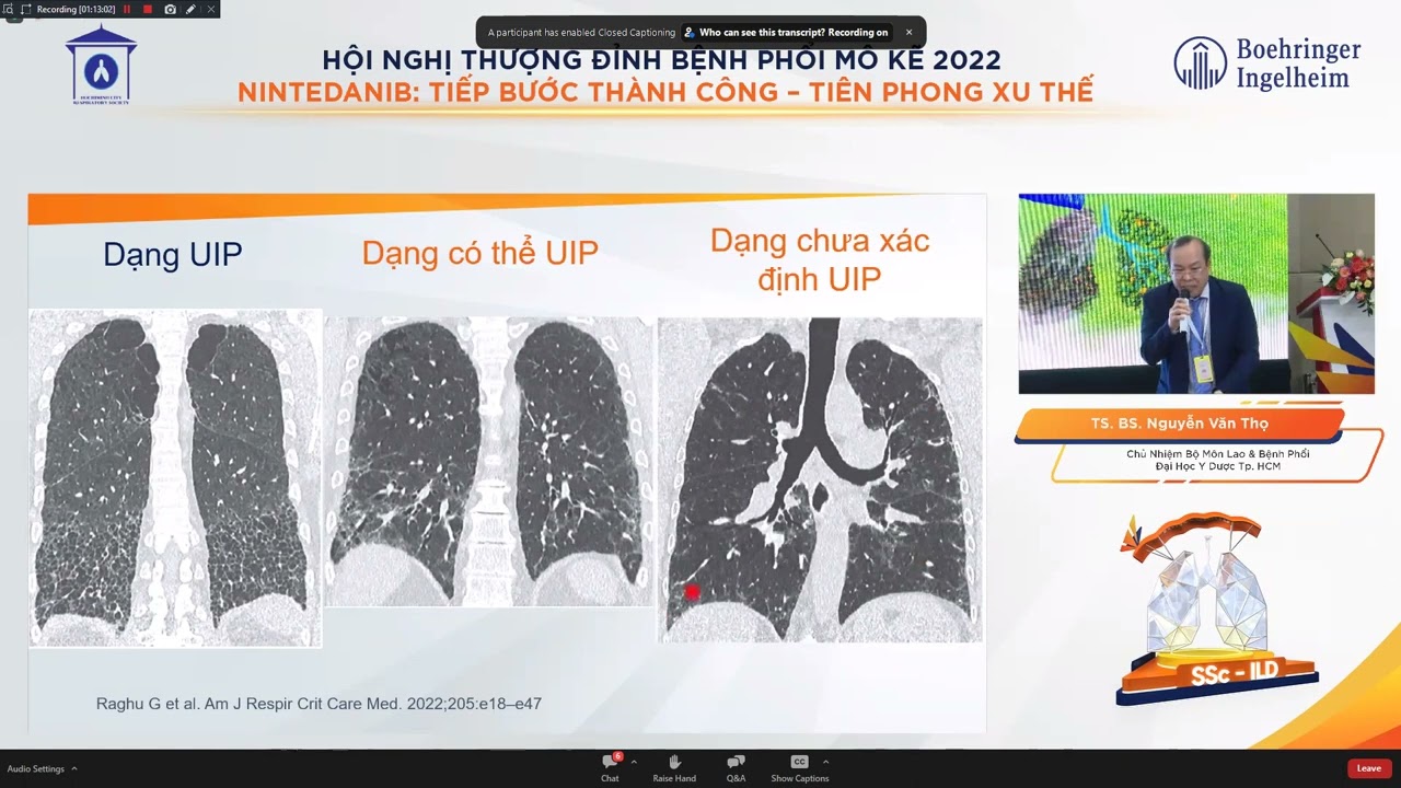
Formulating a differential diagnosis for specific Interstitial Lung Diseases (such as UIP, NSIP, and Sarcoidosis) based on dominant HRCT pattern distributions.
The role of Multidisciplinary Discussion (MDD) in combining HRCT findings with clinical, serological, and histopathological data for a definitive diagnosis.
Utilizing longitudinal HRCT scans to monitor disease progression, evaluate treatment response (e.g., to antifibrotics or immunosuppressants), and identify acute exacerbations.
Introduction to quantitative chest CT and AI-assisted imaging tools for automated scoring of lung fibrosis and emphysema.
37.4K views599likes12:10Original Release: 2019-11-05
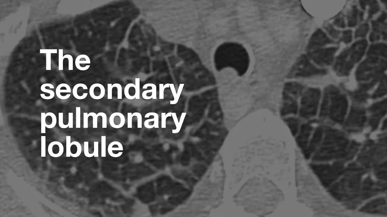
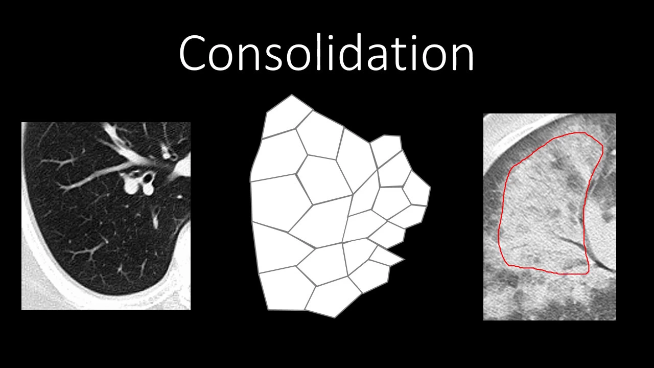
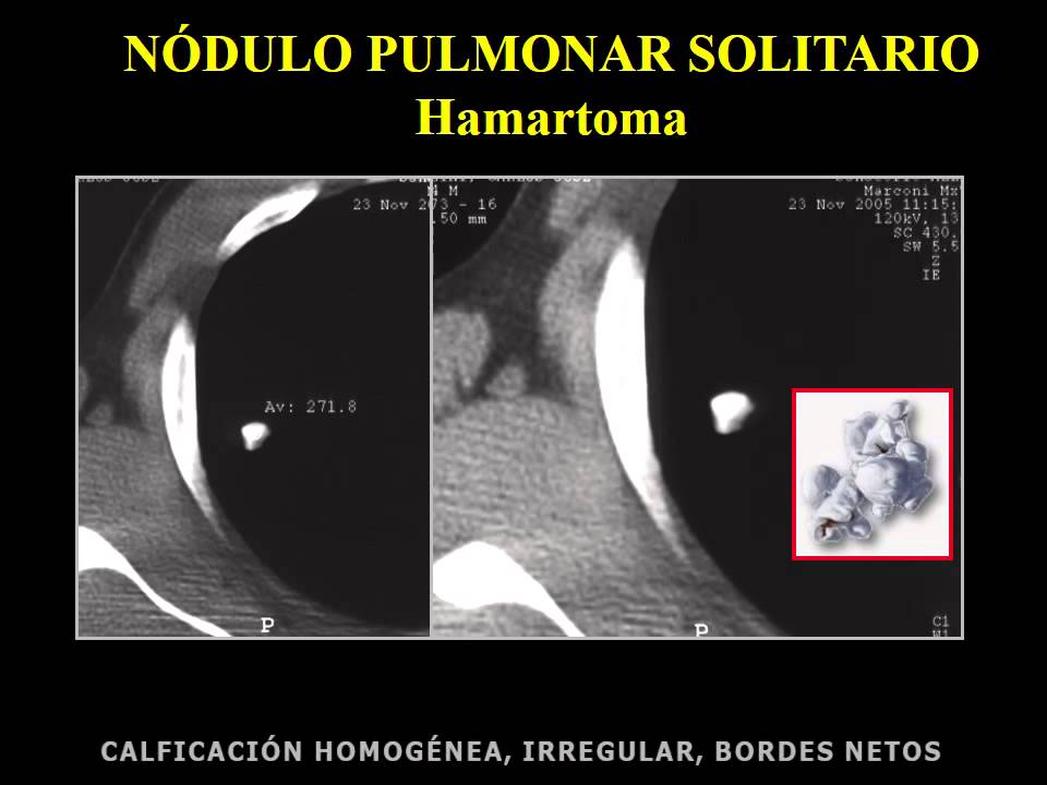
[Music] it is a most important to a good high resolution CT scan you all are aware that it is done in supine position the HR CT in deep inspiration you know the combinations of Quechua when we started CT scan we used to go up so why because he couldn't go look to him today it is available 0.75 1mm collimation is available now why is that yellow so fine and times where you will need to do a prom CT and there are times you will need to do X parenthesis the e construction that is John is it high algorithm depending upon the vendor that is cheap if Siemens Toshiba they are different in Toshiba it can be ed lung or in Siemens it can be be 70 or be 90 so basically these are oniy a lot of them now there is very few centers you retrospective targeted I think it should be modified and they should not be filled like 13 ax on a field because you need to do my new images and the beauty of HR sees your chest findings are you can actually document only 20 other film and the findings are similar as event so you can take a few images of Isis view of the middle part and view of the bases so don't try to make too many images in Montville if you want it with too many images adhesive tumors now the window and lens also is important basically it's a long center that is plus 1600 or 1500 and the level is - 500 - 600 so these are very important basically read forth in inspiration supine position is very very important now before you go on to acquiring the city you must know few it is some detail as I told you the symptoms are similar so at least what's the age is it a female or of the end so some pcs that are common in females smoking because we covering for smoking-related IUD NEX occupational hazard odd even when it comes to subacute hypersensitive pneumonitis whether the patient is near any illusion on whether the patient is near they're feeding the pigeons summation are very sisterly symptoms like tightening of the skin join pins and what is the deviation and profession it's this basic if you know it helps you now what we are going to be is patterns why is that so because you cannot find out this is the sign and this is the diagnosis so you are going to take these patterns for the cheeks offers well and try to come to a diagnosis what do we know is a pathological classification that is is a lymphocyte interstitial pneumonia that it is a respiratory prompt your life is interstitial this is a pathological classification which we give it in diagnosis but what you need to know is patterns patterns whether this less like occurred like in reticular pattern there are no dues he says what these are interstitial changes or other associated airway disease also now they're together or less like pattern as you can see here you can see that these are set to thickening or this is scepter signal basically if you took a lace then this is absolutely mesh or net-like pattern and you actually stop seeing the normal and what you are seeing is sector thickening field intraocular or interlocutor so now this is no noodle type of interlocking or sexual thickening and the white part the paintbrush I call it on this part so this is a second people but it no use with tickle in the lobular sexy thickening and this is intolerable a sexual sticking so this pattern is a rich color factor so this is one pattern now I cannot show you that that that patient had nodular interlock receptacle so why it is important because if you have ro Buehler pattern is more likely going towards a certain BC if it is smooth then it goes in under a needy mom but if it is irregular no it can go into IBM so first pattern is statistically envelope Allah insha Allah now what do you mean by each other below that is 15 the second people attitude of you you get this impression appears complete white first you are not seen in turbulent but 50 sceptile degree is given to another crown glass appearance and this is stuff proven now this is a supine film I told you we are to be crucified but when you get something like that this could be basil depend intensity make the patient prone and it could persist and it is not dependent basil density but this is definitely early I appoint was Vig if that is between the two lawsuits now if you can see this is an ovule which is up these novels are polygonal shape so this is a love you and centrally there is an attitude so this is a typical interlocutor septum you can see thickening that of your septum most of them are subclonal as you can see here I just have a magnified image this is the magnified so you can see this thickened in the lobular section this is thickening the regular septum basically there acts interstitial is either fairly long queue along the bronchi and the business it is in the submarine or this is a in the secondary continually W so there are these three types of interstitial called as actually position the intern ovular septa technique which is seen in the substitute region so we saw intraocular is the lobular and then you get something called as honeycomb B what is honeycombing is this honey coffee no is this honeycomb P know this yes why is that so because by definition whoever described it hard that at least two or three layers of tiny cysts in the solution region one so these are rows of tiny cyst is nothing but honeycombing honeycombing points towards in reversible into system changes next pattern is ground Riaz now what is ground glass is this is black and kingdom where you have this whitish looking area in this is coronal section you can see this is the ground glass appearance our crystal beef consolidation but it is not why you can actually you can see preservation of vascular markings now you have two things you have to remember Congress passed what if this is an ilg and it is a chronic form and you get wrong class there it is exacerbation cellulitis but if you get rowdy us with no changes of funny coping and you don't have traction from cactus then this patient has a reversible IG I hope I'm clear another point of ground glass is it can be seen with hemoptysis basically it means that the interlock length between the two lobules there is either seepage of fluid be mucus or be it live or be it may be blood so this ground has a appearance a little more tighter when it comes to angular village so ground glass appearance can also occur in hemoptysis also 1.12 remember now cystic pattern if it is cyst where do you exist when it started we thought about we saw system sent it over already what are these these are small cyst in the lung maybe some of them have a central white dot but they don't have very well for favori and you have time the lannister cytosis add it to this ah ah di le UI we also an L I D that is in Pacific interstitial pneumonia later comma usual interstitial I have not added but yes you can have a cystic variety of pcp these are the system we heading size subsiding decisis scene in left lower low in guna and one of the SIS must have ruptured and the patient is presently with you want a lot young people another case multiple cysts bilateral pneumothoraces with no news okay we keep this also in mind this is the third type where the whole love is become like a scene they've seen multiple cysts in bilateral man with large bullet there is hockey anymore offices so this is nothing but said over and over which releasing its vocals from sector thickening to ground glass to system equal to no dealer pattern other eleven important yes any total which is more than three centimeters is called a mask so we are dealing with nodules which are pinpoint or and less than three centimeters long written once annual latest says that is it has to be recently sent solidly polymer it no tube is another entity but your we are calling multiple modules and we'll be dealing with pinpoint subsidy because I know you now the distribution is important whether they are very lymphatic please notice if they have any lymphatic look if they are around the fissures then you can say are we dealing with sarcoid are other findings of sarcoid people then if they are land them means their Iman you manage in a spread that can be seen in certain businesses like miliary TB like in silicosis on essential opioid abuse techniques within the second triple burrito Butte which can be seen with high percentage of atlantis tropical nursery so you have no news look at this note you this is a noun he prom can or the bronco vascular area there are no beneficial notice but these are wrong the vessels tiny nodularity around on these bronchus and also around the vessel so this will keep in mind these this is the fisher and you appreciate the fisher this is efficient so these are around the fisher so now those were also on the fairground collision but we have around the fisher so pointing forward towards side point now here these are more central available how are these not use if you remember the society for this is a nice pinpoint a darker white looking here ya go ground glass and they're more sensitive load look at the picture there is no paid official note you look at the prom all cash basis there are no very vascular no use certain types of a decision on this is to have consol but I'm just showing you a consolidation which is actually infective just to understand what a consolidation it's a dense area okay but in this your senior program sometimes you don't see a bronco crimes you have to find out whether the consolidation is released whether it is triangular in shape because there are certain consolidations which are seen in our interstitial some cases for FFP cryptogenic organising memoriam and one more thing that is important when you start if how is the location of all these factors that are should be it no dues be separately be it any form may be a traveler with it's very minor whether it's also an upper low middle upper and middle or only lower or it's by Pope Innocent Radian X it is involving the whole lump but of Isis or equinoxes less and lower lobe is very much them is it we subfloor rule is very high nur all this is important
Up Next
56:43
Lung Nodule Evaluation Beyond Fleischner: A Practical Review
@IcahnSchoolofMedicine
292 views•2024-06-03

48:47
Graphic Medicine: Comics for Collaborative Healthcare Communication
@nationalpatientadvocate
189 views•2023-12-04

10:28
Neuroanatomy: Central and Peripheral Nervous System Divisions Explained
@AKLECTURES
136.2K views•2014-09-20

3:05
Stages of Labor and Vaginal Birth | Childbirth Animation
@nucleusmedicalmedia
52.1M views•2017-08-18
Related Study Plans & Knowledge Roadmaps
Structured learning paths in Medicine
medicine
Abnormal LFTs: Diagnostic Workup & Pathology
01Liver Anatomy and Basic Physiology
02Introduction to Liver Function Tests (LFTs)
03Hepatocellular vs. Cholestatic Patterns
+ 3 more learning modules
12h6 modules
Explore Roadmap
medicine
Analyzing Blood Smears: RBC, WBC & Platelets
01Introduction to Blood Components & Smear Preparation
02Normal Morphology of Blood Cells
03Erythrocyte Pathologies & Morphological Variants
+ 2 more learning modules
12h5 modules
Explore Roadmap
medicine
Mechanical Ventilation: Waveforms & Alarms
01Anatomy and Physiology of Breathing
02Introduction to Mechanical Ventilation & Core Modes
03Ventilator Settings and Waveform Interpretation
+ 2 more learning modules
12h5 modules
Explore Roadmap
medicine
Neuro Exam: Localization & Stroke Syndromes
01Foundations of Neuroanatomy and Vascular Supply
02The Neurological Examination: Step-by-Step
03Principles of Neurological Localization
+ 3 more learning modules
18h6 modules
Explore Roadmap