

















Lung nodule evaluation requires understanding that most nodules are benign granulomas, with malignancy risk increasing from less than 1% for nodules under 6mm to nearly 100% for larger nodules; the updated Fleischner guidelines now consider nodules 6mm or smaller in low-risk patients as benign without follow-up, while emphasizing that nonsolid and part-solid nodules require longer follow-up (up to 5 years) due to higher cancer risk, and that PET-CT has limited utility for these lesions as they often show indolent growth patterns that may never progress to clinically significant disease.
Lung Nodule Evaluation Beyond Fleischner: A Practical Review
Added:Familiarity with the standard Fleischner Society guidelines for the management of incidental pulmonary nodules.
Basic understanding of thoracic imaging modalities, specifically chest computed tomography (CT) and high-resolution CT (HRCT).
Knowledge of lung nodule morphology, including the distinction between solid, part-solid, and pure ground-glass opacities.
Understanding of primary risk factors for lung malignancy, such as smoking history, advanced age, and occupational exposures.
Application of Lung-RADS (Lung Imaging Reporting and Data System) and how it differs from Fleischner criteria in screening populations.
Utilization of quantitative risk prediction models, such as the Brock (PanCan) and Mayo Clinic calculators, for clinical decision-making.
Advanced diagnostic workup options for indeterminate nodules, including PET/CT imaging, navigational bronchoscopy, and CT-guided percutaneous biopsy.
Integration of clinical findings into multidisciplinary thoracic tumor boards for the personalized management of high-risk or complex nodules.
292 views4likes56:43Original Release: 2024-06-03
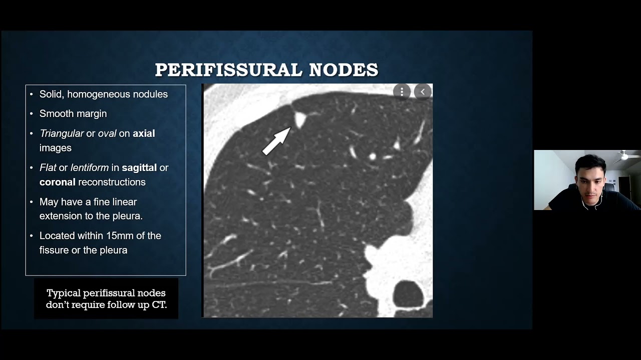
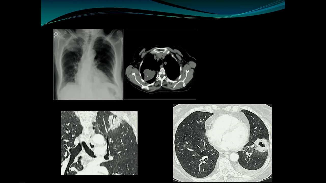
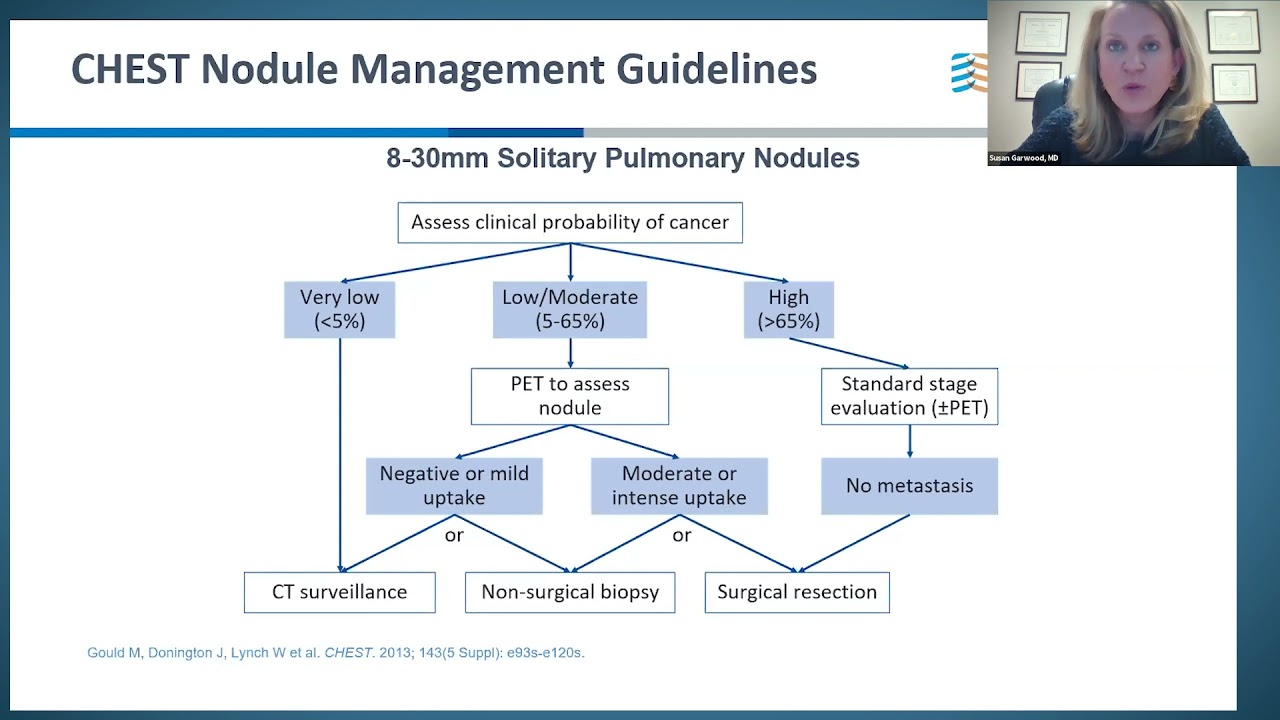
thank you so much for that nice introduction and thank you all for coming um so I'll be talking about this very uh interesting topic that affects us all uh so no disclosures um so the goals would will be to provide a practical review of Chess CS um understand nodal prevalence and ideologies uh talk about the guideline recommendations for followup talk about the different characters of knowledge rules some case and then some important lung cancer screening uh details so just a very quick background about CTS because it just never ceases to amaze me how amazing this uh technology is so it was invented in the 70s by houndsfield who we still use his name in regards to the densities a single slice and this was his brain took hours to get now we can do an entire body within 10 seconds um the CT technology has has drastically changed particularly in the in the 2000s um this is an old number it's probably at least double at this point so 100 million cases a year and of that 10% generally is about is a chest CT um when we look at CTS uh what you're looking at is the density of the body as opposed to like MRI which you're looking at water content and other things so everything is a density so the least dense thing in the body is going to be air with like a minus thousand hfold units then you have fat subcutaneous fat here which is about minus 100 uh then you have uh water which I don't have a picture of here but that's zero to 20 so you see that in like the gallbladder or in the kidneys um soft tissue is in the 50s like the muscle here blood tense to be in the 40s and calcium is uh some of the highest uh handfield units in the body outside of metal or or contrast costs about approximately $500 for the case to do a case and then you guys are all familiar with the general indications um in just a general CT Principle as you know chess CT is way more sensitive for all pathology compared to x-ray um I would strongly advise that even with a negative x-ray you should consider a CT um lung cancer and metastatic workup as you know should not stop with a chest x-ray you have to continue with chest CTS um and then there are certain cases of individuals who we routinely monitor every six to 12 months so people with lung cancer already they get six Monon scans because not only to look for recurrence but looking for new lung cancers which happen average rate about 7% per year and then other known cancers that uh we deal with all the time like liver cell renal cell head or neck they're going to be scanned every six months when we talking about this we're you know obviously something that needs to be discussed as radiation especially when we're talking about following up a nodules at three months six nuts a year so this is is um one of the issues with radiation is the units um the best unit to understand is the Miler which is the uh amount of radiation that actually gets absorbed by the body and we all are exposed to about three Ms per year most of that comes from radon in the ground but you can see other uh things in the sky um other ideologies can can also give us radiation dose so so what does that mean for in regards to Imaging so this is also an outdated slide there is not great new uh new material on this but generally um a chess C I would say depending on the place is going to be from two to five mlion CES these year this time lung cancer screening CTS are less than one million Cs and and an x-ray is less is generally less than 0.1 um so it takes about a you know 100 um x-rays to get to a CT um and you can see also that I mean that that this number is actually quite low compared to what average abdominal CTS do which is routinely about double and the reason is the the chest doesn't have that much generally that much fat to penetrate and so it's uh it can be done at lower doses uh bellies need a lot more radiation because of the increased fat and a principle that we use in Radiology is called thear Principle as which is as low as reasonably possible essentially so our goal is to is to produce an image using the lowest radiation that is still Diagnostic and we're very good at that at Sinai we we take a lot of pride in how low dose we can do it and then just to show you this is at every scan you'll have this information which is get a dose report for the patient and again these units are very bad but essentially if you what we do is we take the total DLP uh which is called the dose length product and you multiply it by a specific K factor that for the chest is 0.14 and you multiply the DLP by that and that gives you the MERS so one chess C in this case is almost approximately one year of radiation and obviously the risk with radiation is potentially um you know cancer development and the jury is out you know there are some people who believe that these CAT scans are going to create lots of cancers in the future that data is really based on on Hiroshima and Nagasaki and other disasters like Chernobyl which is which is much different than what we're doing but still it's something to be super conscious about and um and and and just be wary of it okay so in general most cat skans can be done without IV contrast so for lung cancer nodules pneumonia Ru up Mets all of these things C minus no contrast contrast studies are absolutely necessary for pulmonary emilii dissections Hy adenopathy it's very helpful because there's a lot of action in the central lung Central lung nodules for that same reason so we can tease out what's the artery what's the vein uh absess these avms plural fusions within with out is is is not is almost never indicated um more is not always better and almost all information get can be obtained from the contrast enhanced portion and just to show you that here so this is the C minus this is the C+ here's the aortic Arch here's the enom and vein this is a lymph node so it's a lymph node without with or without contrast it doesn't change much um the definition is a slightly better but we're quite used to what a non-contrast looks like so we we know what we're looking at but interestingly uh if you see at the bottom here you can see there's a much big difference between this and this so you have this paraspinal density here that once you get contrast you can see that it's actually Rim enhancing and this is a par vertebral abscess which we could kind of um potentially un elucidate from the noncon but certainly helps with the contrast all right so um one useful use of non with and without is um is it an aortic dissection in the reason is that if you look at this image which is a contrast study you see this low density in the descending aorta which could be non-calcified plaque but in this case it's intral hemotoma which tends to be bright on the non-con and can be obscured with the contrast so dissections involve non-con and cons to identify this intral hematoma nowadays with a general CT they're all high resolution we do everything high resolution here which just means thin Cuts 1 millimeter Cuts is the routine these days days um inspiratory and expiratory CTS are very useful in regard when we're looking for air trapping and patients with lung transplants to look for o bronchitis obliterans drao malaa right this is an example of an inspiratory and expiratory slic through the trachea in inspiration and expiration you can see there's uh loss of the AP diameter and this frowny face which is indicative of of trium maltia so then the super lowdose chess C is the lung can protocol which generally is sub favorite um it's used pretty much exclusively in lung cancer um but it can be used in a lot of different indications but we like it for lung cancer and you can see here that this nodule is quite easily seen again the chest is not that not that much soft tissue to penetrate and the lungs are very forgiving from radiation but you can see here that if you were to look at the soft tissues you can see how noisy they are and if you would go to the abdomen you would see just how noisy that is and it becomes almost unreadable but the lungs are very readable okay so then just quickly some more Chessy descriptions before we get into the nodule stuff so ground glass is defined as hazy densi in a lung parena that you can still see through you can still see vessels and it has one of the most wide differentials in all of CT so it could be infectious like PJP it can be inflammatory like ards it can look like Hemorrhage here in this patient with diffuse airspace ground glass with peripheral sparing consolid ations are more dense so you can't see through the lung you just see the consolidation you can't see any lung architecture you do see air bronchograms which is a good feature of of consolidations which are typically pneumonia but they OB they obviously have their own differential which can include malignancies like I don't no carcinomas or lymphomas this is septal thickening in the lungs which in this case this is smooth septal thickening which would indicate inter interstitial edema or fluid overload with fiber nodular sepal thickening is more indicative malignancy like lymphangitic spread of disease and honeycombing which is routinely described in in pulmonary fibrosis or uip usual indues from pneumonia honeycombing are these stack cysts in the sub plural lung um it does get difficult to distinguish what's bronchia acusis and bronch and honeycombing in these cases um basically what you would do is connect the broni by going up and down the image and if they connect then brontis and they don't it's honeycombing okay so then just a really quick case here just to understand the utility of Chess C so this is a patient who's inated they have diffuse disease diffuse Aerospace opacities the the CT is really just going to show the exact same thing which is diffuse airospace opacities and the differential remains completely the same um infectious inflammatory ards edema the only clue from the chess C might be that this is a young patient with a lot of coronary artery classifications but honestly that doesn't mean much um differential which is quite large really the only way to know is clinically and time is usually one of the best tellers so you know very typically in these settings right the the intensivist will will we throw a lot of things at the patient uh lasx steroids antibiotics but really the only thing that could clear this dramatically would be edema uh everything else is going to is going to leave some residue at some point and then further on this patient after a Lasix they they they were able to breathe better but we can see in this cardiac MRI that the de sepal and anterior walls of the heart are not moving and this was a an LED infar with a severe systolic dysfunction uh just a couple more descripting descriptors um here is centrolobular empyema which are these lucencies in the central lung with some Distortion of the parena paraseptal lysm is airfill cysts in the peripheral part of the lung pen lobular is when the empyema is at the is at the bases the worst which is unique to things like alpha1 antirion deficiency bull are these large air Fel cystic spaces that tend to occur in the lung apes and can uh lead the patient to get a pneumothorax and then this is Mosaic attenuation so in Inspiration you can see that there is this sort of geographic heterogenity of lung parena here you can see there's this region of lucency next to these areas of gr glass lucency gr glass when they expire this is exe this is more dramaticized so you can see the um the the geographic heterogenity better and this is a whole lecture in itself to understand what's happening here but what essentially what's happening is when the patient's expiring with their breath the lung prancha is going to get more dense because air is being released so the normal part of the lung here is the ground glass the abnormal part of the lung is the loose and that part of the lung is unable to compress because the air is being trapped okay so now on to lung nodule so a lung nodule is defined as any lung opacity that's left than less than three centimeters that has a round shape it's it's infrequently detected on x-ray and very commonly misinterpreted on x-ray as vessels or things of that nature it's unbelievably common in CAT scans of the chest which we'll discuss um almost every nodule that we see is is found incidentally and uh they're way more common as we get older and so I would say I mean we're going to go over these prevalences later in different populations but it it's almost imposs if you're older than 50 the chance of having at least one nodule in my opinion is probably greater than 75% that's just how common this stuff is it's uncommon at Young ages it's uncommon in young patients um you know this was told to me by Lou depalo Who is one of our pul ologist who said that when a patient hears that they have a lung nodule is an emotional tsunami meaning they are very scared they don't know what to do they can go to Google and see what it says and it gets to be very scary um and it's very difficult to impart the sense uh of how common this is and that's really one of the jobs as as a clinician as a radiologist is to is to inform the the the patient that this is actually quite common nodal follow-up is by far the most common indication for follow-up radiologic exam and that's again because almost every single person will have a nodule uh other routine follow-ups in the body are are honestly not that common you know renal cysts can be followed if they're slightly more dense than normal you know sometimes we follow pancreatic cystic lesions but in general you know it is what it is and you don't look at it over time um for incidental lung nodules followup is Guided by the fler society and those recommendations differ depending on the nodule type for nodules detected by lung cancer CT screening it's not incidental right it's what you're looking for and those are reported with lung RADS you actually don't need to use lung RADS and in and in Mount sin here we have the I cap group The Early interstitial early lung cancer Action Program group and they don't use lung RADS they they form their own system which is almost identical to Long RADS except it's not explicitly said what the grade would be for the individual so there's a gigantic differential um almost everything in the lung that's not a cancer is going to be a granuloma whether it's calcified or non-calcified it's almost always a granuloma and that can exist for so many different reasons anything you could inhale can form a granuloma obviously any tuberculosis or B microbacteria type organism that can get in you you know fungal infections every everything in can cause a a granuloma neoplasms um we can differentiate based on the morphology so primary lung cancers tend to have irregular spiculated margins whereas metastatic nodules tend to be smooth carcinoids tend to be smooth homras which is the most Ben common benign nodle in the lung tends to be heterogeneous with different uh sign with different attenuation in the nodule itself hopefully calcium or fat which allows us to identify it so then again in infectious nodules can be from granulomatous or fungal disease abset septic emilii viruses inflammatory nodules and Sarcoidosis rheumatoid nodules the necrobiotic nodules and an theor a pulmonary avms now something that you might have heard about is the the myip images the maximum intensity projection images so we do this on every case and it's essentially what it's doing is it's taking 10 millimeters width of data and superimpose it on to one image and what it allows you to do is at any given instance on this picture right this is a nodule that's a nodule that's a nodule that's a nodule right but we know it's not because when when we scroll back and forth we see it connect to the vessels the my connects the vessels for you because again it's taking 10 millimeters of did and putting it together so what happens is it becomes a lot easier to see the things that don't connect to the tree so finding a 2mm nodule is very is a lot easier than it used to be and we'll discuss how important that is but you can see the you can see almost everything with these myips okay so moving into the nodle C istics which are probably one of the more outside of size is going to be the most important uh feature that's going to distinguish malignancy from non-malignancy so speculated is a are these irregular tags coming from the nodule itself and this is from a fibrotic reaction in the pulmonary insti from the tumor another important factor is going to be the growth rate um this is very important actually so in this case you know let's just look at this one the rest not cancer here this one which uh growing to this size in a year is is about The Sweet Spot that we look for in lung cancer the nodule density so when we see a ground glass nodule we call it nonsolid we're going to go over this stuff right when it has a solid component we call it a part solid nodule so we would measure the whole thing and then the solid component would be measured separately calcifications in the in the lung nodu are almost always a sign of benity and these are the Ben n features of calcification which is either a diffusely calcified nodule a centrally calcified nodu like a bullseye these popcorn or heterogeneous calcifications are also commonly described in in hromas or granulomas and then the location and this I find is very important because lung cancer loves the upper loes particularly the right upper lobes so in and and the lower loes just in general get a lot more junk so meaning they get more infections they get more aaltoes they get they get more of everything the upper loes generally don't get that junky but they they do love cancer so to me I take seriously the long nodules in the upper loes a lot more serious than the ones in the lower loes metastases because their their blood usually hemat um hematologic spread because the lower loes get the bulk of the or get more blood flow than the upper loes the the predilection is for metastases to go to the lower loes so this is uh just a quick review of PM metastases so this is a very dramatic example of these so-called Cannonball metastases they have very smooth borders they're innumerable in this case and this is what Mets typically look like almost all of them almost always look like this spatter cancer is one of the weirder ones that tends to cavitate and look very different uh than most typical Mets so the most common primaries to metastasize still along are essentially the most common cancers that we know breast cancer coloral renal cell head and neck and then there are other less frequent uh primary lung primary malignancies that have a predilection for the lungs melanomas SAR uing sarcomas and thyroid carcinomas um and the lung is by far the most common place to get metastases in in the body when we talk about the the size of a nodule we have to understand sort of what it means in regards to the malignancy risk so you can see here that less than 6 millimeter nodules have a less than 1% chance of malignancy and that's a very good number to remember just as the bulk of nodules that we find are going to be less than six millimeters and the malignancy rate is so minimal um at such a small size as you get larger they get significantly more likely to be cancer so especially once they get to be 2 cmers you know this the these are almost always cancer the malignancy risk goes up when you have emper or fibrosis probably from the uh chronic inflammation that the lung endorses with those States and that causes more malignancy the doubling time which I kind of ref referred to before is an important feature um benign things generally go super grow super slow or super fast right so infections grow fast um benign nodules like homas it takes forever to to grow but malignant nodules have this sweet spot where they double in about a 100 days and that that's very indicative of a primary lung cancer and you know besides what we see and what we say there are these calculators on the internet which allows you to sort of give a better percentage of what this could be and you could see here that the most important ones from this Mayo Clinic one are the age whether or not they're a smoker do they have any other uh cancer the size the shape where it is in the lung and with all that stuff you can can spit out a number of the chances that this is malignant okay getting into fler guidelines there are a couple of really important things to understand about flesner um one is who it applies to so it applies to uh newly detected or a detected nodule in a person who's 35 years of older and it's incidentally found so meaning if you have breast cancer looking for Mets and you see a nodule it's not incidental it's what you're looking for and those play by different R same thing with a head and neck cancer any cancer right but we're talking about a patient who's let's say coming in for shortness of breath for you know interstitial lungies assessment for uh you know rule out PE those sort of ideologies and you find a nodule um where you get a belly CT looking you know somebody has abdominal pain and they find nodules at the lung bases right these are all incidental nodules and um at this point inflation uh you you'll see here that the number magic number was about 4 millimeters and if you had a 4 millimeter nodal or less it did not to be it did not have to be follow up in lowrisk patients which is difficult to exactly what they mean by high risk or low risk but essentially it's a significant smoking history or not um and you'll see here that as the nodules get bigger uh you follow them closer and more and for longer times we'll go over this later on the on the on the updated fleser so again these only apply to incidental nodules that are found uh incidentally um it does not apply to patients with known malignancy um we talked about uh how common metastases are in the lung okay this is the new fler up the new updated fler guidelines and the most important things to know are um that nodules now less than six millimeters not four are considered benign in a low-risk patient so whereas before if you had a 5 millimeter nodule in a in any patient essentially it needs to be followed in a year that's what fman would say and in the current guidelines are if it's it's six millimeters or less does not need to be followed on a low-risk patient in high-risk patients another new thing they say is optional before it was recommended at 12 months now it's optional um that's the words that we use right and as they get bigger you again you follow them more closer actually in this case similar actually the followup guidelines are very similar in lower higher risk patients um uh with a with a single nodule you know it gets more complicated with multiple nodules um but again basically you use the most suspicious nodule as the one that manages uh what happened to the patient next okay so let's just go for a couple more um examples of this so this is um an issue of the the type the exam uh way you do it so thicker cuts things because you can uh uh cut at the edge of them they can look hyper uh look less dense or ground glass in this case but if you had a a real thin slice you would see that it's actually more solid so in today's world you can't really call things solid or non-solid without thin Cuts one millimeter slices which we we already discussed so six is the new four um and this change is based on this data that shows that you know there's such a small risk of malignancy even in patients at higher risk um on the other hand specious morphologies upper location or both can increase the cancer risk to the one to 5% chance even uh even at these small sides so a followup in 12 months is recommended uh so here's an example here of probably an 8 millimeter nodule on the left upper lob so you know things and I'm not you know I'm not going to really get into this from a radiologic perspective and I you know I've been doing this for 15 years I probably read thousands and thousands of chesty tees it's actually a lot more suspicious when you have one nodu and what looks like a clean set of lungs like this um as opposed to if you say a lot of bunch of little over ones uh a junkier set of lungs they become less suspicious but with one just like this in an upper lobe it gets suspicious even at this small size so 8 millimeters this is very suspicious and you could see that in a whatever I don't have the time frame here but let's say in a year or two years it grow it grew significantly right and at this point this is 100 this is almost certainly IM malignancy primary lung malignancy so again we kind of um I kind of talked about this but multiple solid nodules are are are actually a little safer when we see them than a single one um and again a 12-month optional followup is considered for for patients deemed high risk um uh okay let's continue so followup non-contrast chess CS can be followed uh so if you have new nodules um generally uh let's say you're following somebody for whatever reason somebody with a known cancer or somebody that you're looking at nodules or following nodules and then you see new ones uh those typically tend to be inflammatory and you know they can be followed up in short term because very often they'll go away um once a nodal becomes one centimeter or bigger and sometimes even smaller you really you're not supposed to follow it anymore we don't like to follow lung cancer so the question becomes you know what what what do you do um so once the nodules is of a sufficient size then you can start talking about other tests so outside of following it now you could actually do a PET CT you could take tissue either from a broncoscopy or for percutaneous CT guided lung biopsy right so Bronx are generally bigger well you know these days Tim Harkin and and and some of our other pulmonologists they're excellent at getting these nodules and they actually can use a CAT scan as a guidance uh for virtual broncoscopy um or sorry actually CT gued bcopy which which Tim Harkin does so Central and larger lesions um are easier from Bron but but you know they they are becoming increasingly able to get them in the periphery for lymph nodes Bron is really the only way to go for par tral High lymph nodes Bron is the best um CT gued lung biopsy generally is better at the peripheral ones uh but it can we can hit the ones that are more deep in the lung and then there's surgical biopsy or EX so just to go over some pets so what pet does so it's a combination of the CT that's the CT part and the pet uh which uses generally radioactive sugar um when the when the SUV which is the standard uptake value is greater than two and a half which is what with they report uh is the uptake value generally above two and a half is considered maligant um and you can see in this case this is a smoothly marginated mass or nodal and you can see here that there's peripherally high high intensity with with low intensity in the center so this is an necrotic nodule and this was a saroma met so I I personally do not love pet for um as a as a problem solver because generally when we're dealing with a nodule or let's say maybe an il- defined consolidative or and I'll Define nodal density if the differential is infection or inflammation versus cancer the pet is really not going to be helpful because you would anticipate all those ideologies to be active if the if the if the differential was mucoid impaction or a chronic nodule versus malignancy the pet could help right because if it's cold then you could rest ass sht a little better and you don't necessarily need to think about going to tissue analysis at that point but again it doesn't really help you when the differential is something acute or cancer so everything eats sugar almost everything eats sugar um malignant love sugar some some classic malignancies do not pick up sugar like a carcinoid but again uh how often gr can't stress how often these things are granulomas and in the literature if you take out a hyperactive nodule that's not cancer it's almost always a granuloma uh generally one centimeter is that number where the pet can be reliably used and if it's less than a centimeter people can say that uh the resolution is suboptimal for that small besid and you can't really accurately say whether or not this is picking up sugar or not okay so CT guided lung biopsies this is a depiction of it we do a lot here uh Dr en Kitz um is one of the world leaders at this he's able to hit very small nodules in the 5 millimeter range essentially when you hit a nodule you can either use an FNA a asper which is a smaller amount of tissue that you're going to get or corn needle which is a larger amount of tissue it's very good at diagnosing malignancy now this is a very important point when something is malignant uh from a matter primary lung it's really not hard to make the diagnosis because the cells are relatively easy to get either from a final aspirate or from a from a um core the problem is though uh when we are dealing with things that are not lung cancer so for infections or other granulomas it becomes very difficult um uh for the pathologist or the cytologist to say with certainty that this is benign and very often what you get not in this case but when you get let's say a smaller nodle or something that you're not thinking that this is cancer what the pathology comes back is a lot of the time is non-diagnostic or unsatisfactory which generally or at least in some instances does not mean that was the inadequate sample or the fault of the proceduralist it means that no matter what you would get from the nodule it just doesn't have enough cells to make a diagnosis from and yeah you have to remember that the cytologist or the pathologist they only getting a very small piece of this nodule so if they don't uh you know if they don't see cancer they're they're they're they're it's a lot to say that this is a benign nodle and they they generally won't so again it's not a fault of the procedur list if you get an unsatisfactory result sometimes um in these non-cancerous biopsies so generally 10 millim is the size that we can safely biopsy but again we've done very small ones um the new thorax rate is around 10% you know Roger Flores who's one of our Thoracic Surgeons he says that like it's a it's a miracle that every time we do this biopsy it doesn't cause a pneumothorax um chesty placement is less than 5% here you can see there's Hemorrhage in the lung from the biopsy all right so let's go over a quick case so an 85y old with a long smoking history rule out PE so this was the Scout from the P study just to show you how hard it is to find the nodule on an X-ray this is the nodule it's sitting right on a vessel it's Lally impossible to find um on the CT it's much easier to see right you see that it's got a regular margin this is unbelievably suspicious and this eminus patient in the right upper lobe on the pet it's quite Avid um this one I don't even think was bioped because the pneumothorax rate was going to be very high and it was reected and it was an adom carcinoma which is the most common lung mcy primary lung mcy in the lung these days here's another case uh this might have been a World Trade Center patient um I put this here but I I think it was a fake indication you know I'll talk a little about the World Trade Center patients that we see um you know we do routine x-rays on them every year or two uh they're very difficult um you know you basically can say with some certainty that there's nothing crazy going on along but we do see these ill defined things every now and then so here you can see there's this IL defined nodule on the right midline um there and here was the CT so you have this irregular nodular density with some other kind of patchy nodules next to it um so now what do you do right uh do you follow it up in a month to see if it goes away do you check if the patient's sick or not do you give antibiotics and Then followed up should you do a pet should you get tissue should you get surgery consultation so you know this is a young person we have to be uh you know I would say kind of aggressive in a situation like this but at the same time um realizing that very often uh these things can be benign so one good thing about this case is you can see that there's satellite nodules which which actually implies generally that this is not cancer cancers don't like to grow next to each other so I don't have a great answer for what to do next I would probably say do a quick followup because again the pet TT is not going to be very helpful if we think the differential is an infection versus cancer um so so there's no correct answer in my opinion there's not really a perfect answer here um what happened was he was scheduled for a biopsy and when he came back for the biopsy four weeks later it basically had shrunk down no treatment and this is unbelievably common in our chess C world where uh sometimes what we'll do sometimes what they'll do is they'll schedule a biopsy like a month later and uh you know if it's gone it's gone and if it's there they bu up see it um it's also uncommon so in this sort of situation if I were to call the the referer or call one of you guys and say is the patient sick almost always the the doctors would tell me the patient is completely fine no symptoms so it doesn't surprise me at all when people have this sort of in their lung and they don't know it nobody knows it there are no symptoms it it doesn't surprise me at all okay here's another case this is a 32-year-old with chest pain okay so this nodule is a little easier to see even though this is a big blind spot on um x-rays which is the central part of the lung because the higher vessels are approximately this size this is much easier to detect here um but in the central lung it's not as easy so here it was on the CT so again you have this sort of smoothing nodulated nodule with this one little tag and then an adjacent nodule so the decision was made to biopsy this here was the biopsy needle and this turned out to be a crypto caloma which uh is not that common here and we do see it it's you know cryptox and histoplasmosis is super common in the middle part of the country um we don't see this very often but that was the diagnosis okay this one I think the nodule is a little easier to see here in the right upper L but very scaryy location here it is okay so we have a a 10 millimeter nodule on the right upper loobe so at this point again where it's sort of a difficult decision what do you do next the best thing the best thing to have would be an old one a prior right and in fact that could put to beted a lot of these nodules that if you had a 5-year-old prior that looked like this this would be safe to watch then but at this point we really don't know what this could be and priors would be unbelievably helpful so here we do have a one-year prior CT and it looks almost identical or it it does look identical so that should give us some sort of safety in in how we're thinking but at the same time it's in the right upper lob it's it's pretty big I mean it's a sizable nodule it's 10 millimeters um and so the decision can be either to pet it maybe because at this point it would be unlikely to be active if it was just a granuloma or an infection theoretically continued to follow right or potentially put a needle in it so in this case again the the decision was made to needle it um uh and it came back as un non-diagnostic riter because it wasn't cancer it was a it was probably granuloma and unless you get that one granuloma in the in the biopsy they're they're just going to say it's non-diagnostic that's just what they do it's very difficult for them to diagnose pity from a single uh from a single uh pass from an FNA okay here's a case it's pretty old at this point but we see here 11 mm nodule that's smoothly marginated that over two years grew to 16 millimeters so this is a slow growing nodule an atypical nodule for cancer I mean for a met metastases but not crazy for a primary something in the lung uh so it is growing and the decision here was uh here it you can see it better on the soft tissue Windows here was the biopsy and this turned out to be a carcinoid so carcinoids again uh have a different appearance than primary lung cancers which are almost always irregular and much uglier looking carcinoids tend to be very smooth often end the bronchial but sometimes just in the lung and this was a carcinoid okay here is an x-ray for a patient who was who was feeling sick and here we have a pretty sizable nodule on the left lower lobe well circumscribed smooth and round so here's what it looked like on the CAT scan that they use contrast for and you can see it has this peripheral rim of enhancement and more Central lucency its margins are intermediate not um not particularly irregular but not smooth either so there's a wide differential here um over time I think this is followed in a like a month later uh air we can see new air in in the nodule this would be very atypical for a lung cancer or any malignancy actually that hasn't been biopsied to develop air so this is looks like it's an evolving infection more than a Cancer and this was reected and it turned out to be an abscess um with necrotizing and non- necrotizing graning Lum is and no malignancy it is what it is you know okay next young patient for Arenal Stone CT so this is an impossible thing just to remember sometimes that the lung can go under the diaphragm on on a single view like this and then here's a very common situation where get a belly scan and you see a nodule on the lung now this is a big nodule this is probably 20 20 millimeters or 2 centimeters but it has very smooth borders it's incidentally found you know you'd be forced to really work this up in in any person who can be worked up um what happened in this patient I I'm not sure exactly what happened but nothing was treated and in two months it became this scar looking thing and then five years later it became just a scar so we see this all the time on CT where you see these irregular fiber ND densities or very th thin linear densities in the lung and sort of the lung shows all of our Battle Scars through our life and you know just like rings on a tree you can see you know old infections and you know old old problems that have healed in the lung it's it's um it shows its scars here's another case of a 35-year-old with a cough okay so here there's a nodule here here it is right it's in the left lower lobe again it's smoothly marginated in the lower lobes less likely to be a primary lung cancer but certainly within um the spectrum of possibilities it could absolutely be a lung cancer if we could have one thing outside of pathology the best thing would be an old study so here in this case we had a three-year-old x-ray of the belly where you can see this in the left lower lobe right so now we're thinking okay not going to be probably not malignant and then when we look at it closely with the CT we can see that it's very heterogeneous you have these low density areas you have calcifications in it it's got the so-called popcorn calcification these speckled calcifications and this is the most common benign entity in along super common hroma right which is a a benign growth in the lung that has lots of different cell types so they're usually found incidentally Peak age in middle life here in this one you can see there's fat um it's the most common so 8% of all lung tumors in the lung are going to be homas it's the third most common cause of a solitary lung nodule has a bit of a male predominance very often found in the peripheral part of the lung has at least two mes mesical elements in it only a quarter have calcifications the calcifications make it easiest to identify the fat is sometimes difficult and I'm not I don't think I have an MRI of this no I do okay so um some have no obvious fat or calcium like this case uh again it's in the lower lobe it has some benignness features but you can't Clearly say this hmer toen because it doesn't have any other features to it and we are we're getting better at doing this MRI of the lung generally is not a common um common study performed and the reason is that all of our MRIs are basing things on water no long doesn't have water uh or has very minimal water so it's not routinely used but it can be used in a situation like this because MRI is really good for to characterize the tissue and what you do in this case is you can see here that there's some heterogenity there's signal loss from internal fat in this nodule and that's that's a that's a homom okay here is a patient with a history of vver cancer um maybe you can maybe These Are nodules or a nodule on the left lung uh here it was right it grew from this size couple of millimeters to over a centimeter in a year um that's about a malignancy that's that's a good growth rate for malignancy um if it was a met you would might anticipate this to grow quicker but uh this is only four months so this is not this is very if the patient has a known malignancy this is a very good look for for a met there it is it was biopsied and this was a a metastasis okay another example of a patient with chest pain so here we can see this nodule here in the right lower lobe now again what you would want to see is an old x-ray so here's the old x-ray from two years ago and you don't see it there so we cted it and all it was was a a healed Ro fracture this this was the nodule and this is very common where what we misinterpret uh on an X-ray is not a nodule it's actually some something else so I'm going to quickly go over some of these um here's another one this is a very well defined now on the lateral it's very difficult to see in the lungs itself um which you probably would expect to be able to see something this size on the lateral but in fact it's here it's in the patient's skin and we cted it we didn't realize on the first EX this was actually just a big skin tag which gave that appearance of a nodule on the lung so here are some other mimics right the nipple Mark the nipples are big mimics um we can use nipple markers to differentiate them for True nodules like this with nipple markers um cutaneous nodules or warts RI fractures we already talked about fluid or the pseudotumor and the minor Fisher here is a mimic right on the lateral it's very easy now to identify this as being a shadow of feral thickening or fluid right artificial and then overlapping vascular markings or artifacts these are all reasons to incorrectly diagnose a nodule on an X-ray to talk about World Trade Center and long nodules for a second uh because this is very opportune for the group here um this is an uh an actual paper from the 2020 in um the annals of uh American thoracic surgery where they they basically took World Trade Centers uh wa World Trade Center uh responders and they treated them like lung cancer screeners uh and they they graded them like a lung cancer screening CT and what they found was um of these 15600 patients 60% at least had one nodule very common this is probably around the number I would expect for regular humans that weren't exposed it um of those of the 1600 55% uh lung Reds level two finding uh I'm not going to go over the different stages of L RADS but the higher they are the more suspicious they become uh 5% had lung RADS 34 which is basically very suspicious for malignancy so a small group from the scans had something already suspicious and basically one of the comments that were made from this data was that the presence of pulmonary nodules is in this cohort is very similar to the lung cancer screening corat and that maybe uh people with this sort of Occupational exposure should be screened like a lung cancer screening study like a lung cancer screening patient and honestly the jury is out on something like this this has to be studied more I will say that these long cancer CTS have an unbeliev or these lowd dose chest CTS in general have an unbelievably low risk to hurt the patient so we talked about the radiation risk and then another thing is that you know why I would recommend set or having your studies read by at an academic center as opposed to a private group in the community is that you know the act the people who do this umh for a living who just do chest or do their specialty and who can be part of multidisiplinary groups we know best that most nodules are nothing um and so when you we read them we generally don't scare people into saying that this is cancer or scare the patient or cause unnecessary further testing um so I think it's a very safe test that you can do every year to some groups that you same patient groups that you may think are high risk that you want to make sure they don't develop lung cancer and I would say yeah you should probably consider doing a routine chess ET either one every one year or maybe every two years again there's no guidelines for this um okay so then just quickly going over some other nodle characteristics we they can cavitate uh generally the thicker or regular walls more likely cancer smooth walls tend to be benign like infectious or inflammatory ideologies the gr glass Halo is something very commonly described in fungal infections here we have this solid nodal with the surrounding ground glass the so-called ground glass Halo more uh this is very important I think the chess Community has become more confident in understanding this which is that when you have a nodule on a Fisher it is not considered a pulmonary nodule it's called a pericial nodule in almost always these cases this is a lymph node in the in the lung So currently even for lung rat for lung cancer screening CS these are not counted um and these are unbelievably common so up to a third of all reported nodules are probably these fishal nodules which which don't count but again this is why you know you should get to know your radiologist especially if if you're sending a lot of chest C some to to some group uh generally chest Radiologists are much more uh knowledgeable about these entities um they can change in size which gets a little scary for some people but lymph nodes change in size that's what they do um they don't require followup when they're have these benign appearances right the problem is and this is a very common problem in Radiology is that I the most benign thing can also be malignant um and in this case the difference is that these these fishes are studded so there are numerous 10 20 of these nodules now would be atypical for lymph nodes and which be more compatible with plural metastases okay tree and Bud is something that you'll hear a lot so this is what it's representing which is essentially the distal bronchioles as they Branch uh just like leaves on a tree so this is the tree but which is almost always a sign of small iise disease usually of a bronchitis so viral infections microbacterial infections uh those are typically the ideologies for when we see things like this okay so then to go over the difference between nonsolid and solid this is very important and something that really is not intuitive um so instead of calling these ground glass nodules we call them nonsolid and they're basically a new entity with the with the use of CT before this you couldn't you never saw these on X-rays and even in the early beginning of CT the the sections were so much thicker you you infrequently see this so a non-solid Nole is pure ground glass A Part solid nodule has a portion that's ground glass and a portions that's solid these are very suspicious nodules um they're considered when they're at these low stages uh if you took them out there'd be these ideologies called adoc carcinom ins side to or minimally invasive Ador carcinoma and at this point this would be 100% cure rate the problem is is that um is this this is we can start entering the issue of overdiagnosis of lung cancer which is when you reect something like this and it does come out to be an early lung cancer and you feel like we've saved somebody well that's not really the case necessarily because we don't really understand the natural history of these nodules and this probably would never have killed the patient depending on the age um so that's really they pay they die from something else so that's where the overdiagnosis of lung cancer comes into play so here's the sort of recommendations when you're dealing with a ground glass or a part solid nodule and um they say here that they should be for until five years I've seen plenty of lung cancers that look like this that really take off after five years so in my consider in my opinion if you're old enough to have a surgery or old enough to have treatment for a cancer then they just continue to follow them for a year every year forever um and okay so again uh this should not be considered benign with twoyear stability um and as you can see here that three years later in this case so as opposed to most solid nodules that we can say with fle that if they're stable for two years you can consider them benign that's not the case for a part solid nodule um again they recommend up to five years followup I would say indefinite um and you can see here the solid component has increased in size in this nodule which is essentially an adocar uh by definition when you see something like this change over time and even fler at this point would say this can just be reected you don't have to buy Upsy because it's so unlikely to be anything else Psy is a very little value when it's mostly ground glass and that's because these tend to be indolent cancers at that stage and uh they're not going to take up sugar so you'll get a false negative uh from a pet uh or it's not really a false negative it's truly not neoplastic at this time but it shouldn't mean that it's benign again a lung cancer over diagnosis we talked about and you know the actual the most likely malignant nodule is a part solid nodal which is malignant two-thirds of the time right so a nodal like this is going to be cancer two-thirds of the time okay so these adenocarcinoma Spectrum lesions which is what we're describing we used to be termed these Bronco alol carcinomas which is a term that's not used these days uh again here are the path what the pathology would look like depending on the stage uh and if the solic component is less than 5 millimeters is probably a minimally invasive adinal carcinoma we talked about the 100% cure rate okay so then progression with time just to see this one this is a pretty much ground glass not solid nodule in the right middle Lo at this point uh in three years it became solid in the middle so this is very suspicious for an early invasive lung cancer and then in you know nine years from the original scan you can see what happened and this became incredibly more solid more spicula in 100% lung cancer one of the issues is that very often patients have multiple and these are the groups that tend to not be smokers very often Asian women or just women who have these non-solid nodules or part solid nodules like many of them all through the all through the lungs and you know this is a patient has one two three four five six and generally what we'll do and generally there are different stages of their evolution um and again we don't really understand the natural history when this happen happens um but then what we believe in natural history is that they are nonsolid and parts solid and mostly solid and what generally is done is you go after the most aggressive looking one and this is where lung cancer or lung sparing surgeries are very helpful because you know this patient is probably going to need a number of surgeries over their lifetime uh to take these cancers out and another important thing to understand is that when you have most of these nodules even with one aggressive one now these are not Mets it's very unlikely that even a lung cancer non lung cancer will metastasize within the lung itself it it can do lymph engine carcinosis more frequently it can spread to the nodes but it's un infrequent to to to spread to the lung and also these don't look like Mets they're you know they're non-solid they're they some of them can be irregular and they're all primary lung cancers each one all separate primaries in this case that one and this are separate primaries they look like primary lung cancer okay here's another case this is a looks like a cavity but this is in fact something called a pseudo cavity in the setting of these adnoc corom Spectrum Legions and this is because of the lipidic growth which means they grow slowly and they don't invade broni they tether open the broni and that creates this pseudo cavity and this can easily be distinguished as as an early lung cancer based on that all right so two years prior it looked very similar okay this was bioped and it came back atypical right already this was suspicious to to begin with um now we have atypical clustered cells this should be also suspicious for an early lung cancer it was just watched and one year later it has grown and it was reected and it was it was anoc carcinoma okay so just a couple more cases here so here we have a patient with two two irregular nodules one on the right Apex one on the left low these both look irregular and ugly and each one was needled and they were both separate lung cancers separate Ador carcinomas not the same cancer different ones cystic lung cancers are lung cancer that I think historically we've been uh not as familiar with but now we're becoming much more familiar with whereas essentially you have these cystic airos spaces in the lung that are usually the result of a prior infection inflammation causing this uh cystic change that over time you can start to see the walls become more regular more thick right and now it becomes more nodular here and this is cancer this is a cancer in an airfi cystic space this is what it used to be and this is what it became this was needled and it came back lung cancer here are more examples of the same thing this nodule over time this cystic airospace over time having a more solid and larger nodule of the wall and then eventually just obliterates the air filter space and it's just a big tumor here's another one right so just for your knowledge that you know these things should be followed especially with this sort of look with this ill defined wall and you can see over time this becomes Cancer all right I think um I think that's enough to stop for now um thank you and if there's any questions I'll be happy to go over them
Up Next
84:21
Topical Dutasteride and Mesotherapy Efficacy for Hair Loss: A Review
@Kyle_Bu
4.3K views•2023-11-20

48:47
Graphic Medicine: Comics for Collaborative Healthcare Communication
@nationalpatientadvocate
189 views•2023-12-04

10:28
Neuroanatomy: Central and Peripheral Nervous System Divisions Explained
@AKLECTURES
136.2K views•2014-09-20

3:05
Stages of Labor and Vaginal Birth | Childbirth Animation
@nucleusmedicalmedia
52.1M views•2017-08-18
Related Study Plans & Knowledge Roadmaps
Structured learning paths in Medicine
medicine
Abnormal LFTs: Diagnostic Workup & Pathology
01Liver Anatomy and Basic Physiology
02Introduction to Liver Function Tests (LFTs)
03Hepatocellular vs. Cholestatic Patterns
+ 3 more learning modules
12h6 modules
Explore Roadmap
medicine
Analyzing Blood Smears: RBC, WBC & Platelets
01Introduction to Blood Components & Smear Preparation
02Normal Morphology of Blood Cells
03Erythrocyte Pathologies & Morphological Variants
+ 2 more learning modules
12h5 modules
Explore Roadmap
medicine
Mechanical Ventilation: Waveforms & Alarms
01Anatomy and Physiology of Breathing
02Introduction to Mechanical Ventilation & Core Modes
03Ventilator Settings and Waveform Interpretation
+ 2 more learning modules
12h5 modules
Explore Roadmap
medicine
Neuro Exam: Localization & Stroke Syndromes
01Foundations of Neuroanatomy and Vascular Supply
02The Neurological Examination: Step-by-Step
03Principles of Neurological Localization
+ 3 more learning modules
18h6 modules
Explore Roadmap