


























This lecture by Dr. Theresa McLoud explains how to stratify pulmonary nodules based on size, morphology, and clinical risk factors to determine appropriate management. Key points include: nodules under 6mm typically don't require follow-up; the 8mm threshold marks significantly increased malignancy risk (10-20%); benign features include calcification patterns (diffuse, central, popcorn, laminated), fat density, and stable appearance over 2 years; malignant indicators are irregular margins, spiculation, cavitation, and growth; the 2017 Fleischner guidelines provide evidence-based follow-up protocols; and risk prediction models help personalize management decisions. Subsolid nodules (pure ground-glass and part-solid) have different malignancy profiles, with part-solid nodules showing much higher cancer rates (63%) compared to pure ground-glass (18%).
Lung Nodule Stratification with Fleischner Guidelines 2017
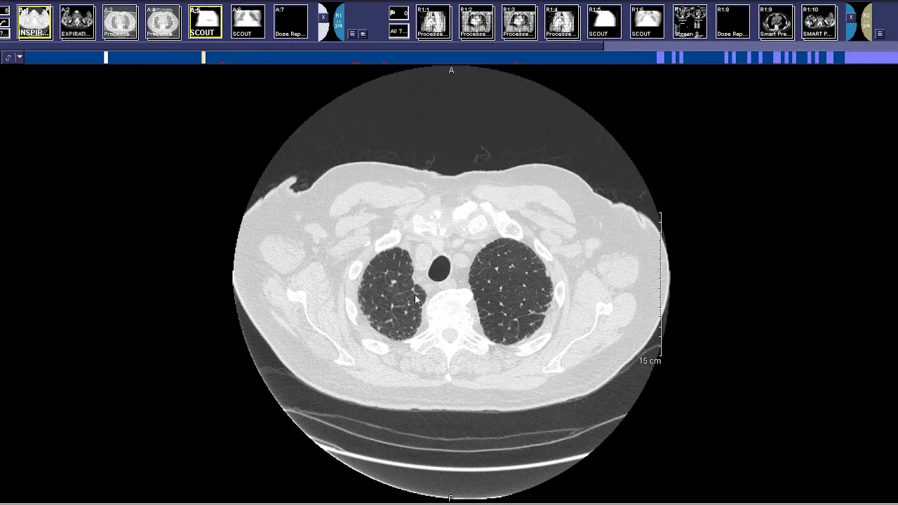
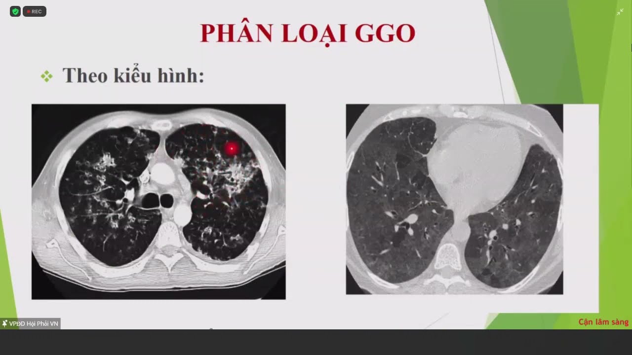
Added:I want to talk about incidental pulmonary nodules and I think that's really the bane of our existence when you're a chest radiologists nodules belong to everyone I think if you have lived a good life and you have any experience and you don't have a nodule in your lung you probably really haven't lived fully so this is a problem that faces all of us and I'd like to give an overall review of the incidental pulmonary nodules and how we manage them and how guidelines and management have changed over the past year the problem that we have of this is small pulmonary nodule and how do we define that was usually a nodule that's less than 8 millimeters in diameter and the very vast majority of these nodules up on I'm so their clinical importance is some less than large non calcified nodules and they're extremely common incidental finding and it's often that patients who are referred for CT for reasons not related to lung cancer are going to have a nodule and often they have more than one nodule so risk stratification is very important and I think sometimes as radiologists we realize we decide on risk we focus on the radiologic and imaging findings such as the size on the shape of the nodule but it's important for us to consider the clinical risk factors I think now it's easy with the current technology we have that we have easy access to patient medical records and we can check for these clinical risk factors if they're not made available to us in the history so they include of course older age history is smoking a history of an extra thoracic cancer within five years for the suspicion of metastatic disease a family history of lung cancer and of course the presence of emphysema COPD in this form of emphysema chronic bronchitis has a high association with lung cancer and then of course we look diligently at the characteristics of the nodule size is important once you reach that eight millimeter threshold the likelihood for malignancy escalates we've been taught since I days and residency the what a characteristics are not infallible but the healthful in determining the risk of whether a nozzle is indeed along cancer the density of the lesions the lack of any calcification the absence of fat brings up the likelihood of malignancy and now we have to deal of course with both solid and non-solid nodules growth is helpful and I emphasize to my residents make every effort to see if there are previous images you can say the patient and yourself a great deal of anxiety and trouble if you can prove that a nodule has been stable over a period of at least two years and then location is important now how do we define nodules nodules by definition are greater than three centimeters in diameter they're surrounded by aerated lung the sub centimeter nodules are the ones who are most concerned about not defined as small nodules less than 8 to 10 millimeters the classic solitary pulmonary nodule that you might pick up under standard chest radiograph usually has to be at least 8 to 10 millimeters to be visible so the fact that you see it on a plain radiograph is is concerning and then they're allotted anar indeterminate where there none of the classic findings for a benign nodule in our calcified there's no fat and unfortunately we have no old studies for comparison so we don't know whether they've been stable over a period of time now there are always to make presumptive benign diagnosis and I think we have to remind ourselves of this fact and that's why it's so important to look to all studies if you can prove that a nodule has been there and it's been stable for 2 years and that's excluding ground-glass nodules or to require further are lengthy a follow-up then we can be pretty well assured that if the lesion is benign the doubling time for malignant nodules can be in the range of 30 to 450 days now calcification we all know about if the calcification is diffused central laminated a popgun we're dealing with a benign lesion the presence of fat can either indicate a localized aspiration pneumonia or lipoid pneumonia more commonly a hamartoma and then those less than 4 6 millimetres are almost invariably benign lesions peripheral nodules have been shown to be benign and in any of these criteria president we just don't have to do any further evaluation or any further follow-up I'd like to emphasize that infection CT is recommended for screening but also for the analysis of these incidental pulmonary nodules and the preferences for thin section CT and the range of one point five to two millimeters now I probably this a very sophisticated audience and I don't have to go through calcified pulmonary nodules but just for the purpose of emphasis here we know the types of calcification that can be seen in benign nodules diffuse central popcorn or lamina and concentric eccentric calcification can be present in malignancies a couple of examples of calcified granuloma a-another calcified granuloma with central calcification this typical of histoplasmosis for a reason popcorn calcification classic for Hamlet Toma and here we can see an example not a calcification but fat density two examples of hamartomas with internal fat now peripheral nodules are interesting there was a paper that was published a few years ago from the group of Vancouver and they found that up to one-third of all nodules that they found in the context of lung cancer screening these are not incidental nodules but in the context of screening where peripheral nodules they usually small around three millimeters and the shape is awfully important they have to be triangular oval and touch the fissure if they become irregular if they cross the fissure or there are other shapes then the likelihood of malignancy can go up they're located most of them below the cry nose so you'll see them usually in the major fissure and the lower half of the lung and they consist of sub floor lymph nodes so they can whack some way and they respond to inflammation or to whatever may be inhaled and they may change over time and this article that was published in 2010 with a seven-year follow-up there were no cancer among all those pair official nodules and here's a classic one showing that nice oval shape now the exception is if that najin were to cross the fissure or become speculated or irregular and doesn't fit the classic description then one has to be suspicious of a lung cancer but in this classic size and shape you can dismiss those without a concern for lung cancer unfortunately in the lung cancer the álvarez which I'm not going to talk about which is in the context of screening pair official nodules still have to be reported and followed but I think it's pretty obvious that they're benign now solid nodules when do we get concerned what about malignancy and what does the malignant potential well it's related to several factors as far as the risk factors from imaging a concern one of course the size location nodules and the upper lobes more likely to be canceled the nodules and the lower lobes the margins and contour are important and then internal characteristics 'center calcification cavitation air programs and then importantly the growth rate if you have the benefit of all studies now it's been shown quite many years ago the small nodules one sort of less than four millimeters there's a less than a 0.5 percent chance of lung cancer even in smokers and we'll talk about the flights no guidelines but it's they've been changed to increase the threshold size of which we begin to get concerned any follow up to 6 millimeters so these very small nodules we can probably forget about and as you know in the National lung cancer screening trial the false positive or over 90 percent that's because the criteria included nodules that will 4 millimeters are greater but those are almost always benign root nodules when you get up to the range of 8 to 10 millimeters a greater have a 10 to 20 percent chance of malignancy even in non-smokers just a couple of studies here looking at the prevalence of malignancy now this varies widely and it depends upon this the group the but in a group of patients having fdg-pet studies and you would imagine that there are likely to be people at risk for lung cancer but they show that nodules 10 to 20 millimeters in size had a 33 to 60 percent chance that's the range for all the studies and nodules greater than 20 millimeters it goes up to 64 to 82 percent so so size is important margins and contour and I think we probably all learned this when we were resonance but lesions that have an irregular contours peculiar margins are this lobulated I have a plural take a floral tape can be fibrous tissue but it may be an indication of extension to the pleural surface of tumor and are maybe a fibrotic response to the tumor and we see an example of that here and a nice example of a speculator nodule on the right hand side okay internal characteristics eccentric calcification cavitation air Branca Graham's and no carcinomas made frequently heavier Branca grams may all be assigned a malignancy and some examples here now these are obviously not small nodules when I'm using this for illustration you see Center calcification this was a lung cancer the presence of air Brock River have speculated margin here another malignancy and a cavity nodule so all these factors make malignancy more likely than not growth rate and again I would emphasize that if you make every effort to see if you have prior imaging studies the absence of growth over two years is generally considered a sign of benignity for solid nodules not non-solid nodules and again emphasize that you need thin sections just to remind you the doubling time 25% increase in diameter is equivalent to the volumetric doubling time and for benign lesions it's usually less than 30 days all greater than 450 days now here is an example of Brotherly short-term follow-up I'm going to show this case later in my second discussion but this measured a little bit less than 6 millimeters which is actually the cutoff for deciding whether this needs any follow-up this patient has had some risk factors and you can see in 6 months not only has the size change but it's become sick and speculated and this was indeed a lung cancer now what's the usual management how do we manage these small nodules that we see all the time as we read CT scans that have done for other reasons the incidental nodule well the fleshly Society came out with recommendations back in 2005 addressed only solid nodules not part solid and not ground-glass nodules and then new guidelines based on revisions from information from other studies for a lung cancer screening and so forth were published in 2017 and dealt with incidental nodules that were both solid and sub solid in nature now the important revision to the guidelines and one is certainly I think beneficial to hustlaz radiologists and of course to patients it's a minimum threshold size size for routine follow-up has increased or greater than or equal 6 millimeters so we can for the most part dismiss those very small nodules that we see frequently in almost all patients the follow-up it was rigid in the first the criteria for follow-up of the standards for follow-up were very rigid in the first of 2005 classification but this gives a range of time based on risk factors and the preferences of the patients up to a point solid and subcellular nodules are addressed and simplified in a table and they're also the issue of multiple nodules which are common finding in many patients is addressed populations not included and this is important to emphasize the fleischer guidelines for incidental nodules do not apply to individuals younger than 35 years those were the known primary lung cancer or immunocompromised patients who may develop nodule or infections now here are the flesh no guidelines and I think it may be difficult for you to read the fine print but I would just like to emphasize a few important points if first of all dealing with a solid nodule and dealing with solid nodule that is truly solitary if the patient is low-risk and there are multiple factors in that neighborhood that you read the paper but generally we're talking about younger patients and we're talking about patients that are former smokers or never smoked you really don't have to follow these up however your allows some degree of discretion and they said some nodules that are less than six millimeters do not require routine follow-up but certain patients at high risk so suspicious nodule and the upper lows you may want to do a follow-up at 12 months and that makes sense you see a speculator nodule it's ugly-looking the patients never had a prior study it may be reasonable to get a follow-up in a year so it allows the radiologists and clinicians some discretion in following the guidelines and I won't go through all these but it becomes obvious that once you get to that critical threshold we talked about which was 8 millimeters then you really have to start getting aggressive and then baby that again you see a patient who maybe has an eight millimeter nodule but it's smooth there's no speculation as in Malolos the patient has never smoked never had a family history of cancer not exposed to anything that's carcinogenic and you may decide that you don't need to follow up yo you can't follow up just a three or six months rather than being more aggressive and doing other studies whether it be a PET CT are actually choosing to do diagnostic in Vaes more invasive physician whether it be percutaneous biopsy or bronchoscopy but actually providing tissue sampling here's an algorithm I think is a little bit simplified here but it helps particularly in dealing with the greater than eight millimeter nodule the nodule that is incidental but is concerning for lung cancer want us to look at the clinical pretest problem of it being malignant the patient's smoking history as we mentioned the presence of COPD history of lung cancer in the family and you can stratify these depending upon not only the clinical factors but also the appearance of the nodule itself into low row without low risk way you can do serial low-dose follow-up at 3 6 12 and 24 months even though it's a little bit above that 8 millimeter threshold most patients will fall into an intermediate category and they recommend then probably doing a PET CT if that's negative case-by-case follow-up but it probably is reasonable to get a follow-up in 3 to 6 months and if it's positive then you have to do some tissue sampling and if the risk is higher than 60% you of the classic smoker 65 years old has emphysema and so forth you probably should have tained of tissue immediately and then consider the patient for surgery now these decisions are usually made by multidisciplinary committees and our own Hospital we have a nodule clinic we look at people come in with suspicious nodules that are likely to be at high risk and there is a decision made by a board includes a thoracic surgeon a thoracic oncologists the radiologists sometimes the pathologist to make a decision as to what to do next okay I would like to emphasize that I've been talking about incidental nodules and if one cancer screening the rules are similar but somewhat different because you're dealing by definition with high-risk patients and of course there are long rods criteria for how these patients should be either followed up or for require immediate diagnosis now subsolid nodules as you know have been identified and represent the spectrum of peripheral and no carcinomas the lung they can be pure ground-glass nodules or they can be a mix part solid pond grout excuse me part ground glass that's a little bit of a tongue twister now some very early study that came from the El Cap lung cancer screening study and some of the numbers are low here but it's inter among nodules that they found that will purely solid the rate of malignancy was low it was only 7% part solid it was 63% the numbers are low it's only 16 cases and the non-solid was in between about 18 percent so basically these statistics have panned out in largest studies except the non solo the pure ground-glass they're often inflammatory in nature now what do we mean by pure ground-glass it's just a hazing increase in opacity and you can see the structure of the lung and the pulmonary vessels through it and the differential is extensive most of ground glass surpass ities that a pure ground glass of benign however that may represent an adenocarcinoma inside to what we used to call the old bronchoalveolar carcinoma that is characterized by the Pittock growth on pathology that is growth along the alveolar walls without filling in the alveolar spaces a typical unknown amidst hyperplasia eh is a pre malignant condition but very very slow and its growth and usually these do not have to be receptive inflammatory lesions and focal fibrosis ground-glass a pass ities a very common and you'll see them in a lot of patients sometimes they're not clearly defined as nice round nodules but there they may be little focal areas of aspiration of whole areas of inflammation and you need to follow them up and they usually go away within three months and here is an example of a mix all of mixed ground-glass Nadja was a large not nigh dose of solid tissue and then the ground glass in the periphery and what this usually represents if it can't if it's a cancer and it's likely to be is that the ground glass component is Allah pitted girls the adenocarcinoma and then the adenocarcinoma is the the solid part is the invasive part so there is a differential this is a study from Oh at all in 2007 well but it's interesting that most of these resolved and he found a regression of resolution in 37 percent of the pure ground glass and surprisingly about 49 percent the part solid nodules just went away on their own now what are the correlations here between the CT features and the pathology well the atypical adenomatous hyperplasia which is the pre malignant it's usually pure ground-glass and so is he I don't know carcinoma in situ as you get too many minimally invasive adenocarcinoma it's usually defined as a solid component that's less than five millimeters a partly solid nodule the chances of malignancy go up and invasive carcinoma is usually at least solid apart solid with a ground glass component that's never if it's invasive pure ground glass so in some solid nodules as we surveil them on CT malignancy is associated with growth that's pretty obvious but also the development and growth of the solid components so that's what we have to look at over time and if it's pure ground glass see if a solid component actually develops so again they emphasize and following these subsets of all nodules if they're incidental and you follow the flesh the guidelines and if you see them on screening there are indications and long rants of how they have to be managed and I won't go into the details here of the long rest categorization but here's a nice example of a pure ground glass nodule and then over a period of about a year it develops a solid component you might continue to watch this for a while because it's less than five millimeters but it needs careful monitoring now in looking at these nodules we wind up doing an awful lot of follow-up CT even though though we don't usually recommend follow-up if they're less than six millimeters in diameter this is very expensive and it's a source of you know great worry for patients as you can imagine when they have to have continuous follow-up so there has been an efforts now to look at multiple factors of risk and to produce calculators or algorithms that allow us to determine the risk and then work with the patient and the clinician so forth to decide on how they should be managed a first group that came out was John Mayo and the group from Vancouver who actually used data from the pan Canadian lung cancer screening program to determine factors which indicate the pulmonary nodules a malignant of all B family not follow up and they integrate both the clinical factors that we talked about there were contributed to risk as well as the morphology of what the nodules that we see on CT since then there have been a number of other models developed they depend on a multi variable logistic regression model format they try to determine the risk of cancer and they use many predictors socio-demographic as well as imaging characteristics and the predictors of malignancy now female may be a little bit of a surprise what we're talking often about non-smokers of people that haven't smoked in quite a while and that's because there is a this identified subset of women particularly Asian women who develop adenocarcinoma in the periphery of the lung and have never smoked family history of lung cancer increasing age larger diameter part solid density lower nodule accounts so if you are doing a CT on someone and they have small nodules and they've got about ten of them scattered in different lobes the chances are they're granulomas if you have two or three then you have to be a little bit more concerned so the more small nodules you have the better off you are in a sense okay Apple law of distribution speculation and the presence of COPD and emphysema all are predictors of malignancy and there is a website address here for this computer-based calculator that takes into account all those variables and now there are other risk prediction models that are available apparently the BOK model is considered one of the best is one that has been developed at sloan-kettering and the worst performers overestimated risk by a factor of two to three and this is an example that for Memorial sloan-kettering of the questions that are asked trying to determine risk the age the gender number of years you smoked at cetera have you quit smoking and so forth and this is integrated with the CT and radiographic findings now there are attempts to make these decisions even better to decrease the number of follow-up CTS that we really need to do on the cost of the patient and the radiation but even the top performers require further refinement to improve accuracy and subpopulations models should be updated to include data from the ACR LCS registry I think what we're learning as we calculate as we determine and tend to screen more people were going to have a tremendous amount of data that will be helpful for us in dealing with incidental modules not just with screening in future iterations should estimate personalized risks incorporating local screening Center accuracy and operative mortality so this is a little bit more addressed to screening but I think also applies to incidental nodules so in summary the solitary pulmonary nodule drives us crazy as well as the multiple pulmonary nodules that we see on chest CTS and patients who are having chest CT x' nothing related to malignancy and we have to make decisions of how they're going to be managed and we need to look at the clinical risk factors and I recommend that you make efforts to go into the electronic medical record it's become much easier now in the days of PACs and the integration of all the information sources we have epic which isn't the easiest to use but it's very easy to search at least for smoking history and age and so forth and put this in your report and take that into consideration when you make recommendations based on the flashiness society so we need to assess clinical risk factors socio-demographic which will be in the medical record but the size the location the morphology classic benign features as I've mentioned and the growth rate make every effort to see if there are previous studies and solidity okay if there are any questions I'd be happy to answer them and that's my email address thank you very much for your attention [Applause]
Up Next
Pulmonary Embolism CTPA Interpretation: Case-Based Radiology Guide
@medisensemeded302
38.4K views•2016-04-18

Graphic Medicine: Comics for Collaborative Healthcare Communication
@nationalpatientadvocate
189 views•2023-12-04

Neuroanatomy: Central and Peripheral Nervous System Divisions Explained
@AKLECTURES
136.2K views•2014-09-20

Stages of Labor and Vaginal Birth | Childbirth Animation
@nucleusmedicalmedia
52.1M views•2017-08-18
Related Study Plans & Knowledge Roadmaps
Structured learning paths in Medicine