







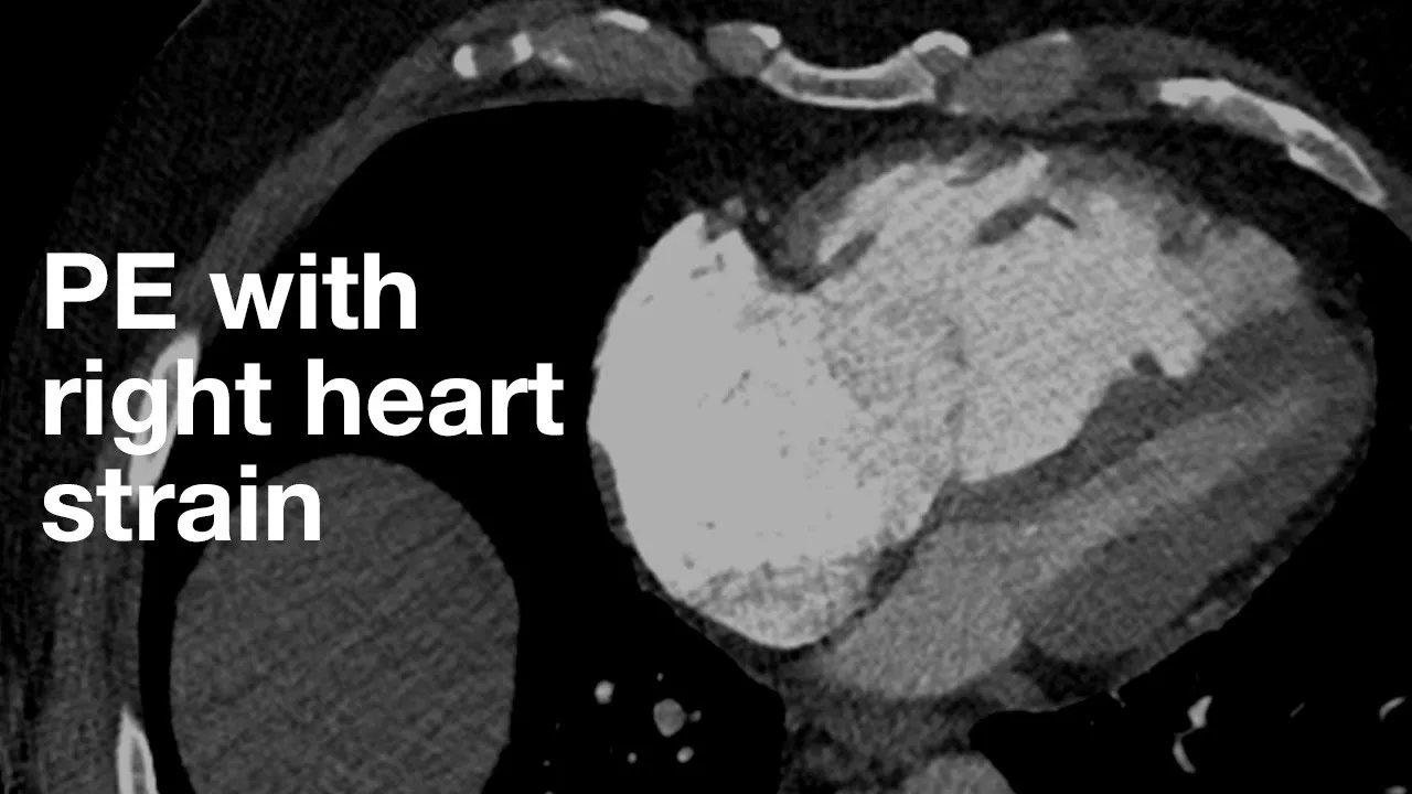

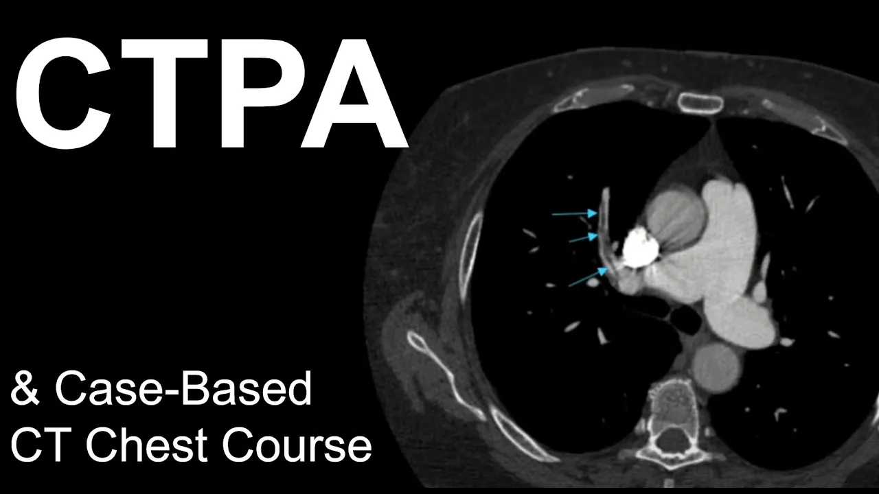


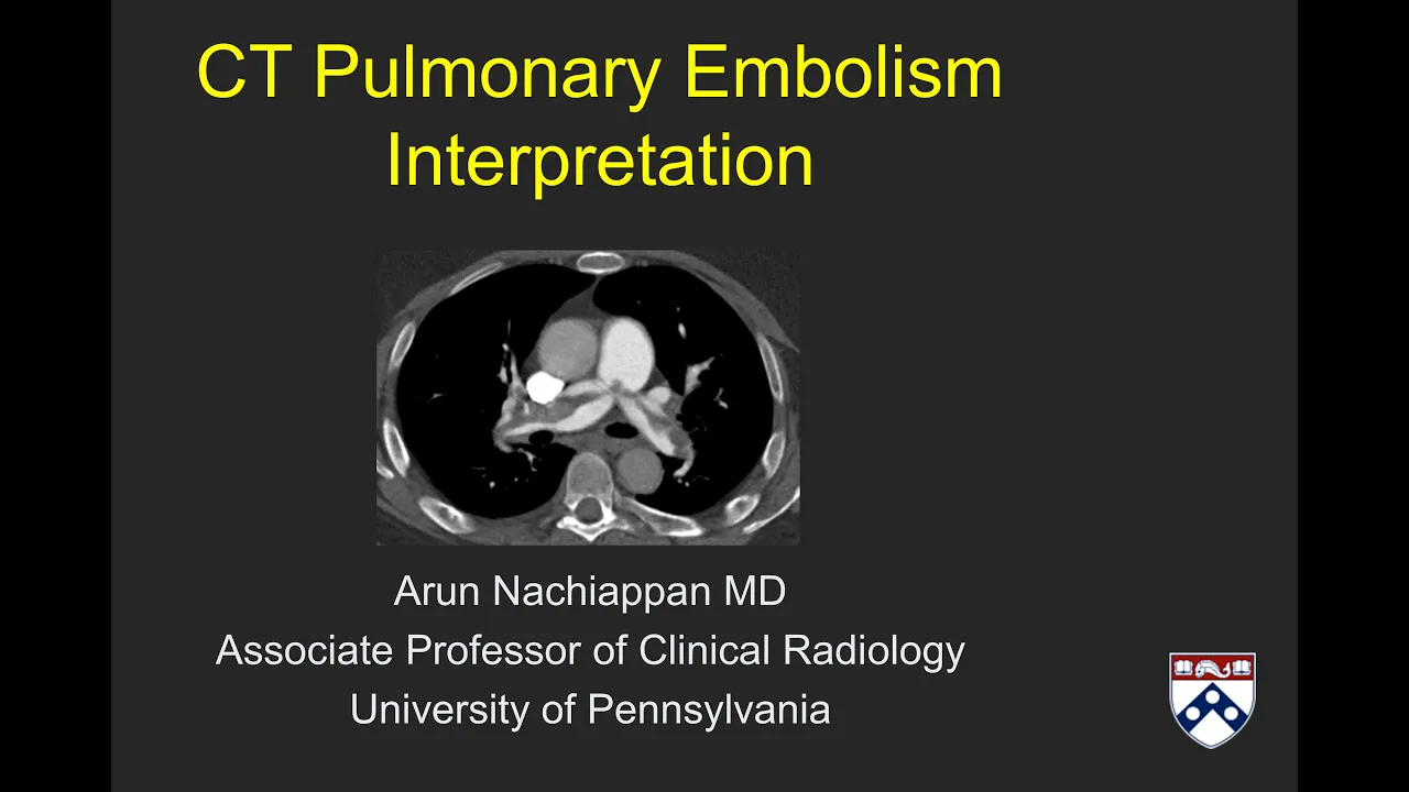




CT pulmonary angiogram is the gold standard imaging modality for diagnosing pulmonary embolism, using intravenous iodine-based contrast to visualize the pulmonary arteries; on the scan, flowing blood appears white (dense) while emboli appear as darker gray material within the pulmonary arteries, allowing radiologists to identify the location and extent of clots in the pulmonary circulation.
Pulmonary Embolism CTPA Interpretation: Case-Based Radiology Guide
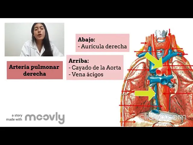
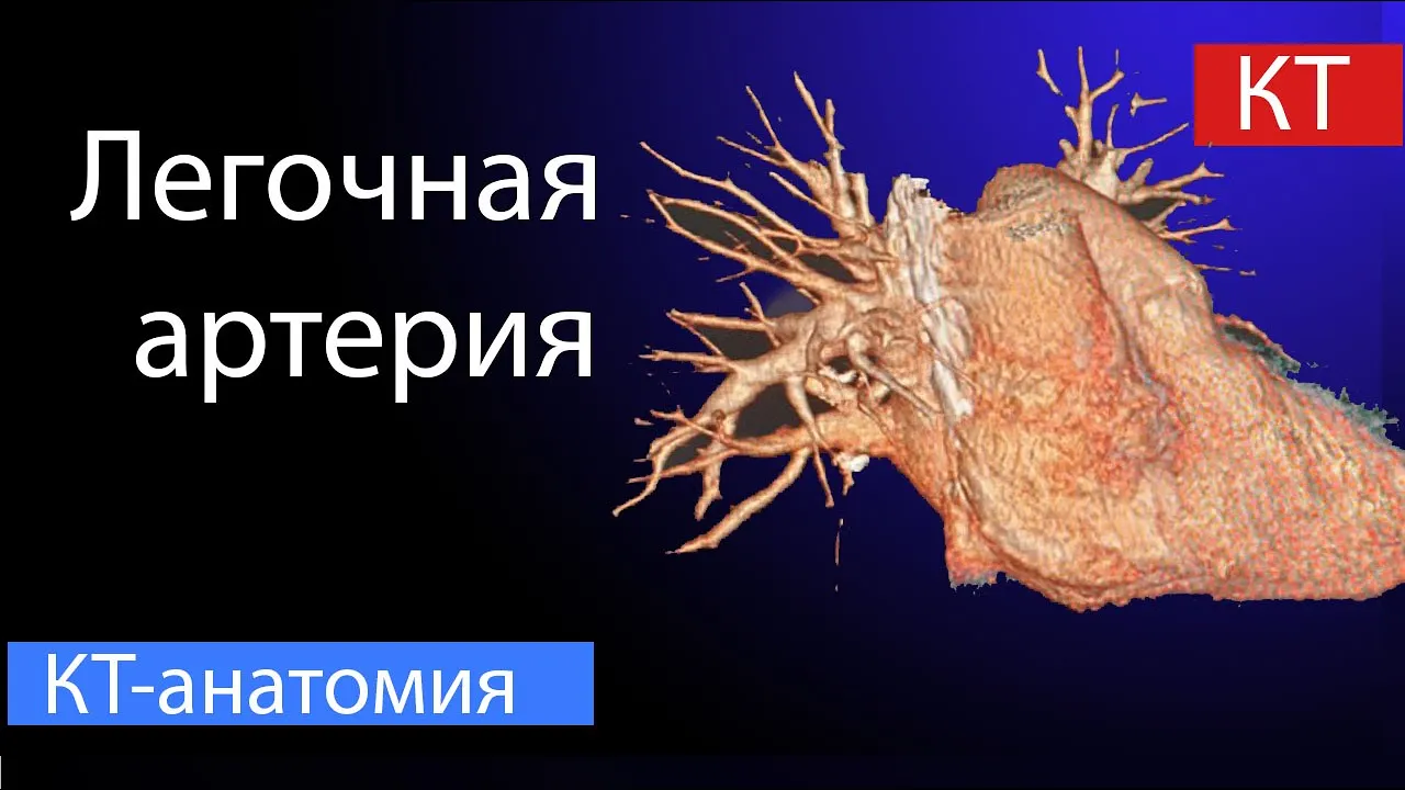
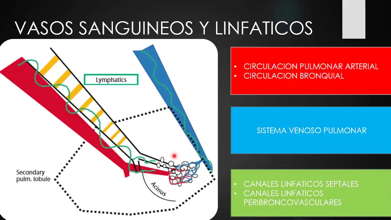
Added:Basic thoracic and pulmonary vascular anatomy, specifically the branching patterns of the pulmonary arterial tree.
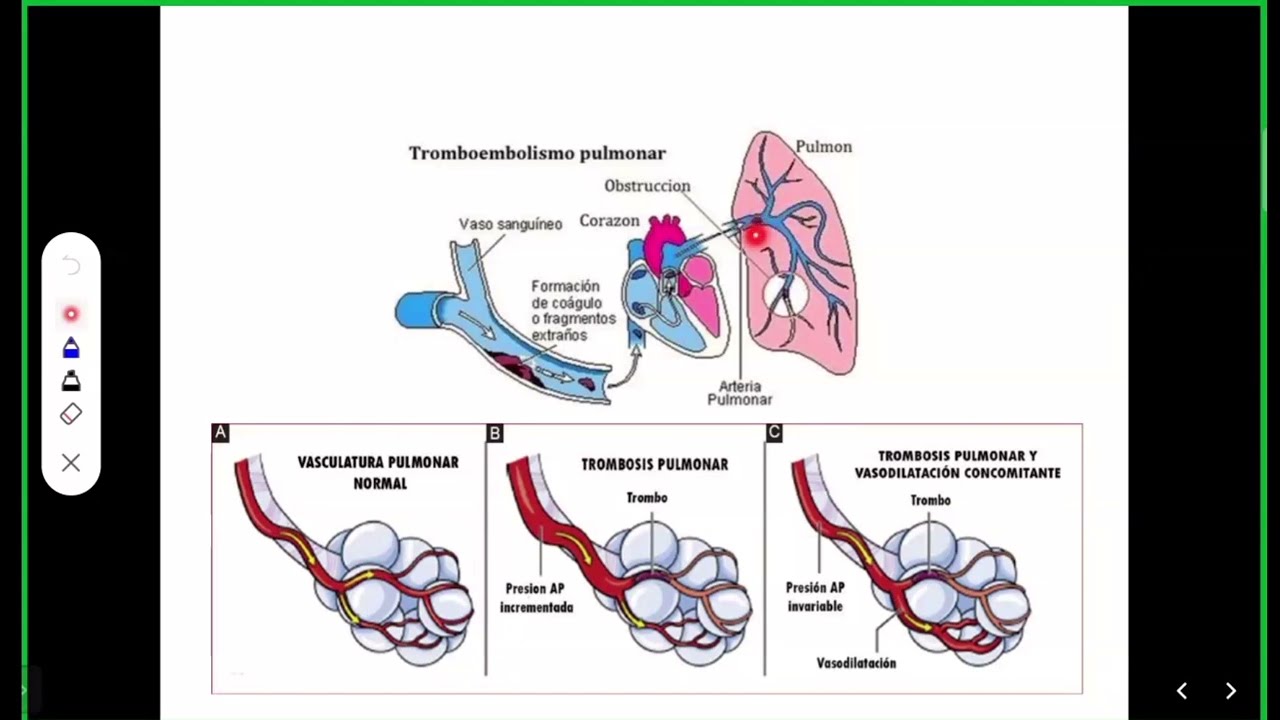
The pathophysiology of pulmonary embolism (PE), including its origins in deep vein thrombosis (DVT) and Virchow's Triad.
Fundamentals of CT imaging physics, specifically how intravenous iodinated contrast media highlights blood vessels (contrast enhancement) on a scan.
Understanding clinical risk stratification tools, such as the Wells Score and Geneva Score, which dictate the medical necessity of a CTPA.
Identifying secondary signs of right ventricular (RV) strain on CTPA, such as an elevated RV/LV ratio and interventricular septal bowing.
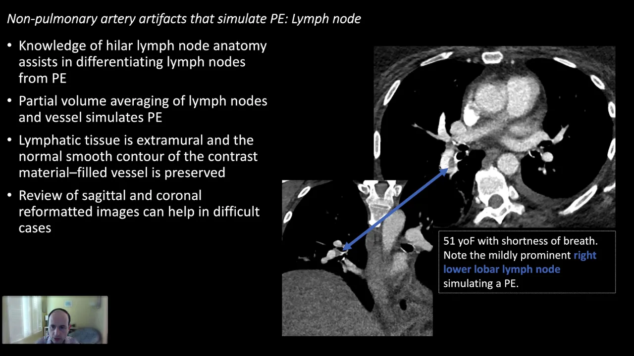
Recognizing common CTPA artifacts and pitfalls that mimic PE, such as respiratory motion artifacts, flow dynamics, and lymph nodes.
Classification and risk-stratification of PE into massive, submassive, and low-risk categories using a combination of imaging and clinical markers.
Exploring advanced management and intervention strategies, including catheter-directed thrombolysis, systemic anticoagulation, and surgical embolectomy.
38.4K views359likes5:39Original Release: 2016-04-18
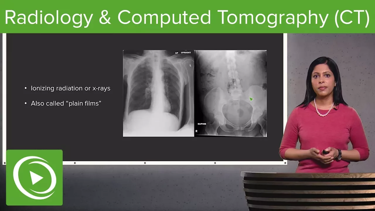
hi there and welcome to 256 shades of grey a video explanation of common radiology problems these podcasts are designed by a team of medical students junior doctors and our resident consultant radiologist dr. Dave Richardson consultant and university hospital north durham we have a 40 year old man who has presented to the accident emergency department with paretic type chest pain answer me mop ptosis when in the exit emergency department oxygen saturations were measured and below he had raised d-dimer's and so the clinical suspicion was of a pulmonary embolus a chest x-ray was performed this shows mild carjam a glee although it is an AP film performed in the resuscitation room of the casualty department the low fields are essentially clear this is in keeping with the permeable asst most people with permeable I have a normal chest x-ray so the next investigation was a CT pulmonary angiogram CT is investigation where we produce cross-sectional images of the body using x-radiation an x-ray tube spins around the patient and on the opposite side is a detector the computer then measures the radiation that passes through the patient and correlates them and produces a cross sexual image through the patient for this particular patient we wanted to image the Permian bly or possible permeable I in the pulmonary circulation so we need to perform the CT scan to show the pulmonary arteries best we want to show my within flowing blood within the pulmonary arteries to do this we need to give a contrast injection the contrast is a complex solution where iodine is the opaque material within the liquid this is injected into the patient's arm and the scan performed when we think the contrast has reached the publicity circulation the idea is to show flowing blood as dense with the ID material within it and the m bliye ask darker material within the flowing blood because it has no my within it so let's show his CT of his chest this image of his CT / me angiogram shows the pulmonary artery dividing the left / me artery is not sure on this image but the rifle me artery can be seen crossing to the right look the white material is the contrast within blood you can see it here in the premier artery and here in the ascending aorta and here in the descending order when we follow the pulmonary artery out more laterally you can see there is a lobulated darker gray material within the poetry this is the embolus in the right pulmonary circulation if we just then move the image to show a different part of the circulation you can see in a lower lower pulmonary artery here contrast around the edge and a darker material in the middle so this is embolus within the lowell of pulmonary artery so this isn't the right look we can do the same on the left lung here we have a crescent of contrast within flowing blood within the left pulmonary artery but they're darker material here showing embolus within the low low battery on the left the computer but can also show the images in a more cook in a coronal plane here we are in a coronal plane showing the main premier artery right pulmonary artery and this shows beautifully the embolus virtually completely obscuring the normal flowing blood in the lower lobe pulmonary artery more ambil Associate here in the upper loop of my artery and here is a nice example of a crescent of embolus in the left pulmonary artery with flowing blood around it so this CT / via angiogram has confirmed emboli within the promo circulation which was a clinical diagnosis confirming the diagnosis of pullman endless this video podcast was produced by medicines you can find more creative ways to learn differently on our website wwjd sense or Guk
Up Next
12:10
Lung HRCT Patterns: Nodules & Septal Thickening | Dr Anagha Joshi
@IndianRadiologist
37.4K views•2019-11-05

48:47
Graphic Medicine: Comics for Collaborative Healthcare Communication
@nationalpatientadvocate
189 views•2023-12-04

10:28
Neuroanatomy: Central and Peripheral Nervous System Divisions Explained
@AKLECTURES
136.2K views•2014-09-20

3:05
Stages of Labor and Vaginal Birth | Childbirth Animation
@nucleusmedicalmedia
52.1M views•2017-08-18
Related Study Plans & Knowledge Roadmaps
Structured learning paths in Medicine
medicine
Abnormal LFTs: Diagnostic Workup & Pathology
01Liver Anatomy and Basic Physiology
02Introduction to Liver Function Tests (LFTs)
03Hepatocellular vs. Cholestatic Patterns
+ 3 more learning modules
12h6 modules
Explore Roadmap
medicine
Analyzing Blood Smears: RBC, WBC & Platelets
01Introduction to Blood Components & Smear Preparation
02Normal Morphology of Blood Cells
03Erythrocyte Pathologies & Morphological Variants
+ 2 more learning modules
12h5 modules
Explore Roadmap
medicine
Mechanical Ventilation: Waveforms & Alarms
01Anatomy and Physiology of Breathing
02Introduction to Mechanical Ventilation & Core Modes
03Ventilator Settings and Waveform Interpretation
+ 2 more learning modules
12h5 modules
Explore Roadmap
medicine
Neuro Exam: Localization & Stroke Syndromes
01Foundations of Neuroanatomy and Vascular Supply
02The Neurological Examination: Step-by-Step
03Principles of Neurological Localization
+ 3 more learning modules
18h6 modules
Explore Roadmap