
















A two-point DVT ultrasound examination is a quick, effective screening method for detecting deep vein thrombosis by assessing vein compressibility at two key anatomical landmarks: the common femoral vein bifurcation (where the great saphenous vein splits off, identified using the 'mickey mouse sign') and the popliteal vein (located behind the knee, with the principle of 'pop on top' indicating the vein sits superior to the artery); this technique has been shown to be equivalent to formal whole-leg ultrasound when combined with D-dimer testing for managing symptomatic outpatients with suspected lower extremity DVT, saving approximately 14.7 hours compared to waiting for radiologist interpretation.
DVT Screening with Two-Point POCUS: A Resident's Guide
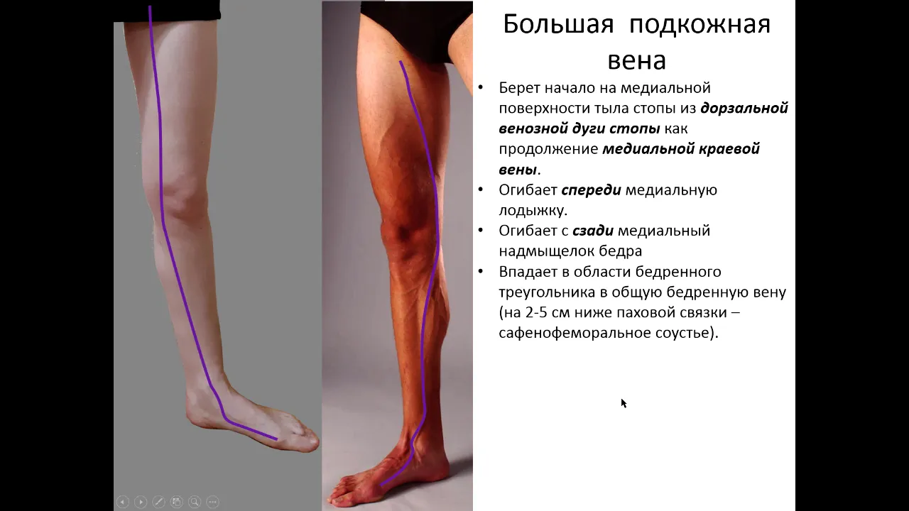
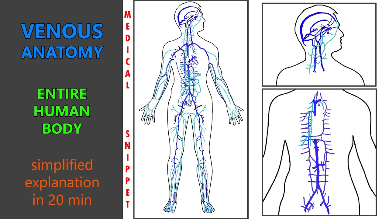
Added:Basic anatomy of the lower extremity venous system, focusing on the common femoral and popliteal veins and their spatial relationship to adjacent arteries.
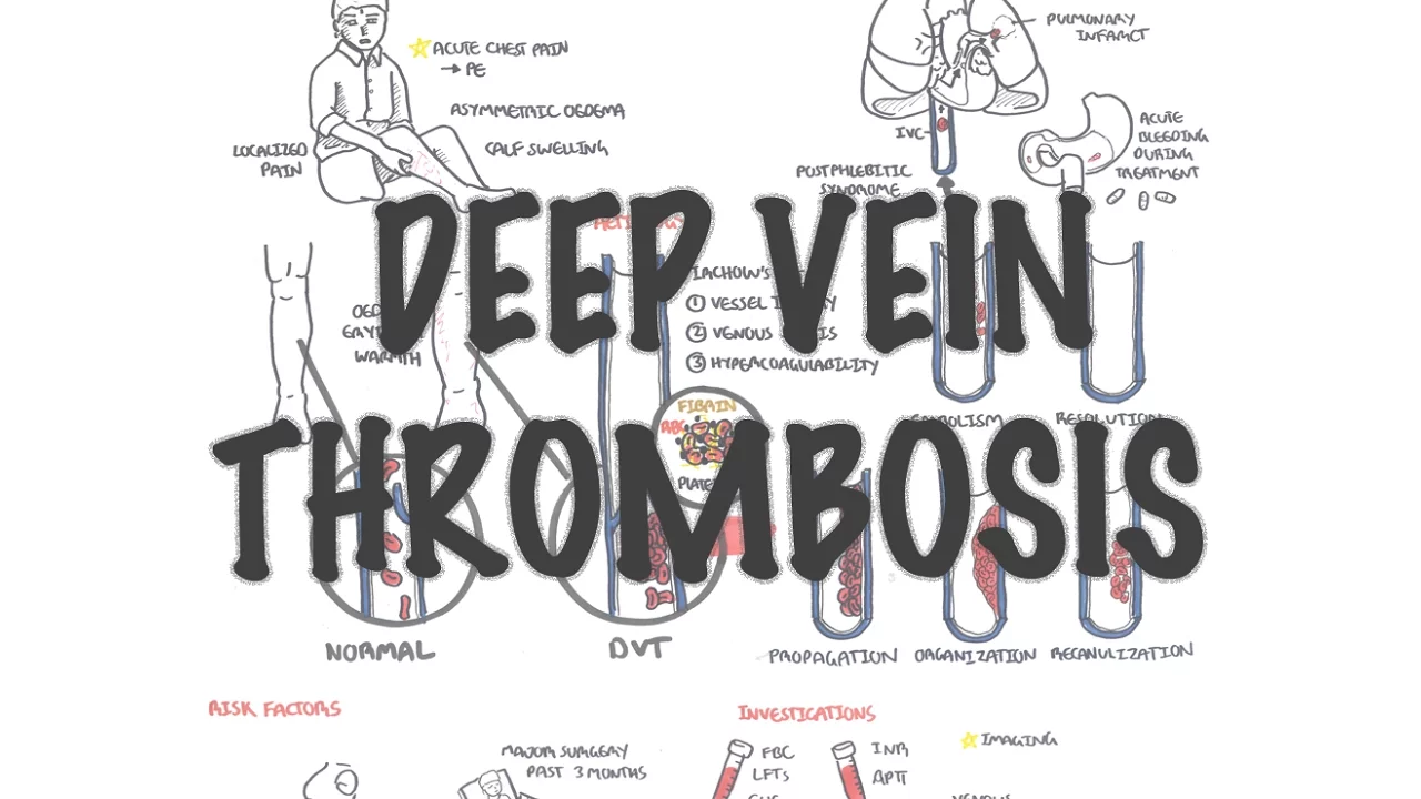
Pathophysiology and risk factors of Deep Vein Thrombosis (DVT), as outlined by Virchow's Triad.
Fundamental principles of ultrasound physics and knobology, particularly the use of a high-frequency linear transducer.
The core concept of compression ultrasonography, specifically how normal veins collapse under pressure while arteries do not.
Advanced venous ultrasound techniques, including three-point or whole-leg compression scans to identify isolated calf DVTs.
Application of Color and Spectral Doppler to evaluate venous flow dynamics, respiratory phasicity, and distal augmentation.
Clinical integration of POCUS findings with diagnostic algorithms, such as the Wells Score and D-dimer assays, for medical decision-making.
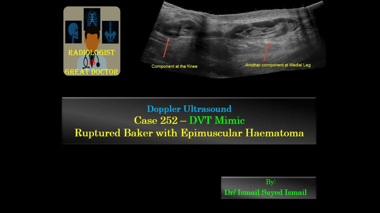
Identification of ultrasound findings for common DVT mimics, such as Baker's cysts, superficial thrombophlebitis, or hematomas.
3.7K views113likes7:54Original Release: 2022-08-06
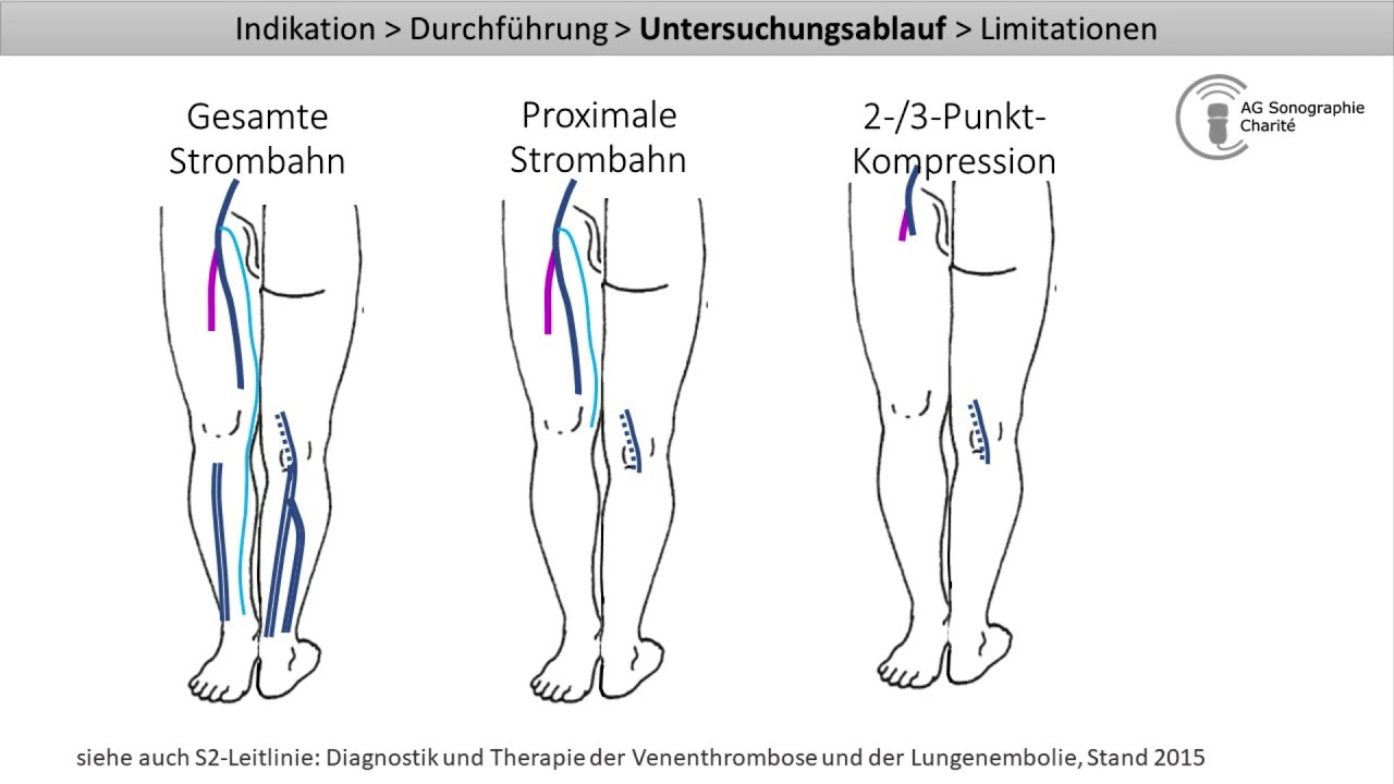
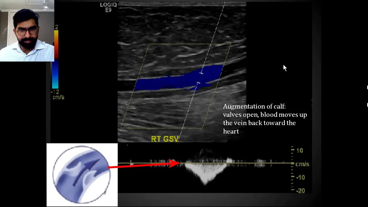
hey guys i just wanted to make a quick video about something that i think all residents should be practicing more often on their ultrasound or pocus exams this is something that i only recently learned about on my icu rotation but honestly i feel like this would be such a useful thing to add to your repertoire and really be able to help you a lot when quickly trying to figure out if a patient has a dvt or pe or not and basically it's what's called a two-point ultrasound there's very different variations of this a two-point dvt ultrasound a three-point dvt ultrasound and then you have to like full scan up and down the leg which is like the formal ultrasound uh but really a lot of the studies showed that a two-point dvt ultrasound is actually pretty good as like an initial screening evaluation for dvt think about how many times you have a patient where you're kind of considering whether or not you have higher or lower suspicion for a dvt or pe and you're not sure if you want to start anticoagulation quite yet right away you know identifying non-compressibility on a point of care ultrasound would be such an easy little test to do especially because all of us are starting to use point of care ultrasound much more frequently and this is something that i think we don't focus on as much we tend to focus a lot more on the cardiac exam and the lung exam lung sliding stuff like that but dvt ultrasound could be a very useful thing to add to your repertoire so one thing that i really like doing is going to this website pocus 101 this is honestly a god tier website in terms of giving lots of guides on how to do all of the ultrasound exams properly and you can see they have one on dvt ultrasound made easy and so first off they go over some of the indications for doing a dvt ultrasound and then kind of talking about first of all you want to put the patient in a frog leg position this is really going to help bring the veins closer to the surface also helps distend them a little bit and make them a little bit easier to to visualize and then we're going to start with the linear ultrasound probe and for the two point dvt ultrasound if you read through this article you're really going to be focusing on two main points here's some important kind of landmarks you should be able to identify but the two points you're going to be looking at is going to be the common femoral vein where it bifurcates into the great saphenous vein and you're gonna also look at the popliteal vein and those are gonna be your two points and so for this first point of the common femoral vein and the saphenous vein what you're really looking for is for uh this kind of mickey mouse sign so you have this mickey mouse you got your saphenous vein here the common femoral vein and then the common femoral artery over here and here is uh basically once you find this structure you're going to start compressing so that the artery slightly gets compressed but the veins should be fully compressed and then obviously if you have a client there you may potentially visualize a clot in the middle of there or you're not going to have compressibility of the veins there and that will be kind of a good screening for if there's a clot the next biggest landmark is going to be the popliteal vein so right behind the knee and uh you're going to basically be looking for compressibility above the popliteal artery so what they really want you to remember is pop on top so the popliteal vein is on top of the popliteal artery and you're just going to scan a one to two centimeter above the popliteal vein and then down the popliteal vein and that's going to give you a good area to test the compressibility and see if there's any clots present what i love about this website is they have all these kind of real-time images that can tell you or show you what an example of an abnormal ultrasound would look like so here's some examples where you see a direct clot this one is just a huge clot blocking the middle this one's a mobile clot that's kind of moving around in there with some turbulence and then they show some uh examples of non-compressible veins so you see here you have the common femoral artery here and then it's they push on it and it starts to collapse a little bit but you don't see any compression of the uh common femoral vein same thing with the popliteal vein here so they are actually fully remember pop on top so they are compressing the popliteal artery and yet the popliteal vein is not compressing at all so that is highly suspicious for a dvt there's a couple of different techniques like augmentation with color doppler but it's not really as recommended as just doing compressibility and what i really like is they have this super super good youtube video that's linked right here uh and it is by pocus 101 and so it is by their own people and you can see that uh you know this is such a great test because it's just going to take 3.5 to 2 to 3.5 minutes or so and just check a couple points and i highly recommend all of you guys just watch this video really quickly it's very interesting watch as well you give some visual examples of how to do it and here they're showing the first point where you're going to be doing the bifurcation of the common femoral vein and the great saphenous vein and you're going to see just in a second here's that mickey mouse appearance with the great saphenous vein here this is the common femoral vein and then this is the artery right here next they're going to be sliding down the femoral vein and just kind of uh looking a little bit uh you know more distally so this is what that view is going to look like you have the superficial femoral vein which is actually a deep vein and then you have the deep femoral vein and you can see both of them are going to be collapsible and then finally you're going to look at the popliteal vein remember pop on top and this is what a normal exam is going to look like you're compressing popliteal artery stays patent and then the popliteal vein will collapse like this and what i really love about this one is this video again it's going to show you these examples of abnormal findings so this is one with a clot in the middle of it here's an example of the artery on top and then the non-compressible vein on the bottom and then finally an abnormal example of the popliteal vein so again pop is on top so pop the teal vein is on top and you can see the artery is compressing but the popliteal vein is not compressing so anyways i just wanted to quickly make this video because this is something i only really kind of learned about a few weeks ago and it surprised me with how easy it was to check for dvts uh just with a simple two-point dvt ultrasound and so i highly encourage everybody to start incorporating this into some of their focus exams especially when they're suspicious of a potential dvt and i think this is really going to you know the more you do it the more you can compare yourself with a formal ultrasound and see if you're doing it correctly or not and really get a good sense for if you're seeing clots or not and i think it's super strong about this you know technique is that uh there is a comparison between two point ultrasonography plus d dimer versus whole leg ultrasonography which again may take like 24 hours or even longer to get done and it showed that the two diagnostic strategies are equivalent when used for the management of symptomatic outpatients with suspected dvt of the lower extremities you know i think you do have to take some of these findings with a grain of salt because um here this study showed that resident performed two-point compression ultrasound is inadequate for diagnosis of dvt and the critically ill but this one was basically they were trained in a two-hour module for checking for dvts and then they found that the sensitivity for finding dvts above the knee was 63 with a specificity of 97 which is honestly not that bad i mean sensitivity you know what i'm really looking for is uh the specificity here because if i see a clot there then i'm going to know 100 that there there's a dvt there and i can start anticoagulation um so sensitivity i'm still going to get that form of ultrasound i'm not going to like not get that form of ultrasound but what you're really saving is that uh time from order of a formal ultrasound to a radiologist's read averaged 14.7 hours so even if you've got a somewhat lower sensitivity if you get a pretty good specificity this can still be a very very useful test that shaves off hours from the formal workup anyways thought i would just bring that to your guys's attention definitely try it out next time you're walking around with your butterfly or your ultrasound and hopefully this is something useful to add to your repertoire i hope you enjoyed let me know what you think down in the comments below thanks for watching i'll see you in the next one peace [Music]
Up Next

13:44
DVT Ultrasound Part 1: Focused Leg Vein Exam Anatomy & Technique
@Sonosite
258.4K views•2011-07-08

48:47
Graphic Medicine: Comics for Collaborative Healthcare Communication
@nationalpatientadvocate
189 views•2023-12-04

8:36
Ventilator Waveforms & Troubleshooting | ICU Basics
@ConanLiuMD
68.7K views•2022-06-21

3:05
Stages of Labor and Vaginal Birth | Childbirth Animation
@nucleusmedicalmedia
52.1M views•2017-08-18
Related Study Plans & Knowledge Roadmaps
Structured learning paths in Medicine
medicine
Abnormal LFTs: Diagnostic Workup & Pathology
01Liver Anatomy and Basic Physiology
02Introduction to Liver Function Tests (LFTs)
03Hepatocellular vs. Cholestatic Patterns
+ 3 more learning modules
12h6 modules
Explore Roadmap
medicine
Analyzing Blood Smears: RBC, WBC & Platelets
01Introduction to Blood Components & Smear Preparation
02Normal Morphology of Blood Cells
03Erythrocyte Pathologies & Morphological Variants
+ 2 more learning modules
12h5 modules
Explore Roadmap
medicine
Mechanical Ventilation: Waveforms & Alarms
01Anatomy and Physiology of Breathing
02Introduction to Mechanical Ventilation & Core Modes
03Ventilator Settings and Waveform Interpretation
+ 2 more learning modules
12h5 modules
Explore Roadmap
medicine
Neuro Exam: Localization & Stroke Syndromes
01Foundations of Neuroanatomy and Vascular Supply
02The Neurological Examination: Step-by-Step
03Principles of Neurological Localization
+ 3 more learning modules
18h6 modules
Explore Roadmap