



















The focused DVT ultrasound examination is a rapid bedside technique for detecting proximal deep vein thrombosis by evaluating the femoral and popliteal veins; it relies on identifying normal venous anatomy (common femoral vein medial to artery, saphenous vein joining above bifurcation, popliteal vein posterior to artery) and performing compression testing where normal veins completely collapse under probe pressure while thrombosed veins fail to compress due to the clot preventing wall apposition.
DVT Ultrasound Part 1: Focused Leg Vein Exam Anatomy & Technique
Added:Basic principles of ultrasound physics, including transducer selection (linear array), orientation, and how tissue density affects echogenicity.
Gross anatomy of the lower extremity venous system, specifically the distinction between the deep venous system, superficial system, and adjacent arterial structures.
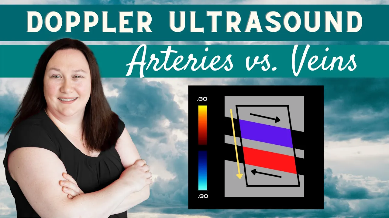
The physiological differences between arteries and veins, such as vessel wall thickness, pressure, pulsatility, and baseline compressibility.
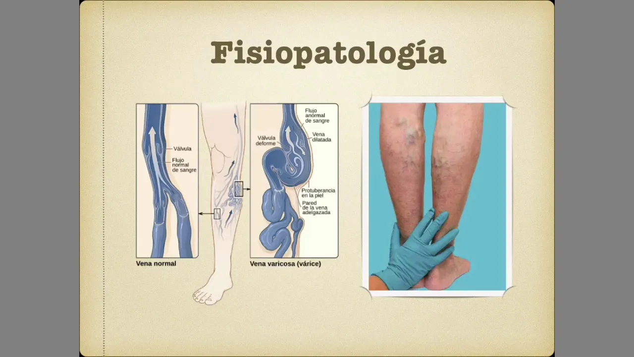
The clinical pathophysiology of Deep Vein Thrombosis (DVT), including Virchow's Triad and common risk factors.
Identification of pathological DVT findings on ultrasound, such as visualization of acute or chronic thrombi and incomplete vessel compressibility.
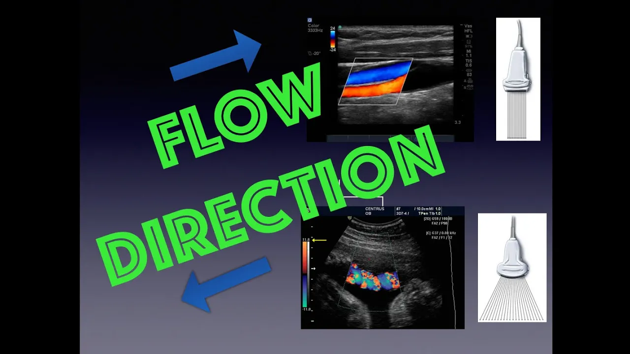
The use of Color and Spectral Doppler to evaluate blood flow dynamics, venous phasicity, and distal augmentation techniques.
Comprehensive lower extremity venous protocols, expanding from a focused point-of-care (POCUS) exam to a formal whole-leg duplex study including calf veins.
Clinical integration of ultrasound results with diagnostic algorithms, such as Wells Score assessment, D-dimer testing, and subsequent anticoagulation management.
258.4K views1.2Klikes13:44Original Release: 2011-07-08
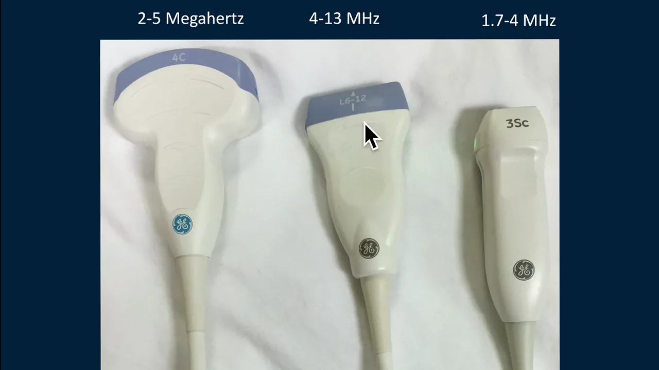
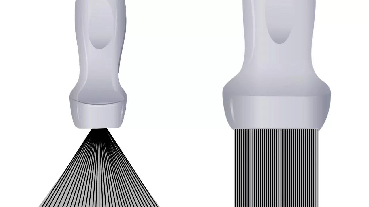
hello my name is Phil Pereira and I'm the emergency ultrasound co-director at the LA County USC Medical Center in Los Angeles California and welcome to sound bites welcome back to sound bites ultrasound in this module we'll learn the focused ultrasound evaluation of the leg for deep Fe is thrombosis now I've divided this module up into Parts one and two in this module entitled DVT ultrasound part one we'll first of all learn the normal anatomy of the leg veins integral to Performance of the DVT ultrasound examination we'll then move on to learn the normal compression exam of the leg veins and how to interpret normal findings on the bedside DVT examination specifically in this module we're going to concentrate on the focused DVT examination the focused or Limited DVT exam allows for increased speed in the performance of the examination we'll concentrate on two specific areas of the leg looking at the femoral region and the pole region this limited examination also maintains excellent sensitivity in the detection of proximal dvts and in fact most Radiology performed DVT examinations screen only down to the poal vessels the caffeine exam is not routinely performed as part of most Radiology performed DVT examination and indeed in the focus DVT examinations we'll skip the examination of the cap veins that leads us into the concept of the focus DVT exam as being an optimal means for evaluation for DVT at the bedside and the focus DVT exam will Begin by examining the femoral vein starting high at the level of the proximal common femoral artery and vein just below the anguin ligament we'll continue the exam of the femoral vein down about 4 to 5 cim through to bifurcation of the vein in into the deep and superficial femoral veins we'll then turn to examination of the poal vein placing the probe high within the poal fossa we'll examine the poal vein about 4 CET within the poal fossa continuing the exam of the poal vein down to trication of the vessel into the calf veins let's now review the lower extremity Venus Anatomy integral to Performance of the focus DVT examination We Begin by identifying the common Fem vein seen here just below the inguinal ligament notice that the common femoral vein is seen just medial to the common femoral artery now the common femoral vein continues down the leg to bifurcate into the deep and superficial femoral veins we note here the Deep femoral vein coursing to the back of the leg and we note the adjacent deep femoral artery we also see here the sainis vein which joins into the common femoral vein above the level of bifurcation now it's important to realize that the superficial femoral vein is the vein that actually continues down the leg to become the poal vein behind the knee and we note The Superficial femoral vein coursing down the leg and accompanied by the paired superficial femoral artery behind the knee that superficial femoral vein will become the pole vein and we see the adjacent poil artery now at the level of trication the papati vein will become three different calf veins and we note here the anterior tibial vein that's going to course anteriorly onto the calf the posterior tibial vein seen posteriorly in the calf and also the perinal vein seen to the lateral aspect of the calf and it's because these cath veins are so small that it's difficult to see them on bedside ultrasonography let's now watch a video and learn how to perform the ultrasound examination looking for a DVT within the femoral vein system We Begin by placing the highfrequency linear array type probe it's the same probe that you'll be using for vascular access in a side to side orientation over the common femoral vein and artery just below the inguinal ligament notice that we're compressing down with the probe and essentially the DBT exam is a compression exam as a normal vein will completely close with pressure down with the probe notice that we're sequentially compressing at different levels along the common femoral vein compressing from the beginning at the top just below the inin ligament all the way down through bifurcation into the superficial and deep femoral vessels now a clot will not completely compress with pressure down with the probe and thus will be identified on bedside examination notice here it's standard to have the marker on the probe going lateral so that we know where we are with regard to the orientation of the probe versus the screen it's best to position our patient slightly up right to distend the femoral vessels for the DVT exam and as shown in this video we actually had the patient with the head of the bed up about 30° we also want to have the leg slightly externally rotated to best Orient so that we can place the probe directly over the common femoral artery and vein here we see the ultrasound findings that will occur when placing the probe as shown in the illustration towards the left note here the probe is placed with the marker dot laterally just inferior to the inguin ligament over the common femoral artery and vein as shown in the picture torial towards the right notice here that the common femoral vein will be seen medial to the common femoral artery and because we have the marker dot oriented laterally or towards the left of the image the common femoral vein will be seen to the right here here's a video showing the actual ultrasound findings of the common femoral artery and vein using colorflow Doppler we Orient ourselves to the image to the left here showing that the common femoral vein will be seen medial to the common femoral artery and we note the ultrasound findings to the right showing Pulsa flow within the common femoral artery located just lateral to the common femoral vein and we see the phasic hum of the blood flow within the common femoral vein seen medial to the artery here while it's very nice to have colorflow Doppler to differentiate the common femoral artery from the common femoral vein we can also discern the two using grayscale or B mode sonography as shown in the video clip here to the right here we note the common femoral artery to the left are lateral to the common femoral vein as shown medially notice that the common femoral artery has more hypertrophic walls and also pulsatile flow within it differentiating it from the common femoral vein as seen medially continuing down the leg as shown in the probe position in the illustration to the left here we see the following ultrasound findings in the illustration to the right we know that the femoral arteries bifurcate at a level above the common femoral vein and here we see the superficial and deep femoral arteries in a location just lateral to the common femoral vein we also see a very important Mark the saffin vein joining into the common femoral vein at this level it's very important to visualize the saffin vein as it's really the only superficial vein in the body that we worry about clot formation within as it goes directly into the common femoral vein and can propagate up into the IVC and Into the Heart here we see a video clip using Color flow Doppler demonstrating the bifurcation of the femoral artery into the superficial and deep femoral arteries and here we see that bifurcation point right there notice that the femoral arteries are located laterally or towards the left of the common femoral vein which we see located medially or towards the right of the image in this video clip we'll note the bifurcation of the common femoral artery into superficial and profundus femoral arteries using grayscale or be mode sonography we knowe the common femoral vein is shown towards the medial aspect of the image or towards the right and here again we see that bifurcation point of the common femoral artery into the superficial and profundus femoral arteries as labeled there and we just remember that point that the artery generally bifurcates at a level higher than the femoral vein in this video clip we're able to get a good look at the sainis vein joining into the common femoral vein and we see the common femoral vein located medial to the common femoral artery note that the sainis vein has the looked often of a little hat on top of the common femoral vein and we note here also the turbulent flow of blood here within the common femoral vein as this was taken in a hypotensive patient now let's turn our attention to the anatomy of the pole fossa we note here the pole vein and the pole artery remember that the poal vein is going to be in an orientation located more posterior to the poal artery which will be located more anterior here's how to perform the focus DVT ultrasound exam looking into the poil faasa it's best to have the patient sitting up to further distend the poal vein and I like to have the patient sitting up with the leg dangling over the bed I can then pull up a chair and move anterior to the patient we'll place the highfrequency linear array probe high within the poil faasa sequentially compressing it levels down all the way down to trication notice that we're using our other hand to stabilize the anterior knee as we press with the probe posteriorly so again we'll start high within that poal faosa compressing sequentially all the way through the levels of the poal arter and vein down inferior there all the way down to trication here's the anatomy that we'll see with a probe placed as shown in the illustration to the left notice that the probe is placed into the posterior aspect of the knee behind the pole faasa again with the marker dot oriented laterally thus we'll see the following images as shown in the illustration to the right note that the poal vein will be located closer to the probe or posterior to the poal artery which will be further away from the probe or more anteriorly located as shown in this image and this image we'll use colorflow Doppler to further differentiate the papal artery from the poal vein and in the video clip here to the right we can see the pulsatile flow of blood within the poal artery as seen anterior or further away from the probe than the poal vein which is seen more posterior than the artery here notice that we see a little bit of phasic flow of blood within the papati vein this video clip employs B mode or grayscale sonography to show the poal vein and poal artery again we can see the poal artery located more anterior than the poal vein and we can see the pulsatile movements of the poal artery differentiating it from the vein and in fact we can see a little bit of turbulent flow of blood within the papati vein here and located more posteriorly than the poal artery when performing the focused lower extremity DVT ultrasound examination we want to First identify the emoral and poal arteries and veins using B mode or grayscale sonography now colorflow Doppler ultrasound can be helpful in differentiating the artery from The Vessel and also making sure that you're looking at vascular structures but is not essential most of our information will actually come from B mode sonography want to apply compression to the vein pressing down with the proe in the short Axis or transverse orientation in a normal examination the walls of the vein will completely touch together conversely if a DBT is present the walls of the vein will not completely touch together as a thrombus within the L of the vein will prevent the walls from completely collapsing here we see normal compression of the common femoral vein and we see here the common femoral vein to the right of the common femoral artery which we see to the left note we're looking in the short axis of transverse orientation pressing down with the probe and note with pressure down on the probe that the common femoral vein completely collapses and that the walls the anterior wall and posterior wall of the vessel meet we also see compression of the sainis vein that little cap on on the top of the common femoral vein so a completely normal exam of the common femoral vein at the level just below the inguin ligament here we're looking a little bit more disty at the common femoral vein at the level of the bifurcation of the common femoral artery into superficial and profundus femoral arteries and we note here complete compression of the vein as we push down with the probe and note again that the anterior and posterior walls completely meet together now let's move down the leg to look at the normal compression exam of the poal vein recall that the poal vein is going to be seen towards the posterior aspect of the image or closer to the top of the image here than the poal artery as we press down we note complete compression of the poal vein and we see here that the artery still stays open so again this would be a normal compression exam of the poal vein with the anter and posterior walls of the vessel completely touching down with probe pressure in conclusion thank you for joining me for the sound bites module going over bedside DVT examination part one hopefully now you understand the focus DVT exam which allows for increased speed with excellent accuracy in the exam performance in this module part one we focused on the basic anatomy and the normal Examination for the DBT evaluation for a normal examination we hope that the femoral and poal veins will completely compressed down with probe pressure unfortunately a Venus thrombosis will prevent the vein from closing and so we'll return in part two going over the positive examination and those findings that you might encounter on the focus bedside DBT examination so I hope to see you in the future as sound bites continues
Up Next

18:45
DVT Ultrasound Technique: Bedside vs Formal Imaging
@atchiem
73.2K views•2013-08-06

48:47
Graphic Medicine: Comics for Collaborative Healthcare Communication
@nationalpatientadvocate
189 views•2023-12-04

24:14
RUSH Exam Part 3: Ultrasound in Shock - Pipes & RV Evaluation
@Sonosite
25.3K views•2013-12-17

3:05
Stages of Labor and Vaginal Birth | Childbirth Animation
@nucleusmedicalmedia
52.1M views•2017-08-18
Related Study Plans & Knowledge Roadmaps
Structured learning paths in Medicine
medicine
Abnormal LFTs: Diagnostic Workup & Pathology
01Liver Anatomy and Basic Physiology
02Introduction to Liver Function Tests (LFTs)
03Hepatocellular vs. Cholestatic Patterns
+ 3 more learning modules
12h6 modules
Explore Roadmap
medicine
Analyzing Blood Smears: RBC, WBC & Platelets
01Introduction to Blood Components & Smear Preparation
02Normal Morphology of Blood Cells
03Erythrocyte Pathologies & Morphological Variants
+ 2 more learning modules
12h5 modules
Explore Roadmap
medicine
Mechanical Ventilation: Waveforms & Alarms
01Anatomy and Physiology of Breathing
02Introduction to Mechanical Ventilation & Core Modes
03Ventilator Settings and Waveform Interpretation
+ 2 more learning modules
12h5 modules
Explore Roadmap
medicine
Neuro Exam: Localization & Stroke Syndromes
01Foundations of Neuroanatomy and Vascular Supply
02The Neurological Examination: Step-by-Step
03Principles of Neurological Localization
+ 3 more learning modules
18h6 modules
Explore Roadmap