












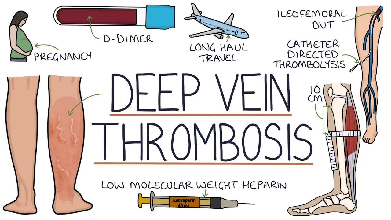

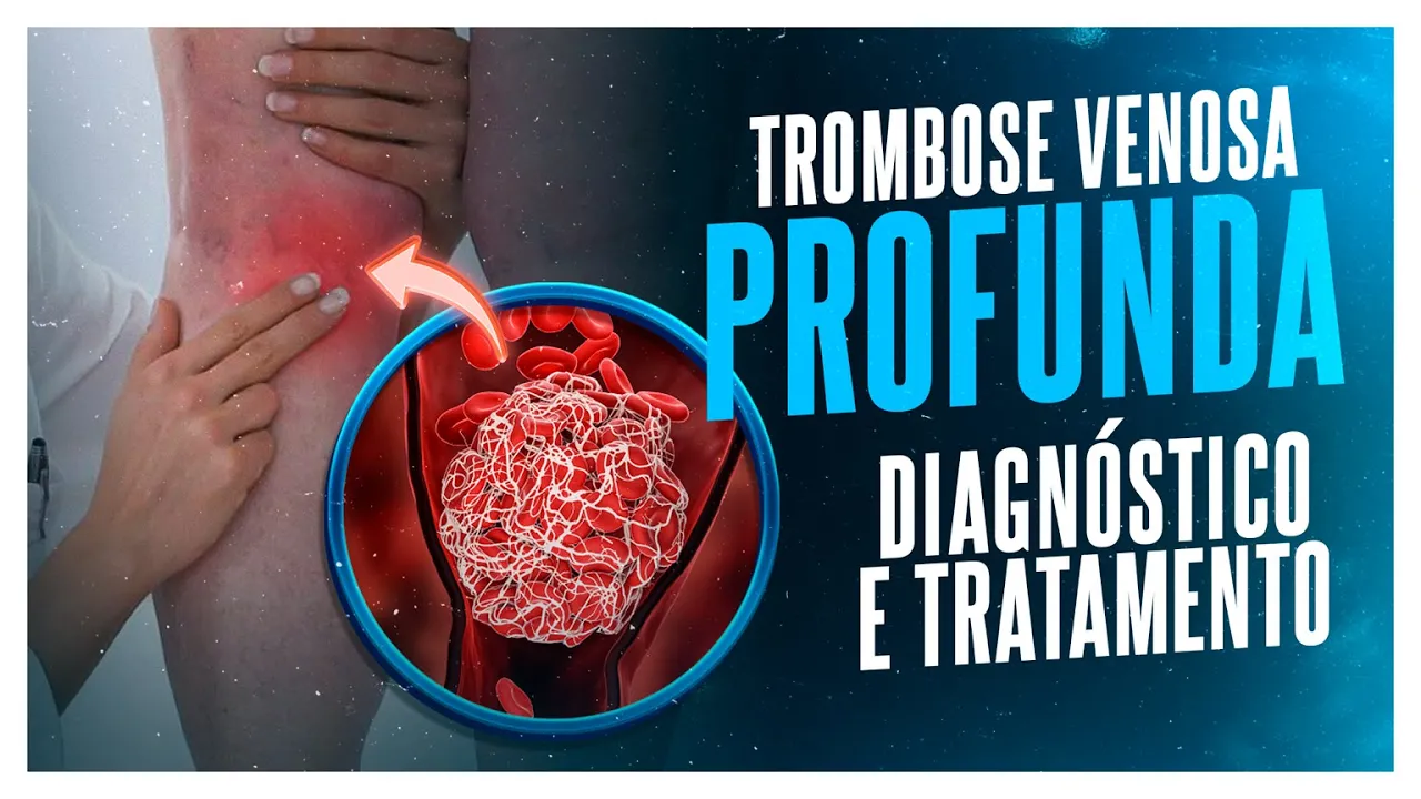





Emergency physicians can perform limited DVT ultrasound with high accuracy (greater than 95% sensitivity and specificity) by systematically examining the proximal veins (saphenofemoral junction, common femoral vein, superficial femoral vein, and popliteal vein) using two-handed perpendicular compression, without requiring long-axis views, color Doppler, or augmentation testing; this approach saves approximately 2 hours of patient length of stay compared to formal radiology-performed studies and requires only a few hours of training with competency achieved after performing approximately 10 examinations.
DVT Ultrasound Technique: Bedside vs Formal Imaging
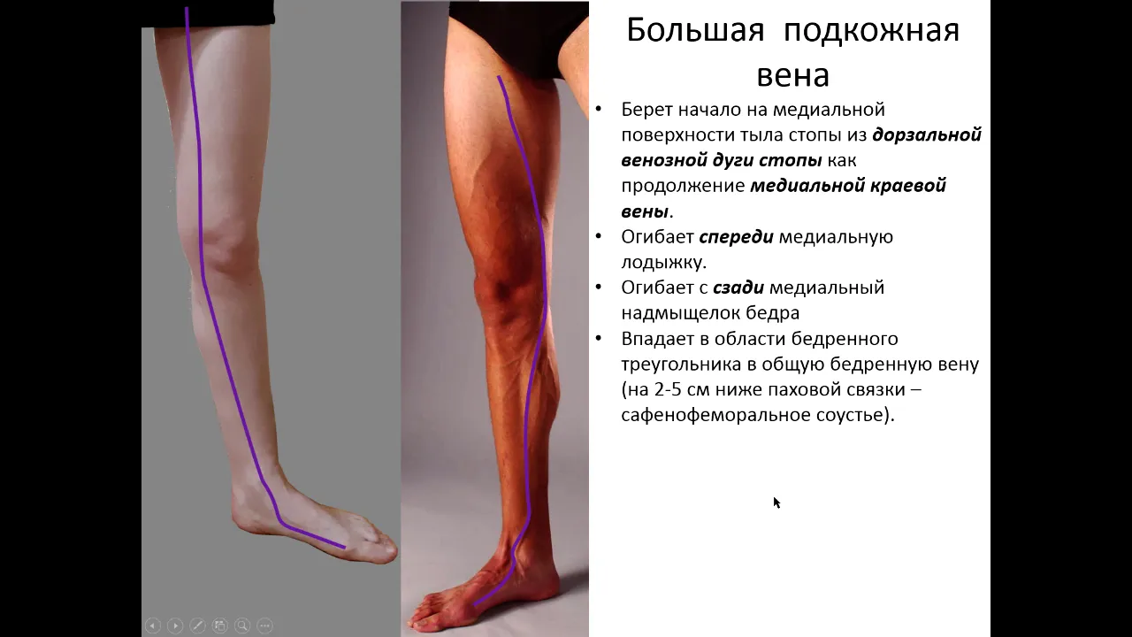
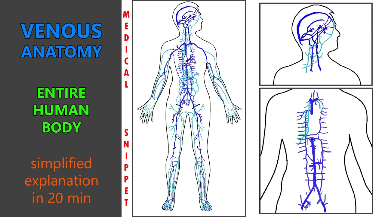
Added:Basic venous anatomy of the lower extremities, specifically distinguishing between the deep and superficial venous systems.
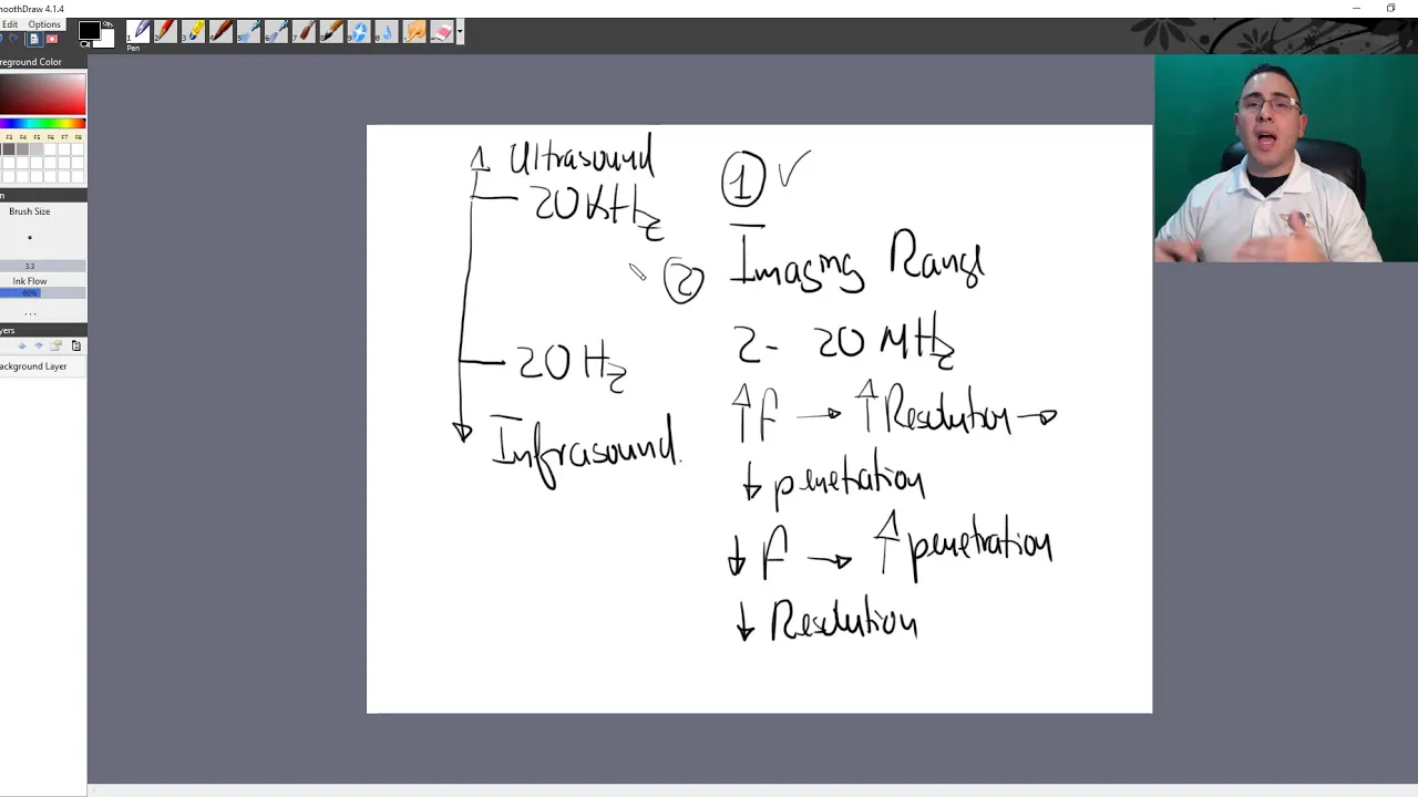
Fundamental principles of ultrasound physics, including transducer selection, image optimization, and the concept of compressibility.
Pathophysiology, risk factors (Virchow's Triad), and clinical presentation of Deep Vein Thrombosis (DVT).
An introductory understanding of Point-of-Care Ultrasound (POCUS) and its role in rapid clinical decision-making.
Advanced vascular ultrasound techniques, including the use of color and spectral Doppler to assess venous flow and reflux.
Clinical management and anticoagulation protocols for acute deep vein thrombosis post-diagnosis.
Diagnostic pathways integrating DVT ultrasound findings with the workup for Pulmonary Embolism (PE), including focused echocardiography.
Quality assurance, documentation requirements, and medical-legal considerations of bedside versus formal diagnostic ultrasound.
73.2K views630likes18:45Original Release: 2013-08-06
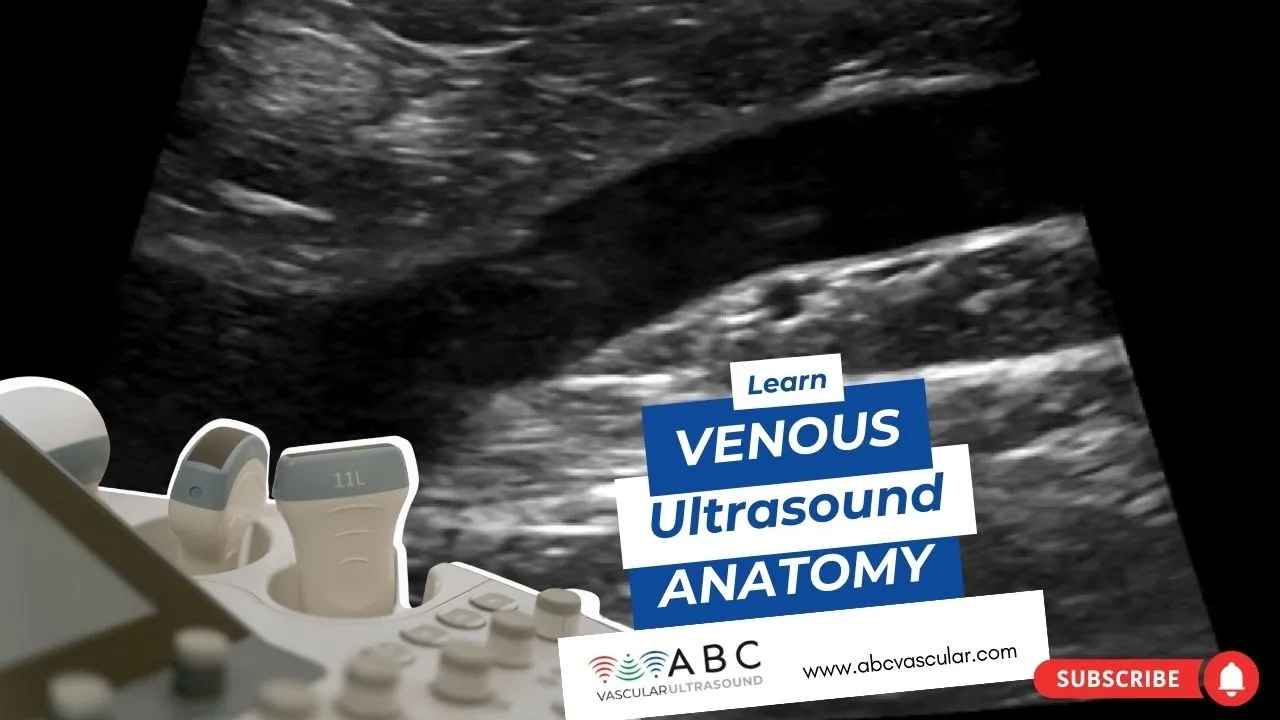
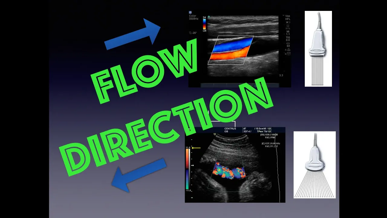
hi welcome to this quick video review on DBT ultrasound remember that you should always have a differential diagnosis it could be cellulitis or absis or it could be systemic congestion from either High hydrostatic pressure is in heart failure or low enoic pressure is in hypo abonia in our liver patients and this is kind of what it looks like you have cobblestoning of the subcutaneous space right here you have a fluid between these fat globules um and it's pretty typical of both of cellulitis as well as systemic congestion you also have lymph adenopathy and it typically manifests as something that looks like it could be a clot but it's actually pretty discreet and spherical so if it goes away uh and you still have your vessels here then you know that that was just a lymph node you also have a ruptured Baker cyst so this is pretty obvious that this isn't a vein you can also put color Doppler on just to make sure there's no flow and this is typically what looks looks like uh and it's also only in the pop seal region then you have your superficial Venus thosis which shouldn't affect your DVT ultrasound and is more likely a clinical diagnosis and then you have your muscle hematoma or myositis where you actually have uh fluid along the actual fibers of the muscle and I'll play it again so there's fluid right here it's not actually in the subcutaneous space is actually along the muscle fibers themselves and last if you had like a killes tendon injury of some sort this is actually a bicep St but it gives you the same idea where the muscle fibers or tendon fibers are actually disrupted and you can notice that it's typically pretty close to the the bone itself so it's not anywhere near any vascular structures uh this is going from a long axis to a short axis view right here so this is a torn fiber and then there's a lot of fluid around there so those should be on your differential for VTE briefly the anatomy so you want to start off at the saffo femoral Junction where the great saffin vein takes off reason is that even though the the saffin vein is not considered a a deep vein if you actually had a clot right here the risk of embolism is still uh rather High then you have your common femoral vein and it will about 2 or 3 cm uh distal it'll bifurcate into your deep femoral vein and your superficial femoral vein this is actually a misnomer your superficial femoral vein is the one that actually Dives posteriorly to become your popal vein whereas your deep femal vein actually just divides into the little tiny veins that Supply the the lateral thigh so again your superficial FAL vein is the one that actually becomes your poo vein and it does this by diving posteriorly into the abductor Canal where it travels has highlighted in the blue section right here and it's in this segment that where if you have someone that has a lot of soft tissue it becomes very difficult to evaluate for a deep Venus thrombosis so your potilo vein travels posteriorly posterior to the poal arteries so on a posterior ultrasound when you're evaluating the poal crease it'll appear to be superficial to the poal artery and this pole vein makes two bifurcations the first one is the B a into the anterior tibial vein so then it'll actually travel deep on the posterior ultrasound and then next it'll bifurcate again into the posterior tibial vein and the fiar or peronal vein it's after the segment as highlighted in the blue that we consider uh the veins calf veins and these calf veins are difficult to evaluate as well as the prior segment of the femoral vein highlighted in blue because the the caffein themselves are really small and it becomes harder to evaluate for clot keep in mind that most of your dbts are actually going to be in the common femoral vein distribution about 3/4 of the time less than 5% is actually going to be in this hard to evaluate spot and then the remaining 20 to 25% are going to be along this Papa teal area before the bifurcation or trication into the caffein the caffein are only responsible for for about 5% of dbts or less so these areas this especially caffeine we we call those segmental areas uh along with this right here and the reason is that uh we can't evaluate these segments very well and there is a risk of thrombosis and a risk of extension of the clot but in we'll talk about that in a second so what is your your diagnostic criteria for gbt the the main criteria is whether the vein is compressible or not if it's non-compressible there's nothing else you need to do this person has a DVT now what are secondary diagnostic criteria you can image the thrombus which is um harder to do in an acute DVT because it's less echogenic and you can also assess for Venus distension but uh you could have Venus insufficiency or valver dysfunction causing the same thing so it's not specific you can assess for a filling defect you could put color Doppler on and see if there's any filling defect uh but it it's neither sensitive or specific for a DVT and you can look for uh loss of augmentation which we'll get to in a second as well as uh respir variation or with Val Salva which also we we'll talk about so what's the difference between a formal SL Radiology performed DVT ultrasound and an emergency department or Emergency Physician performed DVT ultrasound the first is that uh we don't typically do a long AIS view of the vein you typically would want to do one if you saw a DVT and you want to see how far that clot extended or if you had a chronic DBT let's say a patient has a history of of a DVT from a month ago and you want to see if the clot has actually extended uh and you have like a previous measurement then you then you could do a long axis and and kind of measure out that clot the second thing that we typically don't do is a collar Doppler and what that does is it helps you to assess F filling defect sometimes when you're having trouble finding where the vein is usually your Popa teal vein it's it's a good idea to put color Doppler on and then that way you can assess whether that is the vessel of Interest another thing that we don't typically do is augmentation and what augmentation does along with color dopplers it helps you to visualize a clot if it's there the idea behind augmentation is that you have this long vein you apply pressure right here distally and that should actually cause increased Venus return and if you put the probe right here you would actually see an increase in flow on color Doppler now if there's a big obstructive clot you won't see that or you'll see the filling defect now if you had a smaller clot you may actually not see any change with augmentation the last and the most important difference is time time so a formal DVT ultrasound will take close to half an hour to an hour to perform because they're doing all these other things that you don't need to do whereas a DVT ultrasound performed by emergency physicians should take less than 5 minutes to perform you save about 2 hours on a patient's length of stay and it's pretty good too actually uh compared to Radiology perform ultrasound it has greater than 95% sensitivity and specificity keep in mind that there uh studies that that advocate for twoo DVT ultrasound which means just two-point compression versus three-point compression versus just trying to look at those proximal areas uh not the segmental areas that we talked about but in any of those techniques you have high sensitivity and specificity the last point in terms of Ed perform dv2 ultrasound is that it's actually pretty straightforward and very simple to do uh the studies that look at how long it takes to train residents as well as attending to do dv2 alwn it's actually about a few hours of training and as as few as 10 DVT exams will lead you to competency so what are some pearls to DVT ultrasound first make sure to expose your patient sometimes because it involves the groin area or the low extremity some people get bashful but remember that uh you're really trying to R A life-threatening diagnosis and if you counterweigh it with all the patients that we do pelvic exams on because they have lower abdal pain um you get the idea that we should be exposing our patients for gbt ultrasound you also need to touch the patient meaning you can't just use the probe to compress without actually anchoring the probe with the other hand onto the patient's skin or if you have a large patient you need to use your other hand to lift that panis uh off so that you can actually do adequate compression this image actually shows you why it's flawed so when you try to do that remember that you have all this this gel on the probe so when you try to compress you typically slide around and you're not able to evaluate fully if there's compressibility so that's why you should actually do a two-handed approach and what I mean by that is you have one hand this hand your right or left hand Tethered to the patient skin so you know the probe's not moving and then you have the other hand actually do the compression this helps ensure that you're not giving undue uh pressure and unintended compression of of your vein typically with your papal vein it can easily compress like this way so make sure you expose your patient make sure you do a two-handed approach and when you compress you really want to just compress perpendicularly if you're off axis a little bit you may be not Imaging the same area that you're compressing and also uh your vein typically appears to be more echogenic or more visible when you actually are Imaging a plane perpendicular to the vein itself you want to compress in 1 to 2 cm intervals and if you have someone who's difficult you can try to use Doppler and you can also do a reverse Trendelenburg so that the blood is pulled in those veins and it makes those veins easier to see so this is augmentation right here you're squeezing the the Venus circulation distl to where you're trying to examine and you put the probe right here into the femoral area and uh when you squeeze this is what a normal should be right here where you don't see any filling defects that's a normal uh augmentation study and then this is in an abnormal study where you actually see a filling defect in the center so that points towards a clot so in terms of technique you first want to expose a patient this way have the patient ex rotate like a frog leg that helps you to expose a vein along the inul uh crease and you start about right there in the midline and you should identify that saffo femal Junction first right here then uh you go 1 cm compress your common femoral vein then you do another couple centimet until you hit that superficial and deep femoral vein right here and you want to try to compress till you lose that that superficial femoral vein and this is what a normal study looks like again you have that that saffo femal Junction right there you go again there's no lymph node you compress it one more time still compressible you could see the bifurcation into to the superficial and deep right here femoral vein and try to compress one more time and that should be an adequate femoral vein DBT ultrasound this is a superficial femal vein DVT right here notice that it doesn't compress and you could see that maybe there's some echogenic material in here but um remember that the main criteria is that it's non-compressible right here so you're pop the teal vein some people Advocate having the patient dangle the legs down or Flex with the sole on the bed itself I actually like having the patients lay prone or lateral du cubitus because that opens up this area for you so that you can identify the popil vein with ease and also you can do your perpendicular compression so just go about just go above the POA fossil right here and you should identify about right here do you get compression go about 1 cm do another compression you'll probably see a bifurcation into the ENT tubal vein and then go again and then you'll see the bifurcation again of of the fibular or peral vein and the posterior tubal vein and you're done so this is a normal this is your poal vein right here remember the papetal vein is posterior to the artery so it's more superficial on a posterior ultrasound and you're just compressing till you have that vication to the calf veins so this is your poal DVT right here this is the artery it's pulsating but the DV the the poal vein doesn't compress so you have aaal DVT right here so the difference between a proximal versus segmental DVT is in the location and also the yield so segmental dvts are harder to Ultrasound but they also have uh very low yield U remember that your segmental DBT is usually disle to the pop teal vein um when it when you have trication to your caffeinees and also some people like me consider your suici femal vein where it dives into the aductor canal before it merges posteriorly to be your poal vein so why is it low yield well there's only about a 20% chance that a segmental DBT will actually propagate um into a proximal DVT and usually that takes about a week to two weeks and even when it propagates to become a proximal DP there's only about a 25% chance that it'll embolize become a PE so you're really looking at a 5% chance of a segmental DBT even you miss it uh causing something that's life-threatening second in a pretty large study that was published in jamama not too long ago looked at all these patients had normal DBT scans and they follow them for 3 months and your risk of actually having a proximal DVT from a Segal DVT is less than 1% now if you have someone who's intermediate or high risk let's say they have unilateral leg swelling you can't figure out out why or they have a history of cancer or repeat dbts but you have a negative DVT study either you can do a d dier and if it's negative you can rule them out that way or you can have them come back in about 7 to 10 days for repeat ultrasound so some frequently ask questions one how much compression should I need so if you actually see that artery compress a little bit then you know that you have enough compression to compress the vein and if that vein still doesn't compress then that's a DVT second when should you obtain a longitudinal view the only reason you would do that is if you want to evaluate the extent of a thrombus if you have a DVT or if you have uh a chronic DVT and you want to figure out like if there's any resolution third does color Doppler add to the study no not really um only if you are having trouble identifying the veins and another thing is with augmentation it may help but again that's not part of your limited DBT stud the another question is should you ultrasound the contralateral leg especially if it's asymptomatic the answer is no because even with risk factors and a DVT in the other leg you still have only less than a 10 to 15% chance of having bilateral DVT plus the management doesn't change so if you perform a bedside DVT study do you still need to order a formal study that really depends on two things it depends on whether your tending is credential to do bedside ultrasound and it's also comfortable with their DVT study and it also depends on whether the emergency department regularly bills or has an agreement with Radiology to allow for bedside ultrasound now if the policy is that you can perform bedside DVT ultrasound but you need to get a formal DVT ultrasound then you should follow that policy but remember that a lot of emergency departments in the country actually do their own DVT ultrasounds and build for them without actually having to order a follow-up formal study if you actually are going to do that you need to do two things first you need to do the ultrasound and record it and second you need to be able to write that interpretation in the notes because both components are required as part of the quality improvement process as well as for billing so typically aliac V thrombosis uh pregnancy is a risk factor for this the reason is that U when you have a gravid uterus in the third trimester the right iliac artery tends to com press that left iliac vein right here this is called May therer syndrome and this is the reason why most iliac vein thrombosis occur in the left iliac vein and and because you can't compress the iliac vein especially during the third trimester you need to look for secondary signs of a possible thrombosis such as uh change with restory variation so this is a pulse wave Doppler on superficial femal ve where whenever you take a breath you increase Venus turn and and your your flow increases and then when you when you exhale it decreases so you should this is normal so if you actually had an iliac vein thrombosis you would lose this respiratory variation or phasicity now if you really were suspicious about Elan thrombosis you would have to pursue an MRI so the ultimate message for you guys is do it it's really easy to do it's highly accurate and you can save your patients a lot of time by doing your own DVT ultrasound here are some references definitely take a look at these uh they're they're definitely very high yield reading thanks again for listening and uh let me know if you have any questions
Up Next

12:57
Low-Benefit Fiber Types and Better Alternatives
@ThomasDeLauerOfficial
134.5K views•2021-12-16

48:47
Graphic Medicine: Comics for Collaborative Healthcare Communication
@nationalpatientadvocate
189 views•2023-12-04

10:28
Neuroanatomy: Central and Peripheral Nervous System Divisions Explained
@AKLECTURES
136.2K views•2014-09-20

3:05
Stages of Labor and Vaginal Birth | Childbirth Animation
@nucleusmedicalmedia
52.1M views•2017-08-18
Related Study Plans & Knowledge Roadmaps
Structured learning paths in Medicine
medicine
Abnormal LFTs: Diagnostic Workup & Pathology
01Liver Anatomy and Basic Physiology
02Introduction to Liver Function Tests (LFTs)
03Hepatocellular vs. Cholestatic Patterns
+ 3 more learning modules
12h6 modules
Explore Roadmap
medicine
Analyzing Blood Smears: RBC, WBC & Platelets
01Introduction to Blood Components & Smear Preparation
02Normal Morphology of Blood Cells
03Erythrocyte Pathologies & Morphological Variants
+ 2 more learning modules
12h5 modules
Explore Roadmap
medicine
Mechanical Ventilation: Waveforms & Alarms
01Anatomy and Physiology of Breathing
02Introduction to Mechanical Ventilation & Core Modes
03Ventilator Settings and Waveform Interpretation
+ 2 more learning modules
12h5 modules
Explore Roadmap
medicine
Neuro Exam: Localization & Stroke Syndromes
01Foundations of Neuroanatomy and Vascular Supply
02The Neurological Examination: Step-by-Step
03Principles of Neurological Localization
+ 3 more learning modules
18h6 modules
Explore Roadmap