Mitophagy is the cellular process by which damaged mitochondria are selectively removed through autophagy, maintaining mitochondrial quality and cellular homeostasis; this process involves the PINK1-Parkin pathway where PINK1 accumulates on damaged mitochondria, recruits Parkin to ubiquitinate outer membrane proteins, marking them for degradation by autophagosomes that fuse with lysosomes to break down the damaged organelles, while USP30 acts as a regulatory enzyme that deubiquitinates these chains and increases the threshold required for mitophagy induction.
Mitophagy Explained: Mechanisms, Parkin, and USP30 | Vincere Biosciences
Added:Deep Dive
The structure and physiological function of mitochondria, including ATP generation, reactive oxygen species (ROS) production, and membrane potential maintenance.

The Mitochondria Enhancement Debate! - with Alex Kikel and Anthony Castore
7.1K
Mitochondrial health depends on maintaining proper membrane potential (150-200 mV in healthy cells) that drives ATP production. When potential becomes too high (hyperpolarization), excessive proton and electron leakage increases reactive oxygen species production and cellular damage. When potential drops too low (hypopolarization), apoptosis is triggered. The outer mitochondrial membrane is highly permeable while the inner membrane contains voltage-dependent anion channels. Cristae (invaginations of the inner membrane) increase surface area for enzymatic reactions—the larger the cristae, the more functional mitochondria can be. Inside the matrix are mitochondrial DNA, short open reading frames, and enzymes like glutathione and superoxide dismutase. Without proper structural integrity, supplements that stabilize membranes have nothing to stabilize. Measuring membrane potential with JC1 dyes helps determine optimal supplement dosing and prevents cellular damage from overspinning or underspinning mitochondria.
Click to watch →
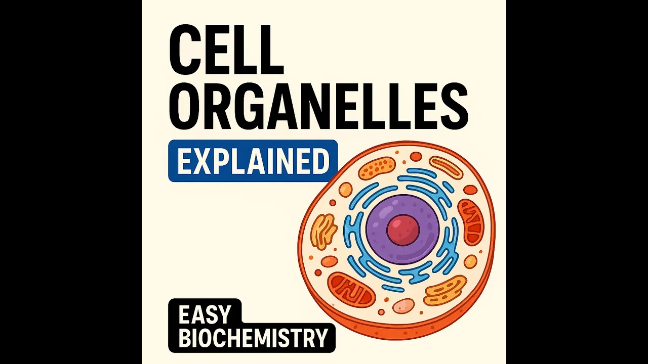
Cell Organelles Explained | Cell Structure & Functions | Easy Biochemistry
165
Mitochondria have outer and inner membranes with distinct protein synthesis: outer membrane proteins from nucleus, inner membrane proteins from mitochondria. The inner membrane contains the electron transport chain and cytochrome P450 system. The matrix contains enzymes for TCA cycle, heme synthesis, and urea cycle. Mitochondria produce ATP through oxidative phosphorylation, regulate apoptosis via cytochrome C release, maintain calcium homeostasis, generate and detoxify reactive oxygen species, and contain maternally inherited DNA with high mutation rates.
Click to watch →

03- Cell structure
322
Mitochondria are membrane-bound organelles with an outer membrane and inner membrane folds called cristae. The cristae contain proteins for the electron transport chain and ATP production. The matrix is the space within the mitochondria where ATP is generated through oxidative phosphorylation. Mitochondria produce reactive oxygen species as a byproduct, which can damage cells and contribute to aging. All cells except red blood cells and urethro sites contain mitochondria, with numbers ranging from hundreds to several thousand depending on the cell's energy needs. Cells with high ATP requirements, such as skeletal muscle and brain cells, contain more mitochondria.
Click to watch →

ANATOPATOLOGÍA - CELULA PARTE 16-03-26
280
Mitochondria have a double membrane with an outer membrane containing porins and an inner membrane forming cristae. They contain their own DNA and synthesize some proteins semi-autonomously. ATP is produced through oxidative phosphorylation using the electron transport chain, which creates a proton gradient driving ATP synthase. Mitochondria also produce reactive oxygen species as metabolic byproducts.
Click to watch →
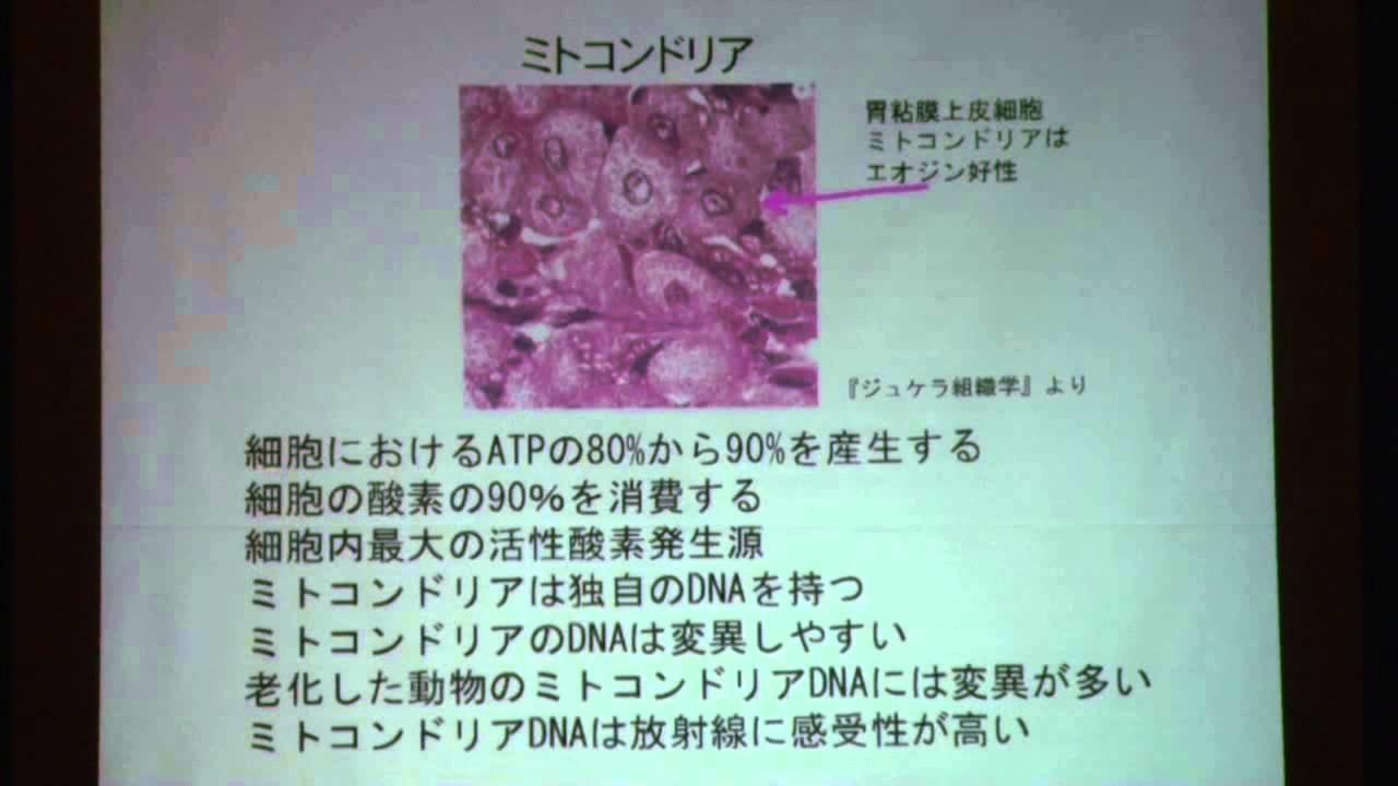
第三回市民科学者国際会議 : 崎山比早子
259
Mitochondria are double-membrane-bound organelles containing their own DNA. They are the primary site of ATP production, generating 80-90% of cellular energy. Mitochondria also produce reactive oxygen species (ROS) as a byproduct of energy metabolism.
Click to watch →
Deep Dive
The fundamental process of macroautophagy, specifically how cell components are sequestered into autophagosomes and degraded by lysosomes.
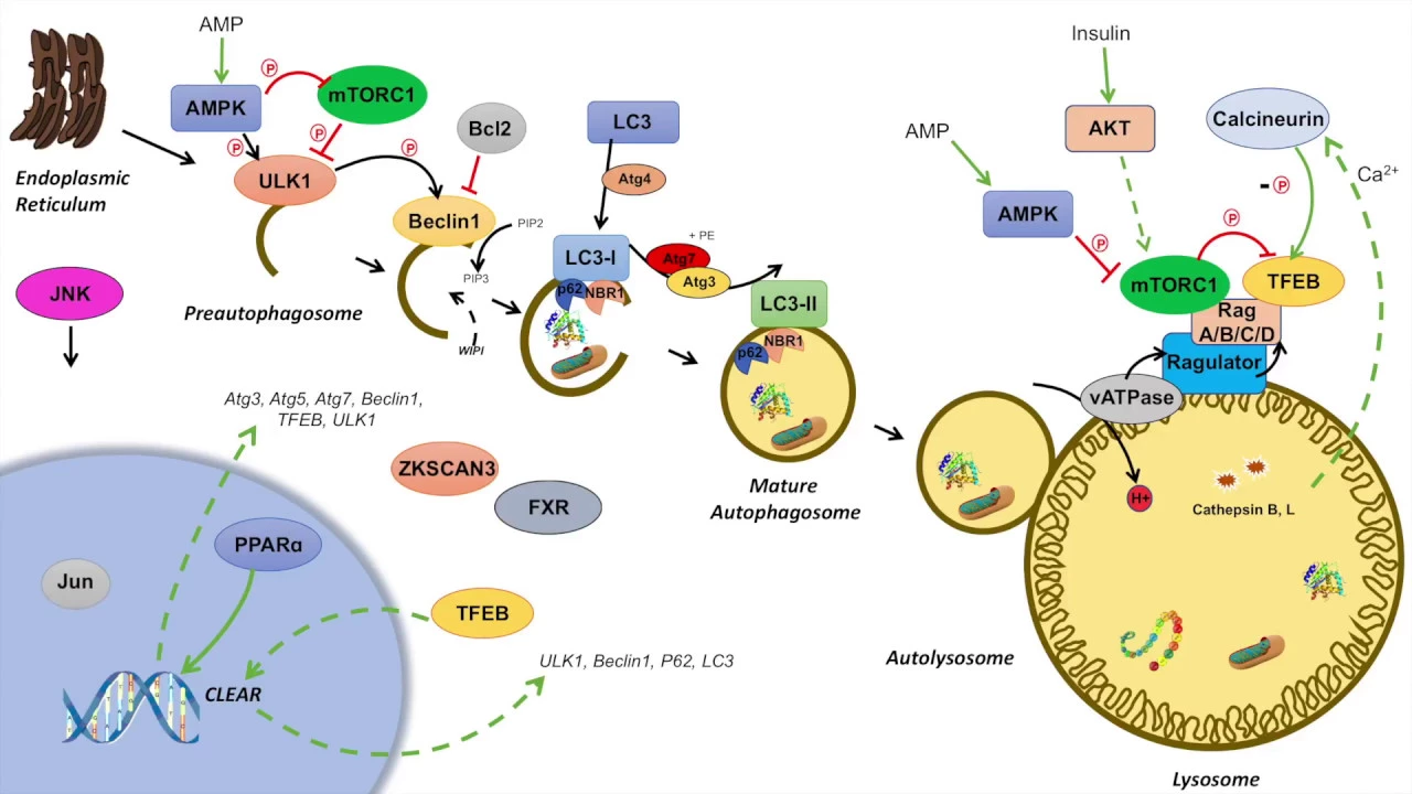
Macroautophagy | Regulation During Feeding, Fasting and Starvation
23.9K
Macroautophagy is a cellular bulk degradation process where cellular components are enclosed in autophagosomes and degraded within lysosomes. During fasting, this recycling generates ATP by rerouting substrates to mitochondria. The process involves autophagosome budding from the ER with Atg1, nucleation via Beclin 1 and PI3K complex forming PIP3, recruitment of p62 and NBR1 cargo receptors, and LC3 processing to LC3-II for autophagosome elongation. Fusion with lysosomes creates autolysosomes where cathepsins degrade cargo.
Click to watch →
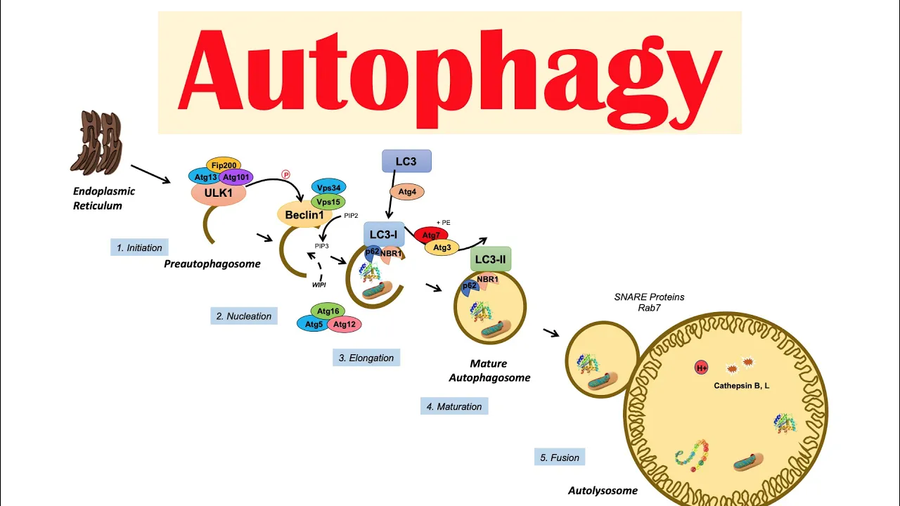
Autophagy | Macroautophagy & Importance in Health
210.6K
Macroautophagy proceeds through five stages: (1) Initiation - ULK complex (ATG1/ULK1, ATG13, ATG101) forms at ER membrane; (2) Nucleation - ULK activates Beclin-1, which with VPS34/VPS15 forms class III PI3K complex producing PIP3 to recruit WIPI proteins; (3) Elongation - P62/NBR1 recognize cargo; ATG5-ATG12-ATG16 targets substrates; pro-LC3 is cleaved by ATG4 to LC3-I, then conjugated to PE by ATG7 to form LC3-II; (4) Maturation - autophagosome completes; (5) Fusion - SNAREs/Rab7 mediate lysosome fusion forming autolysosomes where cathepsins degrade substrates, releasing recycled nutrients to cytoplasm.
Click to watch →

New Research Finds How to Increase Autophagy Without Fasting (6 different types)
196.8K
Macroautophagy is the primary large-scale autophagy process occurring in cells. When proteins aggregate or become dysfunctional, an autophagosome (a specialized vesicle) engulfs these damaged proteins and transports them to the lysosome. The autophagosome then fuses with the lysosome to form an autolysosome, where the damaged proteins are degraded and recycled. This process functions like a barge collecting garbage from the ocean and bringing it back to shore for processing, enabling cells to clear out damaged components while preserving functional cellular machinery.
Click to watch →

The mechanism of autophagy
41.8K
Macroautophagy is a conserved intracellular degradative process where cargo is sequestered in double-membrane autophagosomes. The process involves approximately 20 ATG proteins organized into six functional modules. Autophagosome biogenesis proceeds through five consecutive steps: initiation, expansion, maturation, tethering/fusion, and breakdown/recycling. Bulk autophagy involves random capture of heterogeneous cargo, while selective autophagy targets specific organelles or structures. The initiation step activates the ULK kinase complex, generating a scaffold for phagophore formation through vesicle fusion. The phosphatidylinositol 3-kinase complex synthesizes phosphatidyl inositol 3-phosphate to recruit expansion machinery. The expansion phase relies on ATG2-WP complex lipid transfer and ATG12/LC3 conjugation systems for membrane elongation and closure.
Click to watch →

Autophagy Explained (Fasting Autophagy)
392
Macroautophagy is a four-step cellular recycling process visible under electron microscopy. First, the autophagosome (cellular 'mouth') travels through the cytoplasm, sequestering damaged organelles including mitochondria, ribosomes, bacteria, and viruses. Second, the autophagosome closes to isolate these components from healthy cellular material. Third, the lysosome (cellular 'stomach') fuses with the autophagosome, forming an autolysosome. Fourth, damaged organelles are degraded and their components are recycled to support cellular rebuilding. This process operates through two main mechanisms: demand-driven autophagy triggered by nutrient deprivation that forces cells to consume themselves for energy, and selective autophagy where damaged organelles actively signal their own destruction through specific molecular markers. Different selective pathways target specific organelles—mitophagy for mitochondria, ribophagy for ribosomes, and virophagy for viruses—with damaged components always being consumed first during cellular stress.
Click to watch →
Deep Dive
The biochemical cascade of protein ubiquitination, including the distinct roles of E1, E2, and E3 ubiquitin ligases in tagging cellular proteins.

Ubiquitin and polyubiquitination
15.9K
The ubiquitination cascade requires three distinct enzymes working sequentially to attach ubiquitin to target proteins. E1 (ubiquitin-activating enzyme) activates free ubiquitin using ATP hydrolysis and transfers it to itself. E2 (ubiquitin-conjugating enzyme) receives activated ubiquitin from E1 and carries it to the E3 ligase. E3 (ubiquitin ligase) has two binding regions—one for E2 with ubiquitin and one for the target protein—and facilitates the final attachment of ubiquitin to the target protein's lysine residue via amide bond formation. E3 ligases exhibit remarkable diversity: conventional E3s directly transfer ubiquitin, while HECT domain E3s first transfer to a conserved cysteine before passing to targets. Many E3s contain ring finger domains (zinc-coordinated cysteine rings) that serve as E2-binding regions. The substrate-binding regions vary significantly among E3s, while the gap region connecting domains provides positional flexibility, enabling recognition of diverse target proteins.
Click to watch →

Ubiquitin
123
Ubiquitin is a small regulatory protein (76 amino acids) that acts as the 'fashion police' of the cell by tagging faulty or non-functional proteins with ubiquitin molecules; this tagging process involves three enzymes (E1, E2, E3) in a sequential cascade where E1 activates ubiquitin, E2 transfers it, and E3 ligates it to the target protein, and the proteasome recognizes the polyubiquitinated protein to degrade it into amino acids for recycling.
Click to watch →
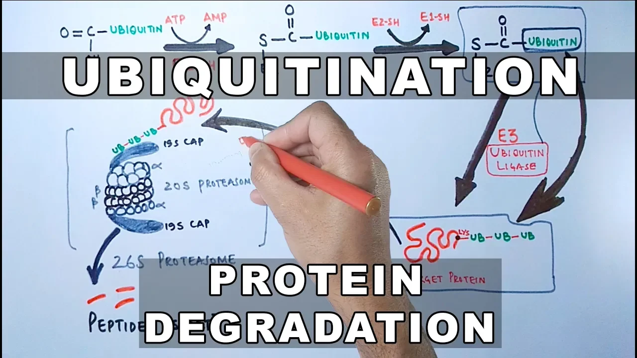
Ubiquitination of Proteins and Protein Degradation
215.4K
Ubiquitination is a post-translational modification where ubiquitin proteins are covalently attached to lysine residues of target proteins via iso peptide bonds, serving as a molecular tag that marks proteins for degradation; this process is catalyzed by a cascade of three enzymes—E1 (ubiquitin activating enzyme), E2 (ubiquitin conjugating enzyme), and E3 (ubiquitin ligase)—which sequentially transfer ubiquitin from E1 to E2 and then to the target protein, after which the ubiquitinated proteins are recognized and degraded by the 26S proteasome complex consisting of a 20S proteasome core and 19S regulatory caps.
Click to watch →

Correzione TEST di MEDICINA SEMESTRE FILTRO 2025
2.4K
L'ubiquitinazione è un processo di marcatura proteica per degradazione. E1 attiva l'ubiquitina tramite ATP, E2 trasferisce l'ubiquitina da E1 a se stesso, e E3 riconosce la proteina bersaglio e lega l'ubiquitina al residuo di lisina. Quando più ubiquitine sono legate, il proteasoma riconosce e degrada la proteina.
Click to watch →

The ubiquitin-proteasome mediated protein degradation pathway | Ubiquitin ligase | Proteasome
37.2K
The ubiquitination process requires three types of enzymes working in coordination: E1 (ubiquitin-activating enzyme) activates ubiquitin in an ATP-dependent manner; E2 (ubiquitin-conjugation enzyme) transfers ubiquitin residues to target proteins with the help of E3; E3 (ubiquitin ligase) coordinates with the target protein and facilitates the transfer of ubiquitin from E2 to lysine residues of the target protein, flagging them for destruction.
Click to watch →
Deep Dive
An introductory understanding of Parkinson's disease pathology, particularly the vulnerability of dopaminergic neurons to cellular stress.

New Clues to Parkinson's Disease From An Unlikely Source
7.7K
While protein trafficking problems affect all cells, dopaminergic neurons are particularly vulnerable because their function depends critically on precise protein delivery to synaptic ends for neurotransmitter release. When trafficking goes wrong, these neurons die first, explaining why Parkinson's specifically targets them. Compounds identified in yeast screens not only rescued neurons from alpha-synuclein toxicity but also protected against mitochondrial toxins that cause Parkinson's-like symptoms, suggesting they address fundamental disease mechanisms.
Click to watch →

The Alarming Rise in Parkinson’s Disease with Dr. Ray Dorsey | The Empowering Neurologist EP. 167
700.2K
Dopaminergic neurons in the substantia nigra are uniquely vulnerable to mitochondrial toxicants due to several factors: (1) The brain consumes 20% of body energy despite being only 3% of body weight; (2) These specific neurons have even higher energy demands because many are not insulated with myelin; (3) Individual axons can extend up to four meters in length, requiring substantial energy for signal transmission.
Click to watch →
Morbus Parkinson – das Rätsel der selektiven Neurodegeneration
1.1K
Parkinson's disease is characterized by the selective degeneration of specific dopaminergic neurons in the substantia nigra, while neighboring neurons with similar characteristics survive. Research reveals that vulnerable neurons respond to toxic stress (such as alpha-synuclein protein aggregates) by dramatically increasing their electrical activity—approximately doubling their firing rate—through a biophysical mechanism involving dysfunction of a 'braking' potassium channel. This compensatory response, occurring years or decades before cell death, may be a key factor in why certain neurons degenerate while others survive, offering potential therapeutic targets for neuroprotection.
Click to watch →

Earliest Signs of Parkinson's Disease with D. James Surmeier, PhD
1.6K
Mitochondrial dysfunction is central to Parkinson's onset, but its role as cause versus consequence was controversial. Dopaminergic neurons possess a unique 'CEO phenotype' controlling vast brain regions through massive axonal arbors, creating enormous metabolic demands. This basal stress, combined with age-related mitochondrial decline, pushes neurons over a bioenergetic threshold. Similar vulnerable neurons share this supervisory phenotype, explaining why multiple cell types degenerate in Parkinson's. Age is the primary risk factor, averaging 60 years at diagnosis, reflecting the decades-long time course required for disease manifestation despite rapid toxin models failing clinical translation.
Click to watch →

4 5 36 Parkinsons disease Pathophysiology non human models 1637
115
Parkinson's disease involves selective vulnerability of dopaminergic neurons due to their unique structural and metabolic demands. These neurons must maintain large amounts of axoplasm and presynaptic proteins while projecting to many brain regions, placing enormous burden on cell bodies maintained for decades. Dopamine's chemical reactivity contributes to vulnerability through reactive oxygen species formation. Environmental factors including pesticide exposure, the 1918 influenza pandemic, and synthetic heroin impurities have been linked to increased Parkinson's incidence. Alpha-synuclein, the best-known defective protein, is intrinsically disordered but forms fibrils and Lewy bodies when mutated, potentially transmitting like prions between cells.
Click to watch →
Prerequisite Knowledge
- Concept 01The structure and physiological function of mitochondria, including ATP generation, reactive oxygen species (ROS) production, and membrane potential maintenance.
- Concept 02The fundamental process of macroautophagy, specifically how cell components are sequestered into autophagosomes and degraded by lysosomes.
- Concept 03The biochemical cascade of protein ubiquitination, including the distinct roles of E1, E2, and E3 ubiquitin ligases in tagging cellular proteins.
- Concept 04An introductory understanding of Parkinson's disease pathology, particularly the vulnerability of dopaminergic neurons to cellular stress.
Subsequent Learning
- Step 01The development of small-molecule therapeutics targeting Deubiquitinating Enzymes (DUBs), specifically USP30 inhibitors, for neuroprotection.
- Step 02PINK1/Parkin-independent mitophagy pathways, such as receptor-mediated mitophagy involving BNIP3, NIX, or FUNDC1.
- Step 03The intersection of mitochondrial dynamics (fission and fusion processes) with the spatial regulation of mitophagy.
- Step 04The clinical trial landscape and translational hurdles for drug candidates aimed at restoring mitochondrial quality control in age-related diseases.
Mitophagy
Therapeutic
Mitophagy
0:00Playing Section
- 1
Explains mitochondrial damage and the need for removal via mitophagy.
- 2
Details PINK1 and Parkin signaling that tag damaged mitochondria for degradation.
- 3
Covers autophagosome formation and lysosomal clearance to restore energy.
The In Vivo Paradox of PINK1/Parkin-Independent Basal Mitophagy
While the PINK1/Parkin pathway is a primary focus of Parkinson's disease research and drug development (such as USP30 inhibition), emerging in vivo evidence challenges its dominance in everyday mitochondrial maintenance. Studies utilizing sensitive mitochondrial reporter mice have revealed that basal mitophagy occurs at normal levels across various tissues—including the brain—even in the complete absence of PINK1 or Parkin. This suggests that the PINK1/Parkin pathway functions primarily as an acute stress-response mechanism rather than the primary driver of constitutive mitochondrial clearance under normal physiological conditions. Consequently, critics argue that focusing therapeutic strategies exclusively on Parkin activation or USP30 inhibition may overlook more physiologically dominant, Parkin-independent pathways (such as receptor-mediated mitophagy via BNIP3L/Nix or FUNDC1). Broadening the therapeutic focus to these alternative pathways may be essential for successfully treating neurodegenerative diseases.
Deep Dive
The development of small-molecule therapeutics targeting Deubiquitinating Enzymes (DUBs), specifically USP30 inhibitors, for neuroprotection.

Targeting Deubiquitinases for Therapeutic Benefit – 20 September 2022
1.1K
Parallel high-throughput screening of multiple deubiquitinases (DUBs) against the same compound library enables identification of selective DUB inhibitors by leveraging differences in compound binding across the DUB family, as demonstrated by the successful identification of selective USP28 and USP30 inhibitors through this approach.
Click to watch →

A hit-finding platform for the discovery of novel inhibitors of DeUBiquitinating enzymes (DUBs)
418
Almac Discovery has developed the Ub Plex platform for discovering novel inhibitors of deubiquitinating enzymes (DUBs), which play critical roles in cancer and neurodegenerative diseases; this platform utilizes the PHERAstar FSX instrument for high-throughput biochemical screening, achieving significant improvements in speed and precision through stacker technology that enables loading of up to 50 plates with reduced reach times to just 2 minutes, supporting the identification and validation of small molecule inhibitors for therapeutic targets such as USP7 and USP19.
Click to watch →
Progress in PD & Movement Disorders Panel @ Sachs_NIF
465
Vincere Biosciences is developing small molecules targeting USP30, an enzyme at the intersection of mitochondria and lysosomes. By enhancing autophagy thresholds, these molecules aim to slow or stop Parkinson's progression. This represents targeting the mitochondrial-lysosomal axis, a key pathway implicated in Parkinson's pathogenesis.
Click to watch →
Slower is Better: Progress Towards Neuroprotection in PD by David K. Simon, MD, PhD
318
Cutting-edge research explores novel mechanisms: USP30 inhibition targets cellular energy metabolism disrupted in PARKIN/PINK1-related Parkinson's, showing dramatic neuroprotection in mouse models. Combined with improved understanding of disease mechanisms, better biomarkers, and precision medicine approaches, these advances suggest we are approaching meaningful breakthroughs in Parkinson's neuroprotection despite past failures.
Click to watch →

Sara Buhrlage - Dana-Farber Targeted Degradation Webinar Series
1.7K
A library-versus-library screening approach using combinatorially assembled covalent active site inhibitors enables systematic identification of selective DUB inhibitors across the entire enzyme family, revealing that selectivity is driven by specific combinations of electrophile, linker, and non-covalent binding module interactions rather than single features, thereby accelerating the pace of DUB probe development and expanding the number of liganded DUBs from a handful to dozens.
Click to watch →
Deep Dive
PINK1/Parkin-independent mitophagy pathways, such as receptor-mediated mitophagy involving BNIP3, NIX, or FUNDC1.
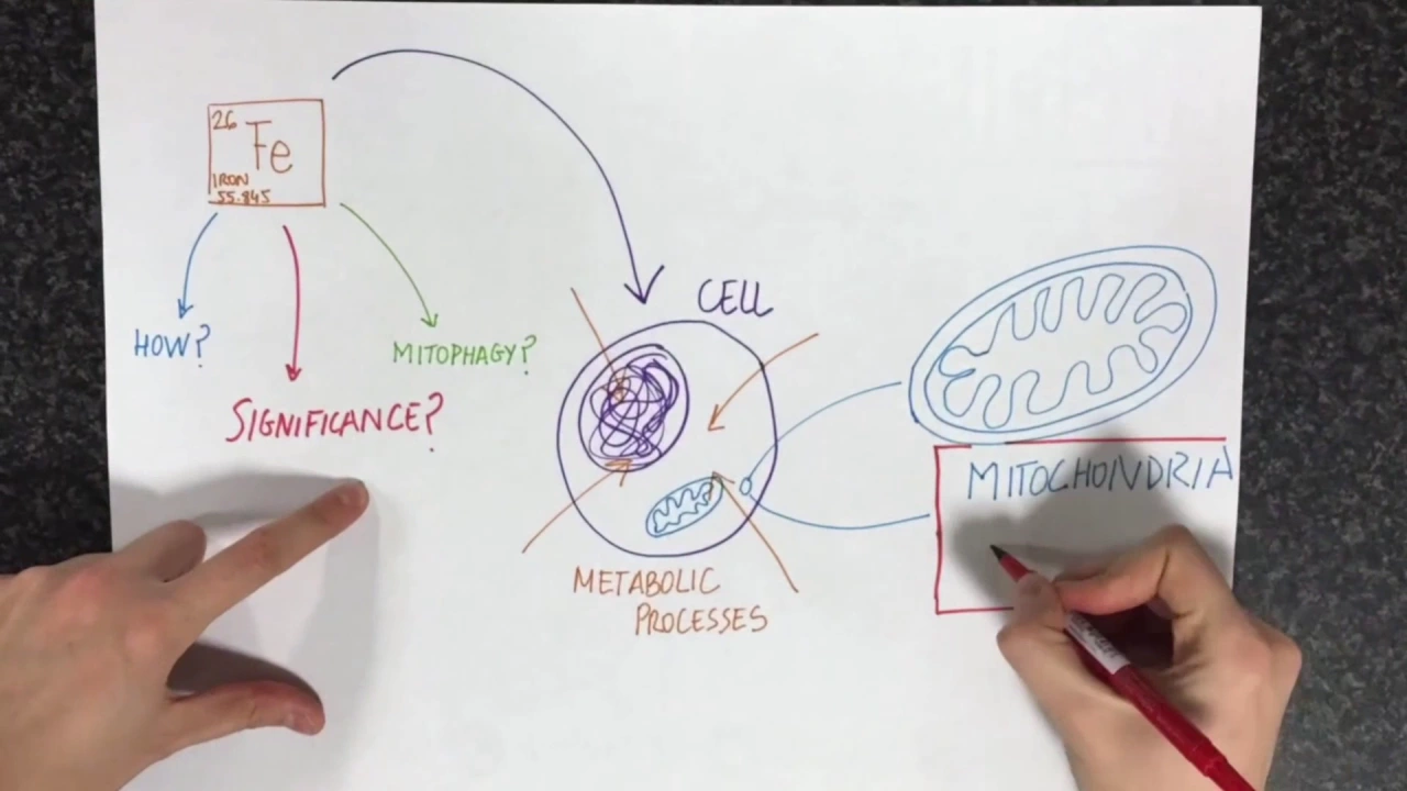
Loss of iron triggers PINK1/Parkin-independent mitophagy
2.1K
Iron deficiency triggers a PINK1/Parkin-independent mitophagy pathway where reduced iron impairs mitochondrial iron-sulfur cluster and heme group formation, causing hypoxia that activates HIF-1α to induce mitochondrial destruction, providing an alternative quality control mechanism for removing damaged mitochondria beyond the classical PINK1/Parkin pathway.
Click to watch →

Targeting Mitochondria for Selective Autophagy
138
This video explains how different types of mitophagy (mitochondrial degradation) require distinct molecular machinery: PINK1/Parkin-dependent mitophagy relies on NIPSNAP1 and NIPSNAP2 proteins that act as 'eat signals' by translocating from the mitochondrial matrix to the surface upon depolarization to recruit autophagy receptors, while PINK1-independent mitophagy (induced by hypoxia or DFP) requires the kinases GCK and PKCδ to recruit the ULK complex to mitochondria; these findings reveal that distinct mitophagy pathways are regulated by different molecular mechanisms and that defects in these processes contribute to neurodegeneration and cancer.
Click to watch →
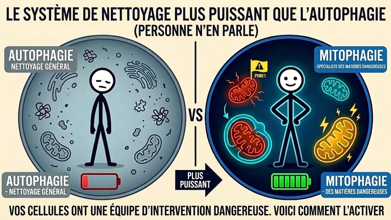
Le processus plus puissant que l'autophagie dont personne ne parle
696
Mitophagy is a targeted cellular process that specifically eliminates damaged mitochondria, unlike general autophagy which recycles all cellular waste. Damaged mitochondria release toxic byproducts that corrupt surrounding cells and drive aging-related diseases including Alzheimer's, Parkinson's, heart failure, and type 2 diabetes. The molecular mechanism involves Pink1 detecting damaged mitochondria through membrane potential loss, Parkin tagging them with ubiquitin, and autophagosomes enveloping them for lysosomal degradation. Alternative pathways involving BNIP3, NIX, and FUNDC1 provide redundancy. Mitophagy dysfunction is a central mechanism in multiple age-related diseases: in Parkinson's, Pink1/Parkin mutations cause hereditary forms where damaged mitochondria accumulate in dopaminergic neurons; in Alzheimer's, impaired mitophagy creates a self-reinforcing cycle with amyloid-beta accumulation; in heart failure, the heart's high mitochondrial density makes it particularly vulnerable; in type 2 diabetes, damaged mitochondria interfere with insulin signaling; in NAFLD, impaired mitophagy prevents proper fatty acid oxidation; in cellular senescence, dysfunctional mitochondria maintain the senescent state through oxidative stress. In cancer, mitophagy has a dual role - suppressing tumor development in normal cells but being hijacked by cancer cells for survival.
Click to watch →

Dinâmica Mitocondrial Glial no Envelhecimento e em Doenças Neurodegenerativas
289
Mitochondrial fusion involves mitofusins (Mfn1, Mfn2) and OPA1, while fission involves MID51/MID49 receptors recruiting DRP1. In senescent astrocytes, both fusion and fission pathways were upregulated, with DRP1 showing approximately 10-fold increase. Mitophagy degrades damaged mitochondria through PINK1-Parkin pathway and receptor-dependent pathways (NIX, FUNDC1, BNIP3). In senescent astrocytes, mitophagy machinery was upregulated but p62 accumulation indicated paralyzed autophagic flux. Despite increased respiratory capacity, senescent astrocytes showed increased susceptibility to rotenone-induced mitochondrial stress. In aging, mTOR pathway activation leads to inflammation and autophagy inhibition. Rapamycin inhibits mTOR, reversing senescence phenotype and activating autophagy. In senescent astrocytes, rapamycin treatment normalized cell cycle proteins, reduced beta-galactosidase activity, restored nuclear lamina, and reduced accumulated lysosomes and mitochondrial density. Critically, rapamycin treatment reversed the increased susceptibility to rotenone-induced cell death, demonstrating that rapamycin-mediated autophagy activation can remove damaged mitochondria and restore mitochondrial stress resistance.
Click to watch →
Lab Med and Pathology Research & Discovery Seminar | Alicia Pickrell
342
Under normal conditions, PINK1 is constitutively imported into mitochondria and degraded by the ubiquitin-proteasome system. When mitochondria become damaged (loss of membrane potential, increased ROS, or DNA mutations), PINK1 accumulates on the outer mitochondrial membrane because import is blocked. As a kinase, PINK1 phosphorylates ubiquitin chains on mitochondrial proteins, recruiting cytosolic Parkin (an E3 ubiquitin ligase) to the damaged mitochondria. This ubiquitination triggers autophagosome formation around damaged mitochondria, directing them to lysosomes for degradation. This pathway maintains mitochondrial quality by removing dysfunctional organelles.
Click to watch →
Deep Dive
The intersection of mitochondrial dynamics (fission and fusion processes) with the spatial regulation of mitophagy.

This Increases Autophagy 3x MORE THAN FASTING | Dr. Tommy Wood
327.1K
Mitophagy (mitochondrial autophagy) is a subset of general autophagy that occurs simultaneously when autophagy is activated. Exercise stimulates both mitochondrial biogenesis (creation of new mitochondria) and mitophagy (removal of damaged ones) through fusion and fission processes. Mitochondria exist as a dynamic, interconnected network rather than static organelles, continuously remodeling and communicating with other cellular components.
Click to watch →
SLU-PP-332 & BAM15 Biohacking Mitochondria for Peak Performance
13.6K
Mitochondrial fission and fusion are dynamic processes essential for maintaining mitochondrial quality control. Fission acts as an early sensor of cellular stress (low calories, oxidative stress, toxins), causing mitochondria to split apart and segregate damaged components for removal through mitophagy. Fusion allows mitochondria to join together, sharing components so partially damaged mitochondria can be rescued and become healthier. This balance between division and merging ensures proper mitochondrial health, supporting energy production through ATP synthesis, cellular repair, metabolic function, and longevity. Disruption in these processes links to aging, neurodegenerative diseases, metabolic disorders, and cardiovascular issues.
Click to watch →

How We Make New Mitochondria (Biogenesis Explained)
60.2K
Mitochondria maintain quality through dynamic processes of fission (splitting) and fusion (merging). Damaged mitochondrial sections are segregated and moved to isolated areas, then released during fission to undergo mitophagy (autophagy), where they are broken down and recycled. Fasting enhances this cleanup process by highlighting weaker mitochondrial parts. Additionally, mitochondria can fuse to combine beneficial traits - one mitochondrion's strength in energy production can merge with another's efficiency in waste removal, creating more effective organelles through shared resource optimization.
Click to watch →

Pourquoi ton corps n’a plus d’énergie (et ce n’est pas l’âge)
1.7K
Mitochondria undergo three critical dynamic processes: mitophagy (recycling damaged mitochondria), fusion (merging to share resources and dilute damage), and fission (dividing to isolate damaged parts). Modern lifestyles with chronic stress and fatigue cause excessive fission without adequate fusion or mitophagy, leading to fragmented mitochondrial networks, reduced energy, and increased inflammation. These processes work together to maintain cellular health and determine overall metabolic function.
Click to watch →

Mitochondrial Cell Biology 2
9.2K
Mitochondria are highly dynamic organelles that continuously change size, shape, and location within cells, ranging from fused spaghetti-like networks to fragmented granules. This dynamic behavior enables mitochondrial function as a single interconnected network in many cells, allowing exchange of matrix components, membrane components, and mitochondrial DNA between individual mitochondria. Mitochondrial fusion (mediated by mitofusins and OPA1) and fission (mediated by DRP1) are essential processes conserved across all eukaryotes. Fusion requires healthy membrane potential and occurs at tip-to-tip contacts guided by the cytoskeleton, while fission sites are marked by the endoplasmic reticulum and require actin/myosin and GTP hydrolysis. These dynamics enable quality control by eliminating defective organelles and maintaining a healthier mitochondrial population.
Click to watch →
Deep Dive
The clinical trial landscape and translational hurdles for drug candidates aimed at restoring mitochondrial quality control in age-related diseases.

Lab Med and Pathology Research & Discovery Seminar | David Marcinek, PhD
1.8K
Mitochondria serve as a central hub connected to multiple hallmarks of aging, positioned at the center or connected to all other aging-related pathways. By understanding mitochondrial changes with age and intervening on mitochondria, researchers can potentially affect many other aspects of aging biology. Sarcopenia, the loss of muscle mass and function with age, represents one of the most identifiable aging phenotypes, contributing to increased fall risk, exercise intolerance, frailty, and poor quality of life. Muscle ages regardless of physical activity level. Elamipretide, a tetrapeptide compound that associates with mitochondria by interacting with cardiolipin, rapidly reverses age-related mitochondrial dysfunction in mice, restoring P to O ratio and maximum ATP production to young levels within one hour. This challenges traditional views requiring protein synthesis for mitochondrial interventions. Treatment improves skeletal muscle function beyond maximum force production, enhancing submaximal force and fatigue resistance. Systemic benefits extend to cardiac function (reduced diastolic dysfunction) and kidney function (reduced pathology). A phase II clinical trial confirmed these findings in older adults with low mitochondrial function, demonstrating successful translation from bench to bedside.
Click to watch →

SENS Research Foundation MitoMouse Q&A Webinar
1.6K
Aging's non-disease classification creates regulatory barriers, though recent developments like the metformin trial have established clinical endpoints for aging-related outcomes. The MitoSENS approach addresses these challenges by targeting mitochondrial diseases first—conditions clearly classified as diseases—before applying the same technology to age-related decline. This phased strategy enables clinical translation while working toward broader anti-aging applications. Beyond nuclear gene therapy, synthetic biology offers potential for designing optimized mitochondrial genomes from scratch, potentially removing faults and adding beneficial features. Selective elimination of mutant mitochondria through small molecule modulators targeting specific pathways could complement gene therapy by actively removing damaged organelles.
Click to watch →
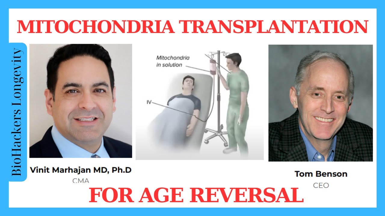
Mitochondria Transplantation Therapy for Age Reversal/ Vinit Mahajan & Tom Benson
1.3K
Mitochondrial restoration aims to replenish the 30-40% of mitochondrial DNA lost by age 80, restoring the ratio of functional to dysfunctional mitochondria. Animal studies have achieved delivering 0.25-0.5% of body mitochondria per treatment, with goals of 2-10% boost. Effects observed include muscle growth, improved strength, enhanced cognitive function, and dramatically improved immune systems. Clinical trials will begin with simple skin injections to assess safety, then progress to systemic injections. Mitochondrial transplantation has already been used in humans for rare diseases, providing existing safety and technology precedent. The therapy can be applied to radiation damage recovery and burn healing, demonstrating that healing and anti-aging effects are connected. Regulatory pathways vary by country - Japan offers a sophisticated pathway where patients can self-report results on websites for efficacy assessment, enabling faster progression. The plan is to begin trials at year-end, with initial regulatory paperwork taking about a year, dependent on funding availability.
Click to watch →

Membrane Lipid Replacement to Improve Mitochondrial Dysfunction with John Casey
669
Human studies confirmed MLR's effects in aging populations. A crossover trial with 70-year-olds vs. 29-year-old controls showed 70-year-olds recovered mitochondrial membrane potential equal to or better than young controls within 8-12 weeks. The rhodamine 123 dye test measured mitochondrial function directly. This demonstrates MLR can restore mitochondrial function in aging populations, allowing them to achieve physical output comparable to younger individuals. The slow washout effect means benefits persist even with inconsistent supplementation.
Click to watch →

Как лечить старение? Всё дело в митохондриях
4.6K
Mitochondria are cellular energy stations producing ATP for all biological processes. As organisms age and develop diseases, these stations malfunction, causing energy deficiency. Mitochondrial dysfunction underlies many diseases and the aging process itself. Over time, mitochondria accumulate damage, produce less energy, and generate more toxic reactive oxygen species, leading to neurodegenerative, cardiovascular, and metabolic disorders. Cells have evolved four natural quality control mechanisms: self-renewal through their own DNA, intercellular transfer via vesicles, fusion to dilute damage, and mitophagy to remove damaged mitochondria. Two critical proteins regulate mitophagy: USP30 (negative regulator) and Parkin (positive regulator). When Parkin function is impaired, as in some Parkinson's disease forms, cells lose their ability to clean up damaged mitochondria. In October 2025, the FDA approved Elamipretide, the first drug specifically targeting mitochondria, treating Barth Syndrome caused by TAZ gene mutations disrupting cardiolipin production.
Click to watch →
Mitophagy
Therapeutic
Mitophagy
0:00- 1
Explains mitochondrial damage and the need for removal via mitophagy.
- 2
Details PINK1 and Parkin signaling that tag damaged mitochondria for degradation.
- 3
Covers autophagosome formation and lysosomal clearance to restore energy.
The In Vivo Paradox of PINK1/Parkin-Independent Basal Mitophagy
While the PINK1/Parkin pathway is a primary focus of Parkinson's disease research and drug development (such as USP30 inhibition), emerging in vivo evidence challenges its dominance in everyday mitochondrial maintenance. Studies utilizing sensitive mitochondrial reporter mice have revealed that basal mitophagy occurs at normal levels across various tissues—including the brain—even in the complete absence of PINK1 or Parkin. This suggests that the PINK1/Parkin pathway functions primarily as an acute stress-response mechanism rather than the primary driver of constitutive mitochondrial clearance under normal physiological conditions. Consequently, critics argue that focusing therapeutic strategies exclusively on Parkin activation or USP30 inhibition may overlook more physiologically dominant, Parkin-independent pathways (such as receptor-mediated mitophagy via BNIP3L/Nix or FUNDC1). Broadening the therapeutic focus to these alternative pathways may be essential for successfully treating neurodegenerative diseases.
The structure and physiological function of mitochondria, including ATP generation, reactive oxygen species (ROS) production, and membrane potential maintenance.
The fundamental process of macroautophagy, specifically how cell components are sequestered into autophagosomes and degraded by lysosomes.
The biochemical cascade of protein ubiquitination, including the distinct roles of E1, E2, and E3 ubiquitin ligases in tagging cellular proteins.
An introductory understanding of Parkinson's disease pathology, particularly the vulnerability of dopaminergic neurons to cellular stress.
The development of small-molecule therapeutics targeting Deubiquitinating Enzymes (DUBs), specifically USP30 inhibitors, for neuroprotection.
PINK1/Parkin-independent mitophagy pathways, such as receptor-mediated mitophagy involving BNIP3, NIX, or FUNDC1.
The intersection of mitochondrial dynamics (fission and fusion processes) with the spatial regulation of mitophagy.
The clinical trial landscape and translational hurdles for drug candidates aimed at restoring mitochondrial quality control in age-related diseases.
55.3K views1.1Klikes3:30@vincerebioOriginal Release: 2020-07-15
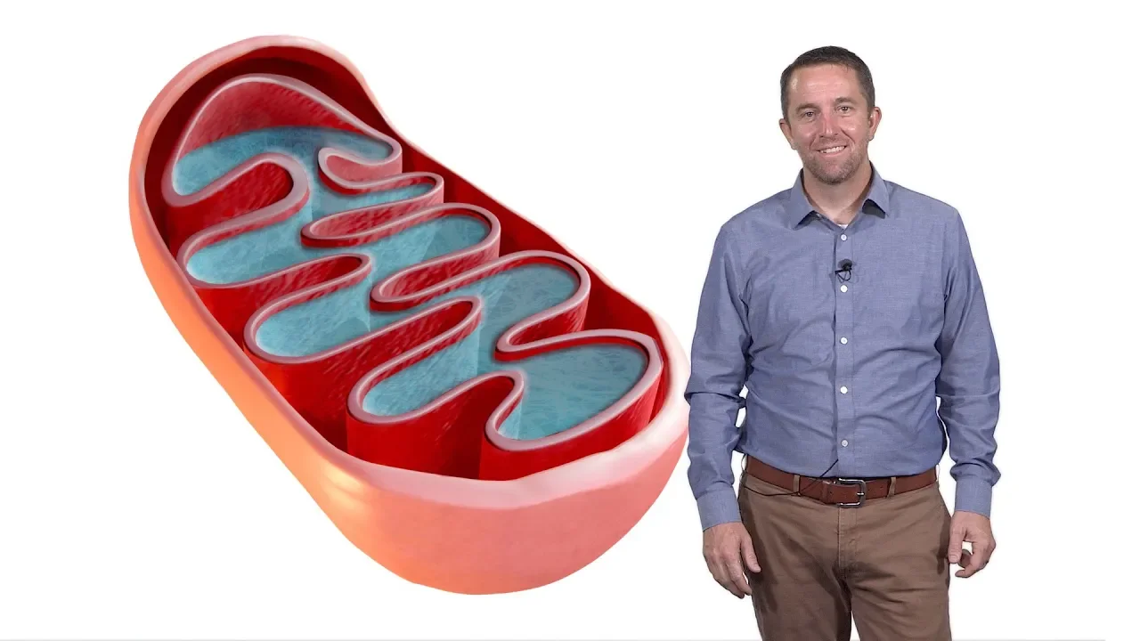
[Music] to understand vin series efforts on creating therapeutic molecules for Parkinson's disease let's talk about mitochondria and the natural removal of damaged mitochondria or my toffee G in order for the body to function our cells require energy which is supplied by mitochondria in the form of ATP like all things in nature mitochondria become damaged frequently causing them to produce less ATP and more harmful reactive oxygen species to fix this damage mitochondrion need to be broken down through my toffee G and replaced with healthy ones through mitochondrial biogenesis so here is how parkin mediated my top g happens an enzyme called pink one is recruited to the mitochondrial membrane when the mitochondria is healthy it removes pink one however when the mitochondria is damaged or depolarized this pink one enzyme accumulates on the outer membrane and starts to phosphorylate outer mitochondrial membrane proteins this in turn leads to the recruitment of another enzyme called Parkin to the outer mitochondrial membrane Park and poly ubiquitinated were adds a chain of ubiquitin molecules to the outer membrane proteins the ubiquitin chains act as a signal for the cell to send its autophagy machinery when in Auto phagosome forms around the whole mitochondria it sequester's it from the rest of the cell so that the reactive oxygen species can no longer call its damage it then transports the mitochondria and fuses with a lysosome where the acidic environment and hydrolysis break down the damaged mitochondria this breakdown then signals to the cell to create new mitochondria in a process called mitochondrial biogenesis the yn2 Parkins yang is another enzyme called USB 30 which serves to D ubiquitinated or cut these ubiquitin chains as parking and other ligase as ad ubiquitin chains on USB 30 keeps taking them off thus slowing down the process of maitake by localizing at the tom complex near pink one USB 30 can also take off ubiquitin even before pink one has a chance to phosphorylate them and initiate my toffee that way USB 30 increases the threshold that pink one requires to induce my toffee G and emerges as a key enzyme able to modulate this process an average mitochondrion in the brain lives for about 30 to 40 days inside the cell before it is cleared and replaced with other healthy mitochondria my toffee G signals for making of new mitochondria restores efficient energy production and helps return the cell to its normal state of homeostasis in brains of patients with Parkinson's disease there's an abnormally high percentage of damaged mitochondria so increasing my toffee G to more quickly clear these damaged mitochondria will in turn increase the number of healthy mitochondria and counteract this key deficit at VIN sorry we are working to make small molecules that can increase my toffee G either by inhibiting us P 30 or activating Parkin with the goal to remove damaged mitochondria and return the cells and the whole system back to health [Music] you
Up Next
30:59
Mitochondria: Cellular Parasite Origins, Metabolism, and Signaling
@scicommlab
191.4K views•2020-01-07

50:12
Bacterial Communication: Quorum Sensing and Biofilm Formation Explained
@JHUAAP
12.6K views•2011-06-28

5:35
Enteric Nervous System Explained: The Gut's Brain | Neurobiology Lecture
@alumniu6029
438 views•2018-09-12

7:08
Bacteriophages: Earth's Deadliest Killers and Future Antibiotics
@kurzgesagt
34.6M views•2018-05-13
Related Study Plans & Knowledge Roadmaps
Structured learning paths in Biology
biology
RNAi: Biogenesis, RISC & Gene Silencing
01Molecular Biology Foundations: DNA to Protein
02Introduction to RNA Interference (RNAi)
03Molecular Biogenesis of miRNA and siRNA
+ 2 more learning modules
10h5 modules
Explore Roadmap
biology
Mycorrhizae: Signaling, Biophysics & Ecology
01Foundations of Mycorrhizal Symbiosis
02Molecular Signaling and Root Colonization
03Biophysics and Bioenergetics of Nutrient Exchange
+ 1 more learning modules
11h4 modules
Explore Roadmap
biology
Circadian Clocks: TTFL, SCN & Periphery
01Foundations of Molecular Biology & Gene Expression
02Introduction to Chronobiology and Actograms
03The Core TTFL: Molecular Architecture of the Clock
+ 3 more learning modules
14h6 modules
Explore Roadmap
biology
Population Genetics: Math, Drift & Selection
01Genetic & Probability Foundations
02The Hardy-Weinberg Principle and Baseline Models
03Mathematical Modeling of Natural Selection
+ 3 more learning modules
18h6 modules
Explore Roadmap