The vagus nerve, the longest cranial nerve connecting the brain to organs like the heart, lungs, stomach, liver, pancreas, kidneys, and intestines, serves as a communication superhighway that activates the parasympathetic nervous system to calm the body, reduce inflammation, aid digestion, and promote sleep. Clinical research demonstrates that vagus nerve stimulation through devices like Pulsetto can significantly reduce stress (39% at 2 weeks, 56% at 4 weeks), decrease anxiety (31% at 2 weeks, 45% at 4 weeks), improve sleep quality, and lower cortisol levels (42% in men, 37% in women) within 4 weeks of daily use.
Vagus Nerve Stimulation: Science-Based Healing for Stress Relief
Added:Deep Dive
The anatomy and function of the Autonomic Nervous System (ANS), specifically the contrast between the sympathetic ('fight-or-flight') and parasympathetic ('rest-and-digest') branches.

How to Recondition Your Mind and Body | Dr. Joe Dispenza’s Powerful Advice in 34 Minutes
175.6K
The autonomic nervous system functions behind the scenes of awareness and regulates body temperature, hormones, blood supply, and blood sugar levels without conscious mind involvement. The sympathetic nervous system is for protection from threats and dangers in the outer environment - think of it as the gas pedal. When there's danger, this system switches on automatically, mobilizing all the body's energy and resources. Physiological functions include: pupils dilate, salivary juices shut off, respiratory rate increases, heart rate increases, blood is sent to extremities, glucose is mobilized, and the immune system dials up and down. This system causes us to run from danger, fight, stay, or freeze and hide. The parasympathetic nervous system is the other branch - think of it as the brake or clutch. This is where the body conserves energy, utilizes nutrients, transports chemicals to cells, and metabolism goes up. This is a time for growth, repair, and restoration. There is a delicate balance between the two systems. When you're feeling constant fear, anger, or suffering and living by the hormones of stress, what was once adaptive becomes maladaptive because stress knocks your brain and body out of balance.
Click to watch →

Nervous system anatomy introduction
133.6K
The autonomic nervous system divides into sympathetic (thoracolumbar origin) and parasympathetic (craniosacral origin) divisions. Sympathetic neurons project from spinal cord to ganglia to targets, enabling rapid 'fight or flight' responses. Parasympathetic neurons project from brain (vagus nerve) and sacral cord to targets, mediating 'rest and digest' functions. Both divisions often work antagonistically—for example, opposing effects on heart rate. This dual control maintains homeostasis through coordinated unconscious regulation of internal organs.
Click to watch →
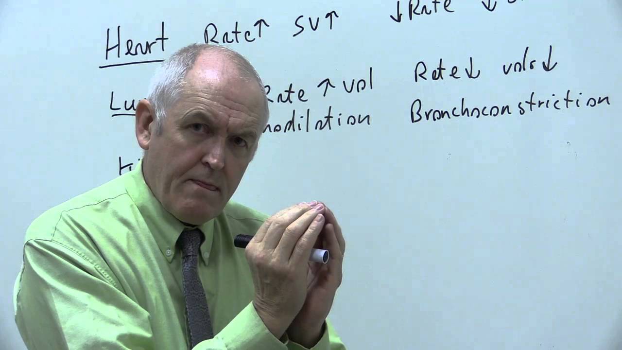
Sympathetic and parasympathetic nervous system
145.1K
The autonomic nervous system, which controls automatic bodily functions, consists of two opposing branches: the sympathetic nervous system (thoracolumbar, T1-L3) prepares the body for 'fight or flight' responses by increasing heart rate, respiratory rate, and bronchodilation while reducing urine production and GI activity, and the parasympathetic nervous system (craniosacral, cranial nerves III, VII, IX, X and sacral nerves S2-S4) promotes 'rest and digest' functions by decreasing heart rate, increasing GI motility and secretions, and promoting glycogen storage and fat deposition.
Click to watch →
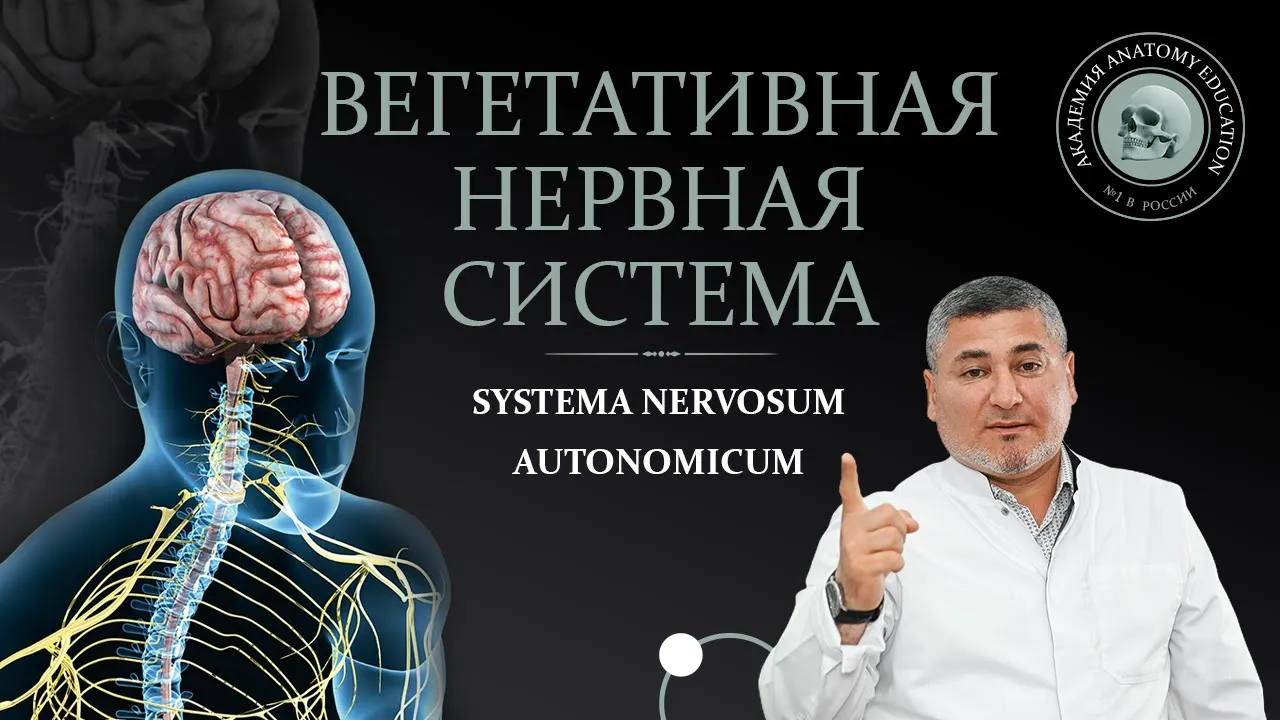
Вегетативная нервная система. Симпатическая и парасимпатическая части
530.5K
The autonomic nervous system (ANS) is the involuntary part of the nervous system that controls internal organs, glands, and smooth muscles without conscious control, divided into sympathetic (fight-or-flight response, originating from T1-L2 spinal segments) and parasympathetic (rest-and-digest response, originating from brainstem nuclei and S2-S4 spinal segments) divisions, which work antagonistically to regulate organ functions like heart rate, digestion, and glandular secretion through a two-neuron reflex arc involving preganglionic and postganglionic fibers.
Click to watch →

الجهاز العصبي الطرفي - آخر محاضرة أحياء 2 ثانوي - تم بحمد الله
163.3K
The autonomic nervous system (ANS) controls involuntary body functions and is divided into sympathetic and parasympathetic divisions. The sympathetic nervous system (SNS) prepares the body for 'fight or flight' responses: dilates pupils, increases heart rate and blood pressure, widens bronchioles, inhibits digestion, stimulates glycogen breakdown, and releases adrenaline from the adrenal medulla. The parasympathetic nervous system (PNS) promotes 'rest and digest' functions: constricts pupils, decreases heart rate, stimulates digestion and salivation, promotes glycogen synthesis, and stimulates bile release.
Click to watch →
Deep Dive
An introduction to Cranial Nerve X (the Vagus Nerve), including its pathways from the brainstem to major visceral organs.
Neurology | Vagus Nerve: Cranial Nerve X
322.8K
The vagus nerve (cranial nerve X) is the most extensive cranial nerve, serving as a mixed nerve that carries general somatic afferent (GSA) fibers for touch/pain/temperature sensation from the ear and neck, special visceral afferent (SVA) fibers for taste from the epiglottis, special visceral efferent (SVE) fibers for motor control of pharyngeal and laryngeal muscles (including the pharyngeal constrictors, levator veli palatini, palatoglossus, palatopharyngeus, and intrinsic laryngeal muscles), general visceral afferent (GVA) fibers for visceral sensation from thoracic and abdominal viscera, and general visceral efferent (GVE) fibers for parasympathetic innervation of the heart, lungs, esophagus, stomach, intestines, liver, pancreas, and gallbladder; it originates from the medulla oblongata, exits through the jugular foramen, and forms important plexuses including the pharyngeal plexus, pulmonary plexus, cardiac plexus, esophageal plexus, celiac plexus, and hepatic plexus.
Click to watch →

Блуждающий нерв X пара черепно мозговые нервы
61.8K
The vagus nerve (X cranial nerve) originates from three nuclei in the medulla oblongata (nucleus tractus solitarius, nucleus ambiguus, and nucleus dorsalis nervi vagi), exits through the jugular foramen, and innervates multiple body regions: the cervical portion supplies the pharynx, larynx, and upper esophagus; the thoracic portion provides parasympathetic innervation to the heart, trachea, bronchi, and esophagus; and the abdominal portion extends to the mid-descending colon, innervating the stomach, spleen, kidneys, and small intestine, while not innervating pelvic organs which receive parasympathetic supply from sacral spinal segments.
Click to watch →
Nervio Vago en 5 minutos | UBA. Museo de Anatomía J.J. Naón
9.6K
The vagus nerve (CN X) is a mixed cranial nerve with motor, sensory, and parasympathetic functions, originating from multiple brainstem nuclei in the medulla oblongata including the nucleus ambiguus, spinal trigeminal nucleus, dorsal nucleus of the vagus, and solitary nucleus; it exits via the jugular foramen and descends through the neck, thorax, and abdomen to innervate the pharynx, larynx, thoracic viscera, and upper gastrointestinal tract, forming plexuses with the esophagus and giving rise to the recurrent laryngeal nerves.
Click to watch →
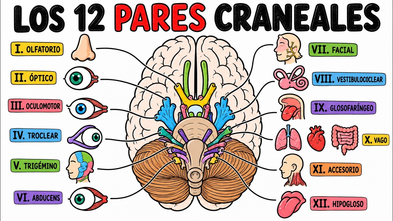
Los 12 Pares Craneales: Qué Hacen y Por Qué Importan (Explicado Fácil)
277
The vagus nerve (X) is the most famous cranial nerve, named for its wandering path from the head through the neck, chest, and abdomen. In the head and neck, it provides sensation to the pharynx and larynx, elevates the palate, aids swallowing, and controls most voice-related muscles. Its recurrent laryngeal branch loops around major thoracic vessels to reach the larynx, so neck or chest problems can initially cause hoarseness. As the main parasympathetic pathway, it regulates heart rate, respiratory control, and digestive functions including gastrointestinal motility and secretions. It is part of a complex autonomic network regulated by the brainstem, hypothalamus, and various body signals.
Click to watch →

Vagus Nerve Functions Explained | How Cranial Nerve X Controls Your Body
1.1K
The vagus nerve (Cranial Nerve X) exits the brainstem through the jugular foramen, travels down the neck sending branches to the voice box and throat, then enters the chest to innervate the heart and lungs, and finally reaches the abdomen connecting to digestive organs, serving as the main controller of the parasympathetic 'rest-and-digest' system.
Click to watch →
Deep Dive
The physiological mechanisms of the stress response, including the role of the HPA (hypothalamic-pituitary-adrenal) axis and the release of cortisol.

(НЕ)ВЫУЧЕННАЯ БЕСПОМОЩНОСТЬ, 50 лет спустя
40.3K
The HPA (Hypothalamic-Pituitary-Adrenal) axis is the neuroendocrine pathway involved in stress response. The hypothalamus releases corticotropin-releasing hormone (CRH), which stimulates the pituitary to release adrenocorticotropic hormone (ACTH), which then stimulates the adrenal cortex to release cortisol. Cortisol increases blood glucose, reduces inflammation, and mobilizes energy resources during stress. While short-term cortisol release is adaptive, chronic stress leads to chronic HPA axis activation and sustained high cortisol levels.
Click to watch →

SEMANA DA SAÚDE E PERFORMANCE CEREBRAL | Aula com ME. Guilherme Lira
4.6K
This segment explains the physiological mechanism of stress response through the HPA axis (hypothalamic-pituitary-adrenal axis). The hypothalamus produces CRH, which stimulates the pituitary gland to release ACTH, which then signals the adrenal glands to produce cortisol. Under normal conditions, cortisol follows a circadian rhythm: high in the morning, decreasing throughout the day, and low at night to facilitate sleep. When cortisol cycles become dysregulated due to chronic stress, individuals experience difficulty falling asleep, lighter and less restorative sleep, frequent awakenings, and waking up tired even after adequate sleep.
Click to watch →

Sistema Endócrino Aula#06 - Adrenal
8.5K
The hypothalamic-pituitary-adrenal (HPA) axis regulates cortisol production: the hypothalamus releases CRH, stimulating the pituitary to release ACTH, which stimulates cortisol production from the zona fasciculada. Cortisol is released in response to stressors including physical stress, emotional stress, pain, and hypoglycemia. Cortisol has negative feedback effects on the HPA axis, inhibiting CRH and ACTH secretion. This axis is crucial for stress response, metabolism, and immune function regulation.
Click to watch →

Why Does Stress Hurt the Body and How Do You Get Rid of It
76.5K
The HPA axis (hypothalamus-pituitary-adrenal axis) represents the body's comprehensive stress response. After initial catecholamine release, the hypothalamus secretes corticotropin-releasing factor, which signals the pituitary gland to release adrenocorticotropic hormone. This hormone travels to the adrenal cortex, triggering the release of glucocorticoids, primarily cortisol. Cortisol, a steroid hormone, increases blood sugar, suppresses the immune system, and aids in metabolizing fats, proteins, and carbohydrates. It also regulates multiple body systems including the heart, lungs, circulation, metabolism, immune system, and skin.
Click to watch →

2034 – A Data Limite. | Maria Pereda P.hD
23.2K
The speaker explains the HPA axis (hypothalamus-pituitary-adrenal) that leads to cortisol release by the adrenal glands. When people are stressed, they either don't sleep or have viruses, bacteria, or any level of stress. Electromagnetic radiation increases cortisol levels, which contributes to insulin resistance, diabetes, and memory loss in the hippocampus.
Click to watch →
Deep Dive
Basic neurobiology concepts, specifically how action potentials and neurotransmitters (such as acetylcholine) facilitate cellular communication.

How to advance your consciousness
2.5K
Dendrites receive input from axons in the form of neurotransmitters - chemicals traversing the synapse. Main neurotransmitters include acetylcholine, norepinephrine, dopamine, GABA, glutamate, and serotonin. Depending on neuron type and transmitter binding, effects differ. Strong depolarization causes action potentials at the axon hillock, occurring in three stages: depolarization (sodium channels open, sodium influx makes interior positive), repolarization (sodium channels close, potassium channels open, membrane returns to resting potential), and hyperpolarization (membrane becomes more negative than resting potential). This occurs all the way down the axon until reaching axon terminals, where depolarization causes neurotransmitter release into the synapse. This process repeats throughout the nervous system.
Click to watch →

Ch. 7 (Nervous System: Action Potential & Neurotransmitters)
822
Action potentials are electrical signals generated at the axon hillock when membrane potential reaches the threshold of -55 mV, triggering voltage-gated sodium channels to open and sodium to rush in, causing depolarization; this all-or-none response ensures reliable signal transmission regardless of stimulus strength, with action potentials traveling down axons via saltatory conduction in myelinated neurons and being transmitted across synapses through chemical neurotransmitters like acetylcholine, which bind to receptors on postsynaptic neurons to either excite or inhibit their activity.
Click to watch →
AP Psychology Unit 2 Summary
284.1K
Neurons communicate using electrical and chemical signals. Resting potential means neurons have more negative ions inside. Depolarization occurs when sufficient stimulus meets threshold, triggering an action potential (all-or-nothing firing). Repolarization returns neurons to resting state. The refractory period prevents immediate re-firing. At synapses, neurotransmitters cross the synaptic gap: presynaptic terminals release them, postsynaptic terminals receive them. Excitatory neurotransmitters increase firing likelihood through depolarization; inhibitory neurotransmitters decrease likelihood through hyperpolarization. Major neurotransmitters include acetylcholine (muscle/action/memory), dopamine (movement/emotion), serotonin (mood/sleep), endorphins (pain), epinephrine/norepinephrine (fight-or-flight), glutamate (learning), and GABA (calming).
Click to watch →

🔥 B. Pharma 2nd Semester HAP-II | 2 Marks Questions Marathon | 20+ Marks Confirm | By Punit Sir ✅
302
Neurotransmitters are chemical messengers that transmit signals between neurons and from neurons to muscles and glands, regulating functions like movement, memory, and autonomic activities. An action potential is a rapid and temporary change in membrane potential of an excitable cell due to ion movement across the membrane. This change is temporary because it returns to the resting potential after the impulse passes, allowing for precise nerve impulse transmission.
Click to watch →

2.1 Neurotransmission
733
Neurons are the basic cells of the nervous system, providing instantaneous communication through action potentials. Neurons vary in size from a few feet to less than a millimeter. The cell body processes inputs and makes decisions before sending signals down the axon. Action potentials are electrical signals that travel unidirectionally from the cell body to synaptic terminals. A nerve is a collection of axon fibers that can be efferent (carrying signals away from the CNS) or afferent (carrying sensory information). At synaptic terminals, action potentials trigger the release of neurotransmitters stored in vesicles into the synapse. Major neurotransmitters include acetylcholine, catecholamines (dopamine, norepinephrine, epinephrine), serotonin, histamine, GABA, and glutamate. The neuromuscular junction uses acetylcholine with nicotinic receptors. Myasthenia gravis involves antibodies blocking these receptors. The parasympathetic nervous system uses acetylcholine with muscarinic receptors on organs. The sympathetic nervous system uses catecholamines with alpha and beta receptors. Cholinomimetic drugs mimic acetylcholine effects and are used in myasthenia gravis, Sjogren's syndrome, glaucoma, and Alzheimer's disease.
Click to watch →
Prerequisite Knowledge
- Concept 01The anatomy and function of the Autonomic Nervous System (ANS), specifically the contrast between the sympathetic ('fight-or-flight') and parasympathetic ('rest-and-digest') branches.
- Concept 02An introduction to Cranial Nerve X (the Vagus Nerve), including its pathways from the brainstem to major visceral organs.
- Concept 03The physiological mechanisms of the stress response, including the role of the HPA (hypothalamic-pituitary-adrenal) axis and the release of cortisol.
- Concept 04Basic neurobiology concepts, specifically how action potentials and neurotransmitters (such as acetylcholine) facilitate cellular communication.
Subsequent Learning
- Step 01The study of Heart Rate Variability (HRV) as a non-invasive biomarker for measuring vagal tone and autonomic balance.
- Step 02Advanced clinical applications of invasive and non-invasive Vagus Nerve Stimulation (VNS) for treating treatment-resistant depression, epilepsy, and inflammatory conditions like rheumatoid arthritis.
- Step 03The Polyvagal Theory proposed by Dr. Stephen Porges, exploring its implications for trauma therapy and emotional self-regulation.
- Step 04The bioengineering and technological design of transcutaneous VNS (tVNS) devices and their current status in medical regulatory frameworks.
Vagus Nerve
Clinical Proof
Vagus Nerve
0:01Playing Section
- 1
Vagus nerve connects brain to organs, regulating calm.
- 2
Stimulation reduces inflammation, aids digestion and sleep.
- 3
Key controller of parasympathetic nervous system.
Methodological Limitations and Commercial Overhype of Non-Invasive VNS
While non-invasive vagus nerve stimulation (nVNS) shows promise, critics and researchers highlight several significant limitations. First, many clinical studies supporting its efficacy for stress and anxiety suffer from small sample sizes, lack of standardized stimulation parameters, and inadequate double-blind sham controls, making it difficult to rule out the placebo effect. Second, there is physiological skepticism regarding whether consumer-grade transcutaneous devices actually stimulate the vagus nerve directly, or if the observed effects are simply due to general sensory stimulation of cutaneous nerves. Finally, the rapid commercialization of wellness gadgets claiming to 'tone' the vagus nerve often outpaces the actual scientific consensus, leading to oversimplified marketing that frames a complex neurological pathway as a quick-fix cure-all for stress and inflammation.
Deep Dive
The study of Heart Rate Variability (HRV) as a non-invasive biomarker for measuring vagal tone and autonomic balance.

Grounding & Heart Rate Variability
656
Heart rate variability is the most effective non-invasive means of measuring how balanced the autonomic nervous system is. The autonomic nervous system consists of the sympathetic nervous system (fight or flight response) and the parasympathetic nervous system (rest and digest response). The balance between these two systems directly influences heart activity.
Click to watch →

Neurology of Cervical Instability:Vagus Nerve webinar - Part 3 - Ross Hauser, MD
475.2K
Heart rate variability (HRV) measures the variation in time between consecutive heartbeats, serving as a practical biomarker for vagal tone assessment. High vagal tone produces greater beat-to-beat variability (HRV values above 70), while stress produces minimal variability (regular, rapid heartbeat). HRV analysis separates signals into low-frequency (sympathetic) and high-frequency (parasympathetic/vagal) bands; sympathetic dominance (LF > HF) increases mortality risk. Dynamic HRV testing evaluates how vagal tone changes during neck rotations, stressful memories, and other challenges, helping identify patients with cervical instability who would benefit from Prolotherapy. This non-invasive tool enables self-assessment and clinical monitoring of autonomic nervous system function.
Click to watch →

Experto en Nervio Vago: "esta es la forma de saber si tu tono vagal es el adecuado (o no)"
35.5K
Heart Rate Variability (HRV) is the gold standard measurement used in scientific studies to assess vagal tone and parasympathetic nervous system activity. HRV measures the variation in time between consecutive heartbeats, reflecting the dynamic balance between the sympathetic (fight-or-flight) and parasympathetic (rest-and-digest) nervous systems. Higher HRV indicates better vagal tone and greater parasympathetic influence on the heart.
Click to watch →

Protocolos Prácticos Para Relajar tu Sistema Nervioso, con Antonio Valenzuela
20.4K
Heart rate variability (HRV) is an objective biomarker that reflects vagal tone and autonomic nervous system function. Higher HRV indicates better vagal tone and greater parasympathetic activity, while lower HRV suggests reduced vagal tone and increased sympathetic dominance. HRV can be measured using simple wearable devices and provides insight into the body's ability to regulate stress responses and recover from physical and mental exertion.
Click to watch →
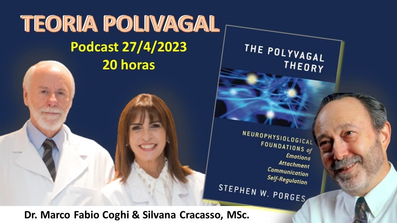
TEORIA POLIVAGAL
8.4K
Heart rate variability (HRV) is the variation in time between heartbeats and serves as a key biomarker for assessing autonomic nervous system function. Higher HRV indicates greater vagal tone and better emotional regulation capacity, while lower HRV suggests reduced flexibility in the autonomic nervous system. Porges used HRV measurements to study how the nervous system responds to stress and social engagement, demonstrating that HRV can predict outcomes in various conditions including trauma, attachment disorders, and developmental challenges.
Click to watch →
Deep Dive
Advanced clinical applications of invasive and non-invasive Vagus Nerve Stimulation (VNS) for treating treatment-resistant depression, epilepsy, and inflammatory conditions like rheumatoid arthritis.

#22 Vagus Nerve & inflammation: the body’s healing reflex with Dr. Kevin Tracey
213
Vagus nerve stimulation offers FDA-approved treatments for depression (50% improvement rate in treatment-resistant patients without immunosuppression) and rheumatoid arthritis (80% clinical benefit in trials). Two device generations exist: Gen 1 pacemaker-like implants with over 30 years of experience for epilepsy and depression, and Gen 2 miniaturized devices (SetPoint Medical's immunoregulator) approved for rheumatoid arthritis. These devices demonstrate potential disease-modifying effects beyond symptom relief, with MRI evidence suggesting joint healing acceleration. However, mechanisms remain incompletely understood, and patient selection criteria for optimal responders require further research.
Click to watch →
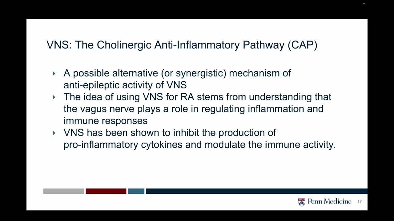
Vagus Nerve Stimulation Mechanisms Epilepsy Rheumatoid Arthritis Clinical Trials- Alnemri
102
Vagus nerve stimulation (VNS) is an implantable device therapy that modulates the vagus nerve to treat neurological and inflammatory conditions; it works through the cholinergic anti-inflammatory pathway where vagal stimulation activates splenic nerve release of norepinephrine, which stimulates choline acetyltransferase-positive T cells to release acetylcholine that inhibits macrophage cytokine production, thereby reducing inflammation and seizures; FDA-approved for drug-resistant epilepsy and treatment-resistant depression since 1997, VNS is now being investigated for rheumatoid arthritis and other inflammatory conditions through clinical trials like RESET RA.
Click to watch →

What is the vagus nerve and can you really hack it?
11.1K
Invasive vagus nerve stimulation is FDA-approved for two conditions: treatment-resistant epilepsy (for patients who continue to have seizures despite medication) and treatment-resistant depression (for patients who remain significantly depressed despite optimal medical therapy). Research is also exploring its use for migraines, cluster headaches, inflammatory bowel disease, and Crohn's disease.
Click to watch →

Vagal Nerve Stimulation in Management of Treatment Resistant Depression
748
Vagal Nerve Stimulation (VNS) is an FDA-approved neuromodulation therapy for treatment-resistant depression, involving electrical stimulation of the vagus nerve to modulate brain activity through projections to the locus coeruleus, amygdala, prefrontal cortex, and other limbic structures, thereby enhancing neurotransmitter release (serotonin, norepinephrine, dopamine, GABA) and promoting neuroplasticity; VNS can be delivered invasively via implanted devices or non-invasively through transcutaneous methods targeting the auricular or cervical branches, with evidence supporting its efficacy in improving mood and quality of life for patients who do not respond to conventional antidepressant treatments.
Click to watch →

Kevin Tracey returns to the podcast to give an update about his work at the Feinstein...
159
Vagus nerve stimulation shows promise for treating various conditions beyond epilepsy, including rheumatoid arthritis, inflammatory bowel disease (Crohn's disease), depression, anxiety syndromes, and other behavioral illnesses. Research continues to explore whether non-invasive devices can achieve similar therapeutic effects as surgically implanted systems.
Click to watch →
Deep Dive
The Polyvagal Theory proposed by Dr. Stephen Porges, exploring its implications for trauma therapy and emotional self-regulation.
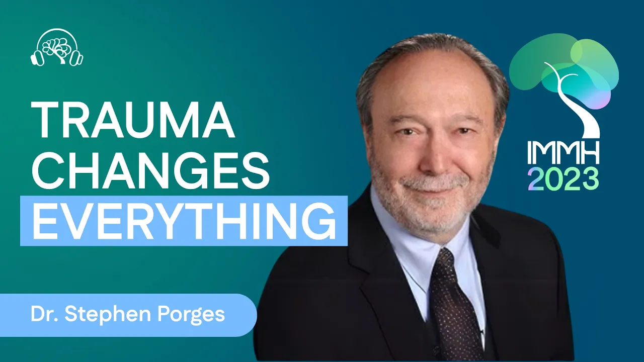
The Impact of Psychological Trauma on Mental And Physical Health | Dr. Stephen Porges
6.6K
Polyvagal Theory provides an optimistic perspective because it assumes autonomic state influences behavior, reactions to the environment, self-regulation, and co-regulation. It assumes a more plastic mechanism where features are naturally occurring adaptive reactions primarily determined by biobehavioral state. The job is to trigger signals of safety to allow the nervous system to return to its normal, optimistic, engaging self. Polyvagal Theory transforms the client's narrative from a documentary emphasizing events and objects to a pragmatic quest wired into us for safety with an implicit bodily drive to survive. It emphasizes feelings rather than events. For trauma therapy to be effective, therapists must first restore a parasympathetic state and reduce the stress response. Nervous systems traumatized find safety or signals of safety risky, triggering vulnerability. The insightful therapist must get the person into a state of accessibility, but the person must be comfortable with being accessible. Medication can downregulate behavior and physiological state but doesn't enhance the neural regulation of calming systems, so it may not be effective in recruiting the nervous system's ability to process signals of safety.
Click to watch →

WEBINAIRE avec le Pr Stephen PORGES : Neuroceptivité, corps en relation dans la thérapie du trauma.
1K
The Polyvagal Theory, developed by Stephen Porges, explains that the vagus nerve regulates the body's social engagement system and that trauma fundamentally disrupts the body's ability to perceive safety and connect with others. Effective trauma therapy requires the therapist to first establish their own sense of safety and co-regulation with the patient, as the body's physiological state directly influences cognitive processing and emotional regulation. The body's automatic detection of safety or danger (neuroception) shapes all subsequent mental and emotional experiences, making somatic awareness and co-regulation essential foundations for healing trauma.
Click to watch →

The Polyvagal Theory. Explained.
212.8K
The Polyvagal Theory, developed by Dr. Stephen Porges, explains how the vagus nerve (the 'wandering nerve') has multiple branches that control our autonomic responses: the dorsal vagal branch causes immobilization/freeze/shutdown responses (low metabolism, low heart rate), while the ventral vagal branch enables social engagement, connection, and calm states; trauma can trap people in either hyper-aroused sympathetic (fight/flight) or hypo-aroused dorsal vagal (freeze/shutdown) states, and healing requires activating the ventral vagal social engagement system to restore healthy nervous system regulation.
Click to watch →

Polyvagal Theory with Dr Stephen Porges
4.4K
Polyvagal theory gave those who had been traumatized a physiological explanation for their experiences, so they no longer felt crazy because they were shut down. This theory welcomed Porges into the trauma world without him being a stakeholder, allowing him to be a welcomed guest who could contribute without competition. The theory provided an explanation that validated trauma survivors' experiences. In academic worlds, people never give up their defenses, which means the level of communication is always contained. The creativity and synergism that comes from truly learning in an accessible mode gets compromised. Porges learned from clinicians worldwide for 20 years, developing a take on trauma that is different from his friends who are leaders in the field, but without vested interest in a brand.
Click to watch →

CÓMO APLICAR LA TEORÍA POLIVAGAL EN TERAPIA
3.5K
The polyvagal theory, developed by Stephen Porges and expanded by Deb Dana, explains how the nervous system responds to safety and threat through three hierarchical states: the ventral vagal state (social engagement and safety), the sympathetic state (fight or flight response), and the dorsal vagal state (shutdown or immobilization). In trauma therapy, understanding these states helps therapists create neurobiologically regulating environments that allow patients' nervous systems to experience safety, enabling them to move from dysregulated states (dorsal vagal or sympathetic) toward the ventral vagal state of connection and regulation. This somatic approach recognizes that trauma responses are automatic survival mechanisms, not conscious choices, and that the nervous system never assigns moral meaning to survival responses.
Click to watch →
Deep Dive
The bioengineering and technological design of transcutaneous VNS (tVNS) devices and their current status in medical regulatory frameworks.
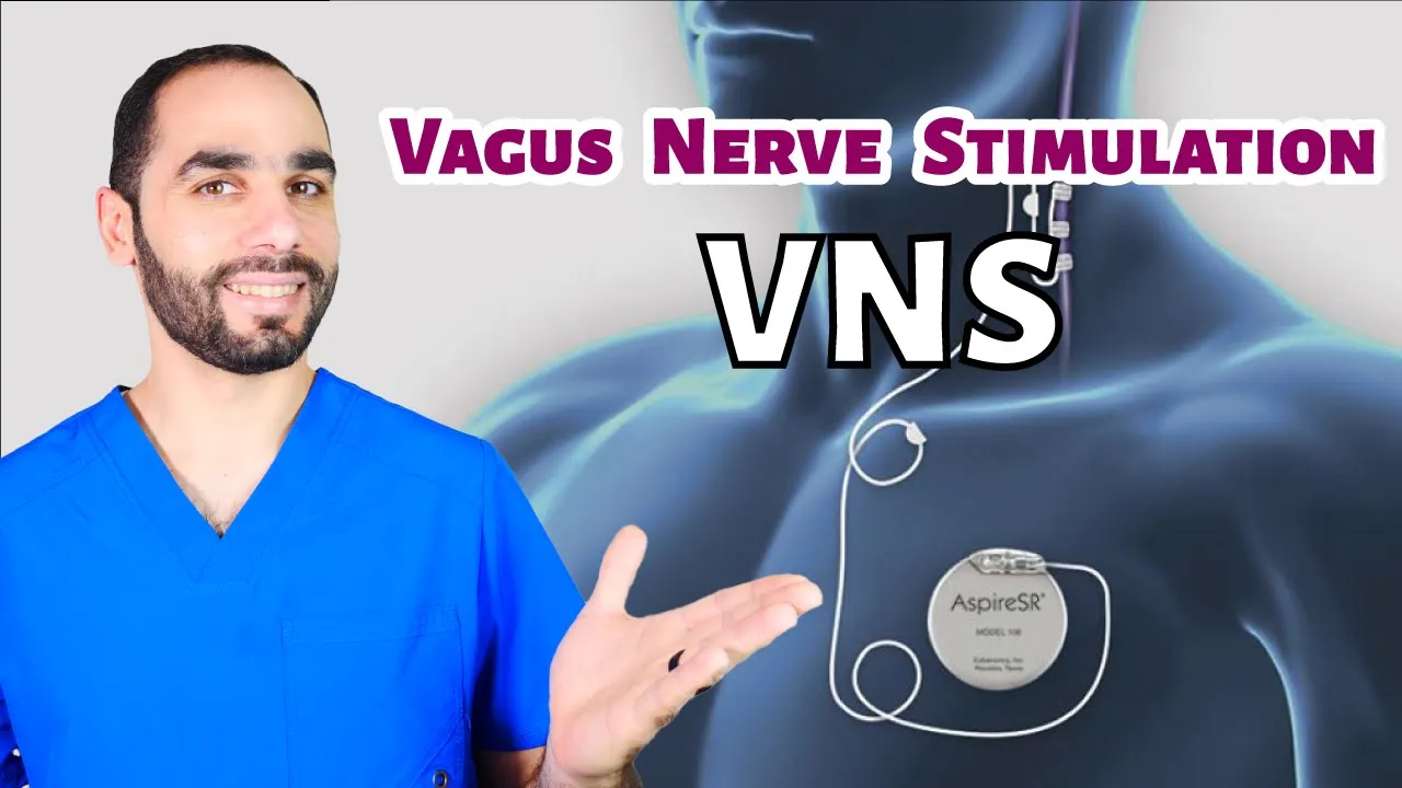
Vagus Nerve Stimulation (VNS) for Epilepsy
39.8K
VNS cannot be remotely controlled or hacked; only specialized clinic programmers can access it. Modern devices feature heart rate monitoring that automatically detects seizures (occurring in 80% of cases) and delivers additional stimulation. The magnet can be used during seizures to deliver extra discharges for approximately one minute. Newer iPhone models (12 and 13) contain magnetic fields that may interfere with VNS, so phones should not be placed in pockets near the device. Transcutaneous VNS (tVNS) stimulates the auricular branch externally through the ear, showing promise in European studies but not yet FDA-approved in the US. VNS represents one of several treatment options for drug-resistant epilepsy, with costs varying by insurance and country.
Click to watch →

Novo tratamento traz mais qualidade de vida para quem tem epilepsia
519
The VNS device is a small, lightweight implant weighing only 15 grams. The device consists of a generator implanted in the chest and an electrode placed around the vagus nerve. The electrode sends electrical impulses through the nerve to the brain. Since January, Brazil's National Supplementary Health Agency has regulated the use of this device, making it mandatory for health insurance plans to offer the treatment. However, the treatment has not yet been approved by the public health system (SUS), though many patients have gained access through legal court orders.
Click to watch →

Home-Use Devices | Linda Carpenter | Interventional Psych Consortium
131
VNS clinical trials for depression initially failed to meet primary endpoints at 3 months, but long-term follow-up showed increasing efficacy as physicians adjusted settings. A large Medicare-sponsored trial is underway to establish efficacy in randomized sham-controlled trials over one year, which would influence insurance coverage. Transcutaneous VNS (tVNS) offers alternatives without surgical implantation, stimulating either the vagus nerve in the neck or the auricular branch in the ear. The auricular approach allows easy sham stimulation for controlled trials.
Click to watch →

An interview with Prof. Armin Bolz on tVNS Therapy
411
Transcutaneous vagus nerve stimulation (tVNS) is a non-invasive medical technology that delivers small electrical pulses to the auricular vagus nerve (located in the ear) to stimulate the vagus nerve, which helps the brain emit neurotransmitters and promote neuroplasticity, thereby improving conditions such as epilepsy, depression, inflammation, anxiety, Parkinson's disease, and stroke without the risks associated with surgical implantation.
Click to watch →

Dirençli Epilepsi ve Beyin Pili Tedavisi (VNS-TVNS)
445
Vagal nerve stimulation (VNS) is a brain implant therapy for drug-resistant epilepsy, approved in Europe (1994) and the US (1997). The vagus nerve connects the brain to vital organs (heart, lungs, kidneys, intestines), coordinating autonomic functions. VNS works by reducing electrical activity and altering neurotransmitter levels to control seizures. VNS involves surgical implantation of a device with a battery below the clavicle, sending electrical impulses to the brainstem. TVNS (transcutaneous VNS) is a newer non-surgical alternative (since 2010) using an earpiece device. Both treatments have variable effectiveness: VNS reduces seizures below 50% in 45-75% of patients, while TVNS achieves 30-60% reduction. Treatment selection depends on seizure type, frequency, and family considerations.
Click to watch →
Vagus Nerve
Clinical Proof
Vagus Nerve
0:01- 1
Vagus nerve connects brain to organs, regulating calm.
- 2
Stimulation reduces inflammation, aids digestion and sleep.
- 3
Key controller of parasympathetic nervous system.
Methodological Limitations and Commercial Overhype of Non-Invasive VNS
While non-invasive vagus nerve stimulation (nVNS) shows promise, critics and researchers highlight several significant limitations. First, many clinical studies supporting its efficacy for stress and anxiety suffer from small sample sizes, lack of standardized stimulation parameters, and inadequate double-blind sham controls, making it difficult to rule out the placebo effect. Second, there is physiological skepticism regarding whether consumer-grade transcutaneous devices actually stimulate the vagus nerve directly, or if the observed effects are simply due to general sensory stimulation of cutaneous nerves. Finally, the rapid commercialization of wellness gadgets claiming to 'tone' the vagus nerve often outpaces the actual scientific consensus, leading to oversimplified marketing that frames a complex neurological pathway as a quick-fix cure-all for stress and inflammation.
The anatomy and function of the Autonomic Nervous System (ANS), specifically the contrast between the sympathetic ('fight-or-flight') and parasympathetic ('rest-and-digest') branches.
An introduction to Cranial Nerve X (the Vagus Nerve), including its pathways from the brainstem to major visceral organs.
The physiological mechanisms of the stress response, including the role of the HPA (hypothalamic-pituitary-adrenal) axis and the release of cortisol.
Basic neurobiology concepts, specifically how action potentials and neurotransmitters (such as acetylcholine) facilitate cellular communication.
The study of Heart Rate Variability (HRV) as a non-invasive biomarker for measuring vagal tone and autonomic balance.
Advanced clinical applications of invasive and non-invasive Vagus Nerve Stimulation (VNS) for treating treatment-resistant depression, epilepsy, and inflammatory conditions like rheumatoid arthritis.
The Polyvagal Theory proposed by Dr. Stephen Porges, exploring its implications for trauma therapy and emotional self-regulation.
The bioengineering and technological design of transcutaneous VNS (tVNS) devices and their current status in medical regulatory frameworks.
239.9K views15.5Klikes2:47@DrWilliamLiOriginal Release: 2025-07-27
You might know me from my work in food as medicine, but today I want to share another powerful way that your body heals itself. And that's through your vagus nerve. What's the Vegas nerve?
Well, it's the longest cranial nerve in your body, which means that it comes from your brain and runs through your neck and chest, touching your heart and your lungs, and then goes through your diaphragm into your belly, where it connects to your stomach, your liver, your pancreas, kidneys, and intestines.
Think of the vagus nerve as a communication superhighway between your brain, and your other organs. Now, why is it so important? Well, the vagus nerve is the key player in your parasympathetic nervous system. That's a system that counterbalances your fight or flight response. The vagus nerve actually helps to calm your body, reduce inflammation, aid digestion, and promote sleep. Now, here's the exciting part.
You can stimulate your own vag nerve to turn on these calming effects. One device I've seen and used is called Pulsetto. You wear it around your neck just like this and it delivers gentle electrical pulses through the skin to activate the vagus nerve. It's been shown to reduce stress, ease depression, and improve gut function and gut health.
So now, of course, let's look at the science. Researchers conducted a clinical trial in Lithuania. They studied 40 adults using Pulsetto twice a day, eight minutes each session for four weeks. They measured stress, anxiety, and sleep. They did it at the start, at 2 weeks, and again at four weeks. And they also measured cortisol levels, your body's main stress hormone, by testing hair samples collected before and after four weeks of pulset use. So, what are the results? Well, stress symptoms dropped by 39% at 2 weeks and by 56% at four weeks. Anxiety dropped by 31% at two weeks and by 45% by four weeks. Many participants reported better sleep within two weeks and 70% of users had lower cortisol stress hormone levels measured after 4 weeks. The cortisol level dropped by 42% in men and by 37% in women. That's a measurable drop in a biological marker of stress just from stimulating the vag nerve. So here's the bottom line. Your vagus nerve is a built-in healing pathway and now you can tap into it from the comfort of your own home. If you're curious to try it yourself, comment pulsetto and I'll send you a link with a 10% off code already applied.
Up Next
6:00
CBT Explained: How Cognitive Behavioral Therapy Works
@sprouts
423.7K views•2021-09-15

48:47
Graphic Medicine: Comics for Collaborative Healthcare Communication
@nationalpatientadvocate
189 views•2023-12-04

10:28
Neuroanatomy: Central and Peripheral Nervous System Divisions Explained
@AKLECTURES
136.2K views•2014-09-20

3:05
Stages of Labor and Vaginal Birth | Childbirth Animation
@nucleusmedicalmedia
52.1M views•2017-08-18
Related Study Plans & Knowledge Roadmaps
Structured learning paths in Medicine
medicine
Abnormal LFTs: Diagnostic Workup & Pathology
01Liver Anatomy and Basic Physiology
02Introduction to Liver Function Tests (LFTs)
03Hepatocellular vs. Cholestatic Patterns
+ 3 more learning modules
12h6 modules
Explore Roadmap
medicine
Analyzing Blood Smears: RBC, WBC & Platelets
01Introduction to Blood Components & Smear Preparation
02Normal Morphology of Blood Cells
03Erythrocyte Pathologies & Morphological Variants
+ 2 more learning modules
12h5 modules
Explore Roadmap
medicine
Mechanical Ventilation: Waveforms & Alarms
01Anatomy and Physiology of Breathing
02Introduction to Mechanical Ventilation & Core Modes
03Ventilator Settings and Waveform Interpretation
+ 2 more learning modules
12h5 modules
Explore Roadmap
medicine
Neuro Exam: Localization & Stroke Syndromes
01Foundations of Neuroanatomy and Vascular Supply
02The Neurological Examination: Step-by-Step
03Principles of Neurological Localization
+ 3 more learning modules
18h6 modules
Explore Roadmap