















![[REPLAY] Le cinéma intérieur : projection privée au cœur de la conscience](https://i.ytimg.com/vi/OZro7Jq-b5A/maxresdefault.jpg)




Visual information travels from the retina through the optic nerve to the optic chiasm, where approximately 60% of axons cross to the contralateral side and 40% remain ipsilateral, then proceeds through the lateral geniculate nucleus (LGN) which separates visual processing into magnocellular (movement/contrast) and parvocellular (color) pathways before reaching the primary visual cortex (striate cortex), where neurons exhibit orientation selectivity and are organized into ocular dominance columns and orientation columns; beyond V1, visual processing diverges into two major streams—the dorsal stream (where pathway) for spatial location and motion control, and the ventral stream (what pathway) for object and face recognition.
Neuroscience of Vision: The Visual Pathway Explained
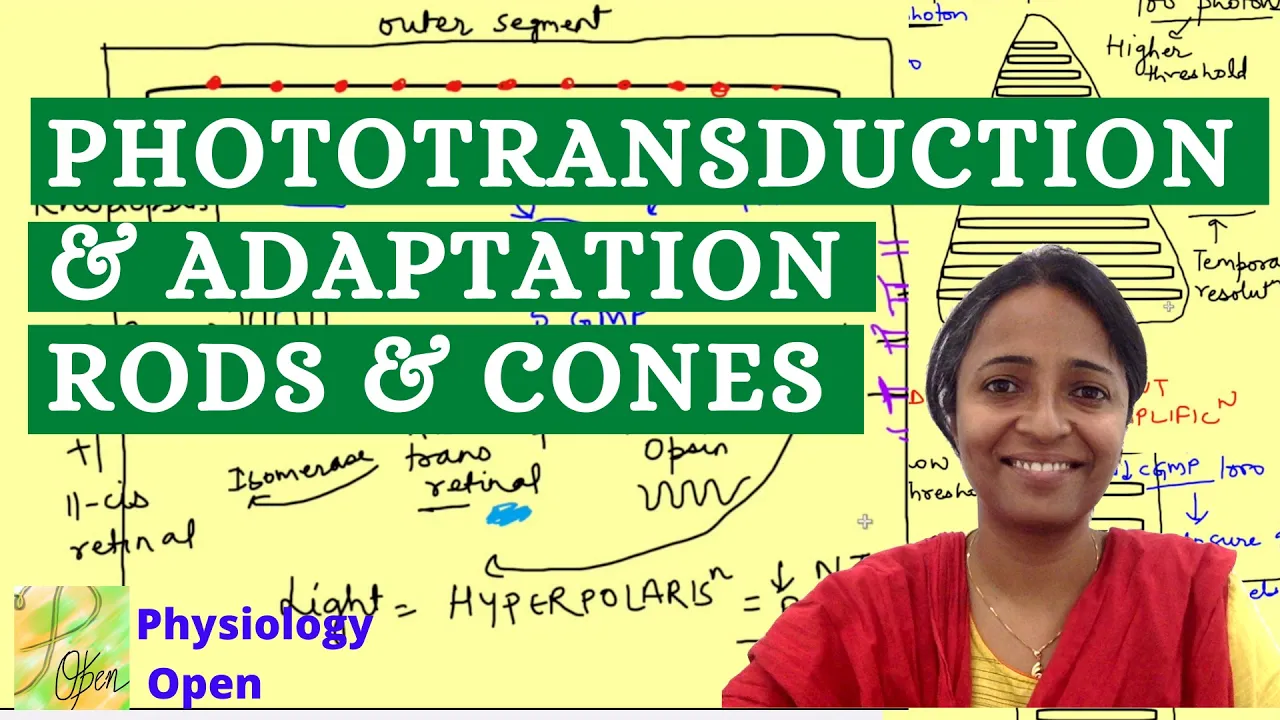
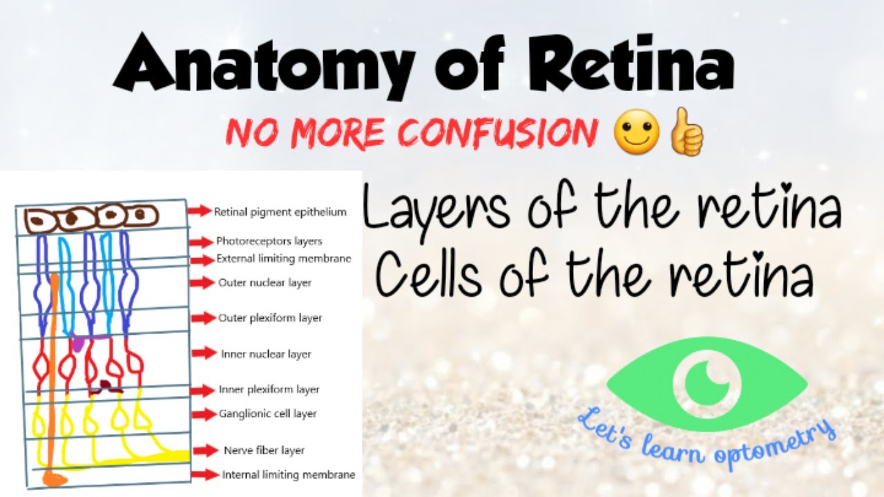
Added:Basic anatomy of the eye, including the function of photoreceptors (rods and cones) and retinal ganglion cells in phototransduction.
The principles of neural communication, including how action potentials travel along axons and cross synapses.
General brain anatomy, specifically the location of the thalamus and the occipital lobe within the cerebral cortex.
The concept of visual fields and contralateral representation, where information from the left visual field is processed by the right hemisphere of the brain.
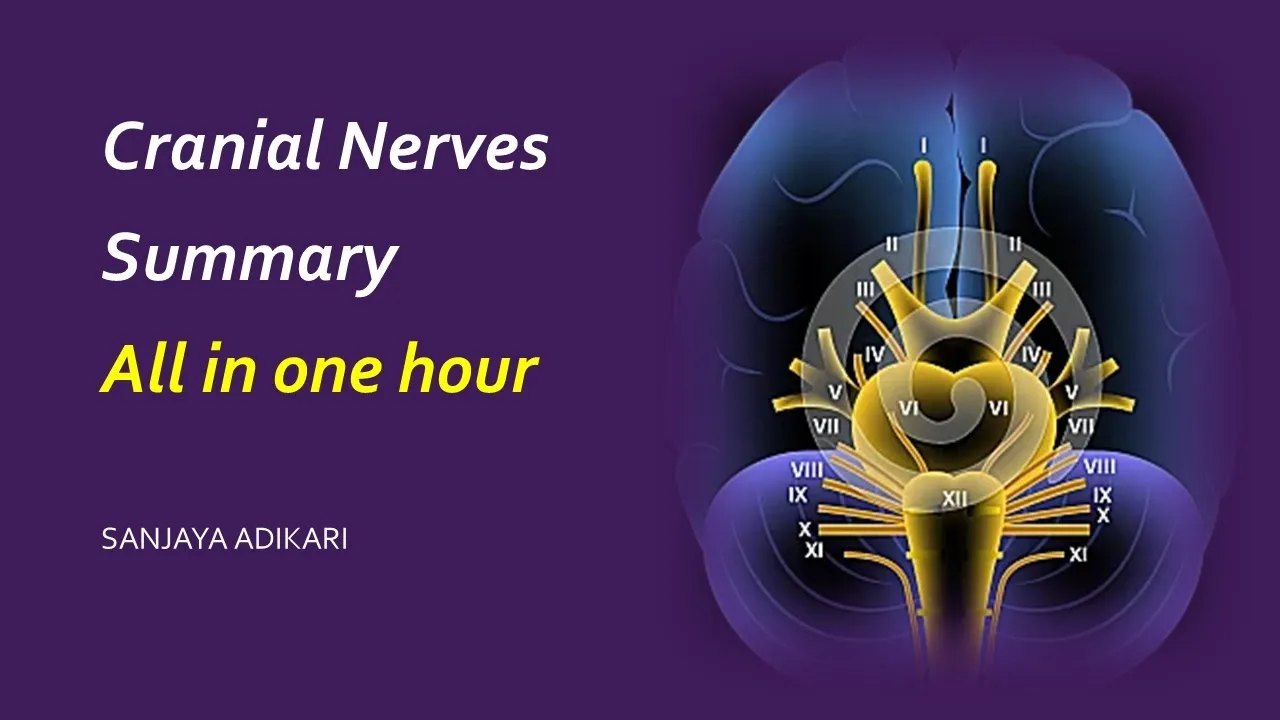
Advanced cortical processing in extrastriate visual areas, such as V4 (color constancy) and V5/MT (motion detection).
Clinical neurological deficits resulting from pathway damage, such as visual agnosias (e.g., prosopagnosia) and optic ataxia.
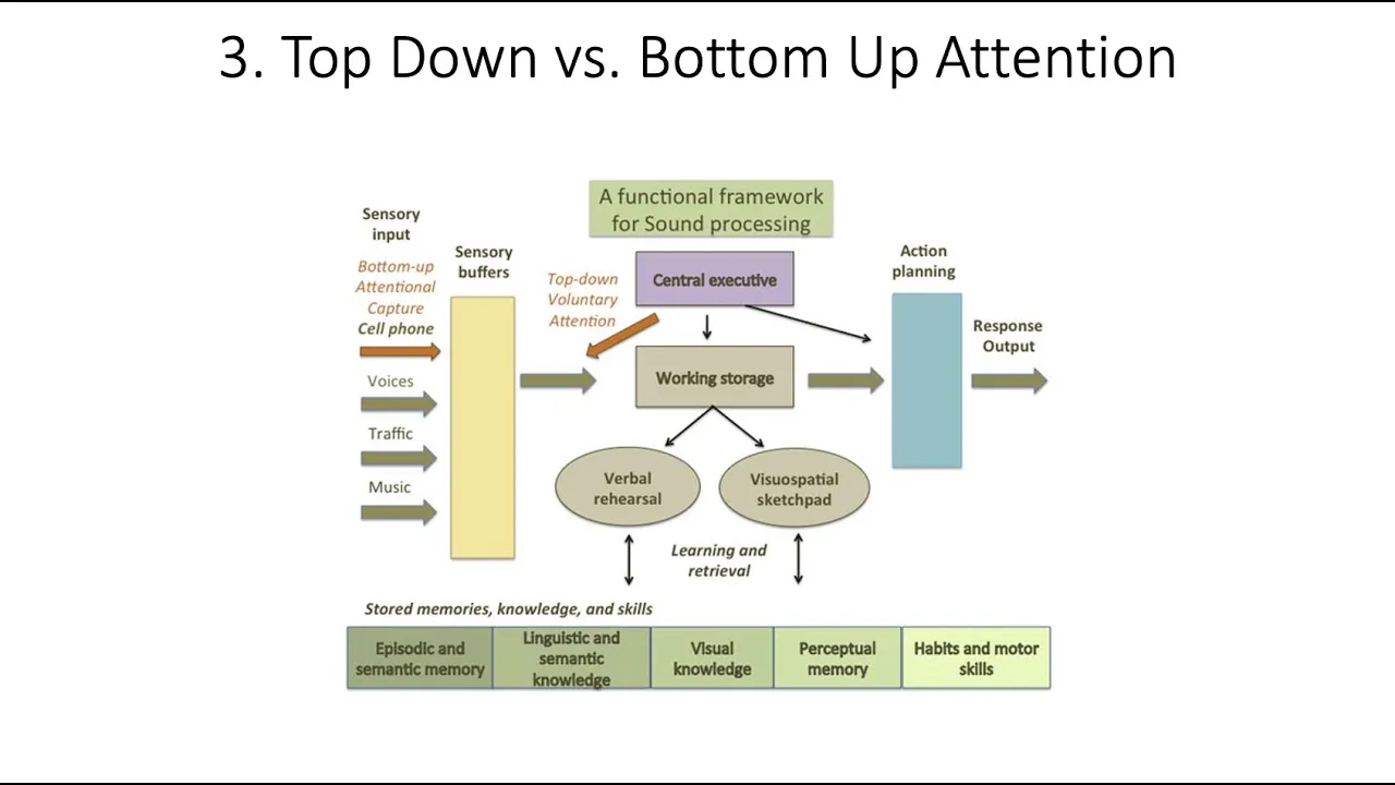
The cognitive neuroscience of visual attention and how top-down processing influences bottom-up sensory inputs.
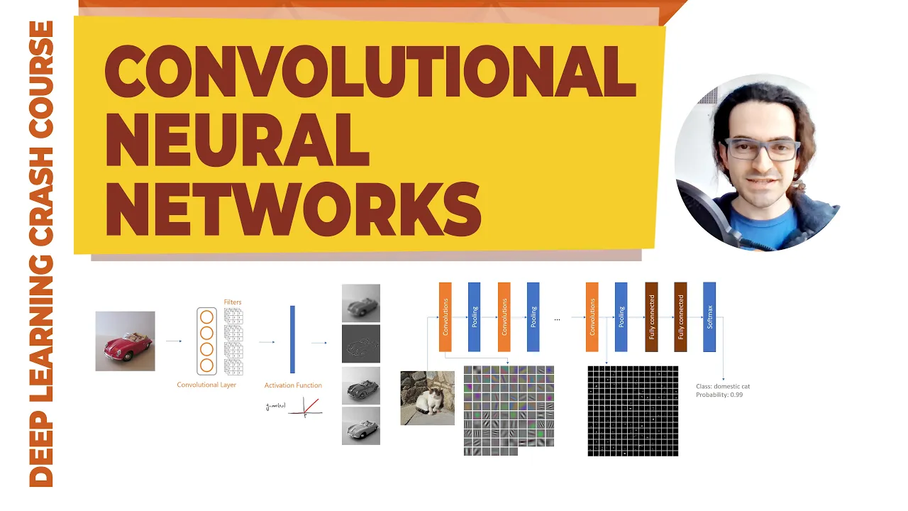
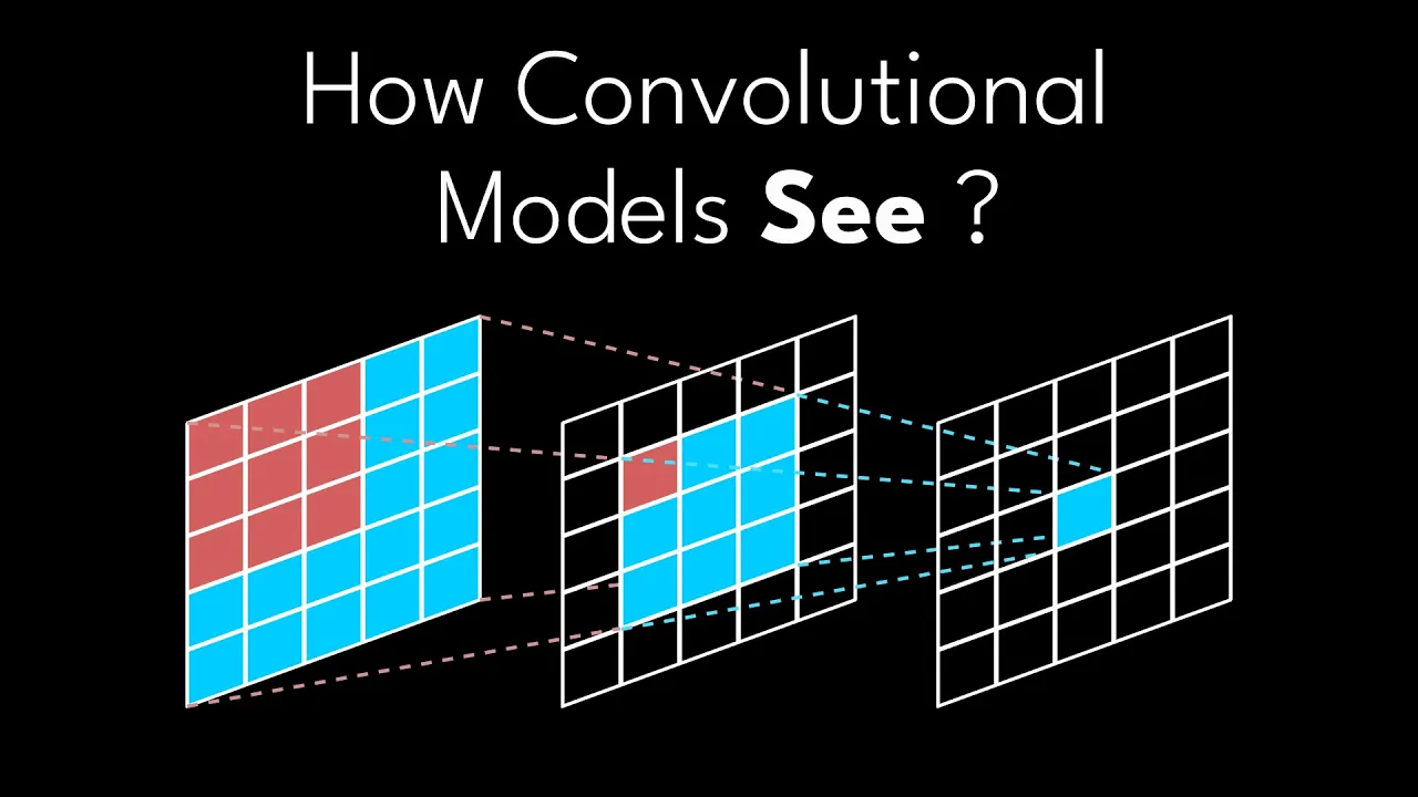
How biological visual pathways and V1 receptive fields inspired artificial intelligence, specifically Convolutional Neural Networks (CNNs) in computer vision.
3.6K views58likes1:15:14Original Release: 2020-09-01
so this lecture is going to be focused on understanding the other the rest of the visual system so in the first lecture we focused on what the processing that happens in the eye and now we're going to be looking at things that happen past the eye as we take that light information from the eye and uh uh before we move on i want to just kind of give an overview for how most basically all sensory systems operate uh sensory pathways will generally involve four steps so the first step will be sensory reception and that's where involves the use of sensory receptors that are located in the periphery that detect a stimulus and in the case of the visual system this is the photoreceptor cells the cones and the rods so light enters the eye and then is going to be entering and interacting in the photoreceptor cell so that's the recep sensory reception part the next step is sensory transduction and that is taking that is the step in which the physical world that is including the relevant stimulus as being translated into something that leads to a neuronal membrane change a potential change and so in that case that's the transducer pathway where the light interacts with the opsins in the cones rhodopsins and rods causing that molecular change that leads to the change in cyclic gmp and then sodium and so that is the sensory transduction portion of the visual system today we're going to be talking about uh transmission which is how action potentials are propagated from the periphery into the central nervous system and then perception how an organism becomes aware of the stimulus when the brain receives sensory information and so we're going to be covering these aspects these two aspects we already covered in the first lecture okay so the visual system uh is made up of several sub pathways there's one main pathway that we're going to focus on today there are a couple of other smaller pathways but still are very important and we will be discussing those at later points so the first part is the retina we talked about that on monday or in the first lecture the uh the retina has ganglion cells that have axons that go into the optic nerve right so those ganglion cells run up to the optic chiasm chiasm is a word that means cross it comes from the greek word kai you know which is kind of an x shape so that's what we have here it's cross now the ax the axons from retinal ganglion cells in the eye make their way to the optic chiasm and interestingly only sixty percent of those axons decision they go to the other side of the brain so in this case the left eye going to the right side of the brain forty percent actually stay on the same side so another way to phrase this is that uh sixty percent of the axons go to the contralateral side of the brain the other side of the brain whereas forty percent stay on the ipsilateral side of the brain so they stay in the left-hand side once they're past the optic chiasm then those paths are referred to as the optic tract so this is the optic nerve and now we're talking about the optic tract because we're essentially in the brain nerves are outside of the brain um the main visual pathway that we're going to be talking about involves the uh the great uh the ganglion cell axons terminating in what's called the lateral genic nucleus so 40 of the epsolateral ones ending on the ipsilateral lateral geniculate nucleus 60 ending up on the contralateral lateral geniculate nucleus or the lgn then from there the lgn projects to the primary visual cortex also known as striate cortex so and this is known as the optic radiation so there's a big projection that comes from the lgn and then and then projects out into the visual cortex there are other visual pathways as i mentioned that are also important um one is the uh the pre-tectum so there are projections to an area of the brain called the pretectin and this is what allows the brain to control reflex control of the pupil and lens so all those little minor adjustments to make the lens look close or far or adjust the amount of light that's coming in through adjusting the iris and changing the size of the people the pre-tectum is involved with that we will not be discussing that there is a projection from uh the eyes to the superior colliculus colliculus it's a very interesting part of the brain it helps with orientation of movement of the head and the eyes and we will be talking about that in a couple of weeks when we talk about uh motor systems so i'm not going to talk about that today and then there's one last pathway that's important which is the eyes project to areas of the brain in the hypothalamus for the regulation of circadian rhythm i want to just briefly cover this because i want you to have an appreciation for uh the fact that there are different kinds of retinal ganglion cells we only talked about two main types on and off retinol ganglion cells and there's lots of subcategories of those but there's another kind of retinal ganglion cell which actually doesn't receive any input from the cones and the rods so the the retinal ganglion cells that we talked about are kind of depicted here in gray these are the ones that have the cones and the rods and horizontal cells play a role and then we have the bipolar cells there's amacrim cells those are the retinal ganglion cells that we will be mostly focusing about uh today and and what we talked about uh exclusively on uh the previous lecture but there's another kind that i want to mention which are called the intrinsically photosensitive retina ganglion cells so they do not receive inputs from rods and cones and instead they have their own photopigment so these are neurons or retinal ganglion cells that have uh their own photopigment which is called melanopsin and it is sensitive to blue light and these intrinsically photosensitive retinal ganglion cells um when you shine blue light on these cells they start to fire and they have a very slow time course so this is the change of electrophysiological change in a cone here's the light intensity here you can see this is over minutes and it takes minutes before these cells start to fire so they're very very slow compared to a cone which is within milliseconds right so um and then we'll maintain its membrane change with that light intensity whereas with these cells they just start to fire a whole bunch anyway these cells project to the hypothalamus and they help us regulate sleep and we will talk about them in more detail later in the semester but since they are part of the visual system i want to make sure that we cover that as well because you know that's also testable material so the visual field is split in the brain all right and i'm going gonna walk through this figure in some detail um so pretend that in front of you let's say it's christmas time and you're looking at something that's red on the left side and green and the right side so that is the visual field in front of you you got right over here and green over here now your eyes are not in the exact same position there's a little bit of space between them the right side of the visual field will be sending photons to your eyes and they come at slightly different angles right because your eyes are in different positions but for both eyes as long as the object is far away enough that light on the right hand side will end up on the left hand side of each eye right so that's it's shown here in green so the green light is ending up on the left hand side of the eye remember because the eyes create a reverse image on the back of the eye on the retina and that means of course the red part that's in the left hand side ends up in the right hand side of each eye now importantly the eyes are not identical they are mirrors of each other so there's a nasal side and a temporal side to each eye the nasal is the part of the eye that faces the nose and the temporal side is the side that faces your temple on the outside of your head so this green region projects to the left hand portion of each eye but because they are mirrors the green region is on the temporal side of the left eye and the nasal side of the right eye so functionally different parts of the eye even though it's the same side left hand side so if we continue to follow this green region the temporal retinal ganglion cell axons they go down to the optic chiasm and those are the axons that stay ipsilateral see so it's the temporal axons that stay it's lateral and project to the ipsilateral uh lateral geniculate nucleus the nasal retinal ganglion cells they stay they actually cross so those are the ones that decosate and they cross the optic chiasm and end up on the contralateral lateral geniculate nucleus and so what this means is that even though we have axons that are splitting and and going from two different the the the axons are from the eye are going to different regions of the brain once some are going ipsilateral and some are going contralateral the visual information the green information is ending up on the same side of the brain but down slightly different pathways so the left eye information is maintained separately from the right eye information even though they are seeing the same green thing and then the same is true for the red right so then the red uh at the left-hand side deca states and ends up in the right lgn and then the temporal side stays on the ipsilateral side okay so the visual field is split and then from there this lgn will project to the primary visual cortex or striate cortex and this is then all right visual field on the left hand side of the brain and then the opposite is true for the right hand side of the ring all right now we're going to talk about so that's an overlap overview of just the pathways uh now we're going to talk about the lateral geniculate nucleus of the thalamus so the lgn is a major relay station of the visual system and why do i call it a relay station well that's because the thalamus kind of serves as the brain's sensory relay station so so let's talk a little bit of the thalamus so in a coronal section this is where the thalamus is located right here and um it has somewhere between 30 and 40 subnuclei so you can break it down into into several different brain areas and and each serving as a critical relay station for the cortex and other brain areas so the the thalamus is organized so that each nucleus is responsible for specific functions and we can break down these functions into three major subdivisions okay so uh first is sensory relay stations and there are different sensory relay stations that process auditory information somatosensory information but also visual information such as the lateral geniculate nucleus there are other modalities uh and subnuclei that process that act as for motor relay including the basal ganglia as well as the motor cortex we'll talk about those later when we talk about motor information and then there are portions of the uh thalamus that are very important for processing emotionally relevant information and we'll talk about that in the much later portion of the class but today we're going to focus on the lateral geniculate nucleus the thalamus is a very critical part of the brain all right so the lateral geniculate nucleus of the thalamus is specialized to process visual information we have the right and left lateral geniculate nucleus and they receive input from two optic tracts which synapse here so that the third order neurons carry visual information onto the primary visual cortex this is a close-up of a human lateral geniculate nucleus the the primate or human lateral genetic nucleus is made up of six major layers and unfortunately um you know neuroscientists have decided to call them one two three four five six with one starting at the bottom and six out at the outer region and this almost kind of looks like layers it's almost like a volcano with the layers of like striations like uh like layers of lava that's kind of been poured on top of them um the central portion of the uh lateral geniculate nucleus um it represents the center part of the visual field generally and then all leading to one side so there are different layers of the so these different layers actually now get more and more complicated so layers one and two uh comprise the magnocellular uh layers of the lateral geniculate nucleus and layers three through six comprise the parvocellular layers of the lateral geniculate nucleus so these layers of the lateral geniculate nucleus uh actually have segregate segregated inputs from the different eyes and the different visual fields so this figure's complicated and i don't expect you to be able to replicate the the the schematic from memory there may be questions about some of these details especially since the test will be open book so um we're going to walk through this a little bit in some detail so that you can have an appreciation for the complexity of how the visual system works and how different light information getting to the eyes is kept separate so in the schematic we have the left eye we have the right eye here is the optic chiasm the right the left visual field would be blue over here and the right visual field would be red over here that's not shown but we can tell that's the case because the left side of each eye is red and the right side is blue so there's something blue over here and there's red over here okay so we've got red going down staying ipsilateral on the left hand side and on the right eye it it decasates and goes to the left lgn so this is a schematic of the lgn and here here we have the six layers one two three four five six the left eye projects to layers two three and five the right eye which is dark red here projects to one four and six so the information of course you know it's the nervous system so it can't be completely logical in order you've got one and two and then three four five and six where they're interspersed this is because layers one and two provide different kinds of light information to the brain than layers three through six these two pathways are known as in green the is the parvo cellular pathway and purple is the magnocellular pathway the names magnocellular and parvocellular refer to the size of the cells in the lgn so the magnacellular cells are relatively large that's why it's called magno so these cells are pretty big in layers one and two whereas layers three through six they're relatively small and they're known as parvo cells now the kinds of light information that receives that the brain receives from these two pathways is actually pretty different the magnacellular pathway that comes from one and two is largely responsible for movement the light information there is help helps what regulates movement and to some degree contrast but not color information even though there's some cone information that's received by layers one and two the kinds of retinal ganglion cells that that come from the eyes to layers one and two are mostly sensitive to contrast and and they help with the organization of of movement layers three through six are a separate pathway and this is where a lot of the color information is regulated in the parvo cellular pathway the way the visual system processes color is really super interesting but it's pretty complex and it's beyond the scope of what we're going to cover in this class so we're not really going to be talking too much about what's happening in the parvocellular pathway if you want to learn more about it i highly encourage you to read online you can even go to wikipedia but certainly the textbooks associated with the course also provide nice detailed information about how this pathway works it's really cool and interesting the way it's organized but from here on we're kind of going to be talking about what's happening within the magnacellular pathway okay so to re uh to to go over this once more we have the visual system pathway the visual field going to one side of the brain from the left eye and the right eye ipsilateral and contralateral but they're going to separate layers of the lgn so the information from each eye is still segregated so that the brain can process the information from both eyes separately uh in order to provide things like binocular vision and that kind of thing um but then we also within the lgn have two outputs which is the magnacellular pathway which is really important for movement contrast and the parvo cellular pathway which is really important for color as well as a bunch of other uh visual processes but color primarily is the most important one so the output of the lgn is to the visual cortex and they project to the primary visual cortex which is the first station of the cortical visual system we refer to this there's a few names in which the primary visual cortex is referred to so primary visual cortex is one of the main ways it's referred to striate cortex is another name for the visual primary visual cortex and then we we briefly talked about in the very first lecture in this class about broadband's areas and this is brodmann's area 17.
so it's also sometimes referred to as area 17 as shown in this figure here um i will be using primarily primary visual cortex or striate cortex um just not gonna refer to area 17.
so this is showing what the visual cortex looks like the primary visual cortex looks like in two primates the macaque monkey which is an uh a primate that had been heavily used for studies on the visual cortex and how it regulates how it controls visual perception and then this is showing where it's located in humans so if you're looking at the lateral side in humans there's a little bit that you can see but if you're looking at the mid sagittal plane of the brain the that the the primary visual cortex goes along this this figure here where you can see it kind of as this long stripe in all mammals the primary visual cortex is located here in the occipital lobe neurons in the cortex the mammalian cortex are organized in layers you learned about this in neuro one so i'm going to briefly but i'm going to briefly cover this uh here again because it's a very important concept that you guys need to understand all cortex has six layers so layer one is largely cell free then we have layers two and three which in some parts of the cortex can't really tell the difference but in other parts you can then you have layers four which is broken down in this three sub sections a b and c then we have layers five and layer six that is the cellular portion well layers 2 through 6 are is the where most of the neurons almost all the neurons are located within cortex striated cortex or visual cortex has the same overall organization what we're looking at here is just a small section of cortex in a in a section a brain section so someone had sliced i think this is human so a human brain um sliced it on a what's called a micro tone to make very thin sections sort of like almost like salami and then these sections were then put onto glass slides and then they were stained with something called nissel and nissl actually stains all cells but because of the way it stains you can tell the difference between neurons and glia and um and it shows up kind of as a purple ish or bluish color and and so it shows the cell bodies of cells and neurons are included in that and you can see the different layers so it's a really easy way of seeing where cells are located within neural tissue it's commonly used and we're going to be seeing nissl stain over and over and over again so you should know what nissel or cressel violet that's another name for it is called so there are different neurons that reside in different layers uh the the major type of neuron that you find in the cortex is referred to as pyramidal neurons and they are called that because their cell bodies kind of have a pyramid shape and kind of see it pretty well in this one and you'll find pyramidal neurons in multiple layers there also are spiny stellate neurons as well you're going to find these different kinds of neurons in different layers the lateral geniculate nucleus projects to these layers of area 4 primarily there's tiny little projections to other areas but most of the input comes to layers 4a and 4c and this is generally true for most areas of cortex that are receiving inputs from the thalamus the projections go to layers four so layer four is the primary input for most areas of the cortex particularly from the films then from there in a given area of cortex going to find and we're talking about a relatively small area you're going to have these columns where the neurons then project to different layers so there's there's interlayer communication interlaminar communication within a section of cortex with the outer surface here the inner surface here and you're going to find neurons going up and down across those layers so there's inputs coming to here and then they project in and out then there is output so there's input to layer four then there's processing that happens within the cortex these are referred to as cortical columns and then there's output so in the visual system we have uh some processing that happens within that part of the cortex and then these layers layers 2 and layers 4b the neurons there they project to other visual cortex areas and other areas as well layers five actually send projections to the superior colliculus and then layer six actually sends reciprocal product projections back to lgn so it turns out that there's an additional connection vectoring here so we will talk about we'll talk about this projection when we talk about superior calculus so that's gonna be a little bit later uh we're actually not gonna talk about this at all but now we're gonna sort of refer to some of the well actually in a few slides we will be talking about what is happening from the output of uh visual uh primary visual cortex first we gotta talk about what's happening within primary um yeah before i go on the um and so this is this isn't showing exactly that so we're showing layer layer 4 here that receives the input from the lgn and what you can see is that there's right then left then right and then left and right and then left and then you have this interspersing of the eye so the eye information even though it's seeing the same part of the visual field we're still maintaining segregation between so this illustrates the data that they had seen in this experiment the scientists didn't work they presented bars of different orientations so here's one that is horizontal and then they rotated it more and more and more and then they had vertical and they rotated it more and more and more back to horizontal so they could just rotate over and over and over again and they find the particular part of the visual field that the neuron is sensitive to so it might be the upper right or the upper left or lower right it kind of depends on so if they're looking at the left cortex uh which part of the visual field would they be looking at them answer that question that's a question for you guys so they're recording visual cortex which where would they have to put the bar in the visual field on the left or the right i'm a legend okay so looking in cortex i put the bar in either left or right depending upon which part of the cortex they're looking at which side and then if they rotate it they find that in some orientations you know so they're presenting their the the stimulus right here between what seconds one and two so nothing then they present it and then they turn it off to get a baseline and then see what happens afterwards if they present a horizontal bar they don't see anything no spikes if they start to rotate it now they start to see some spikes and as it gets to the vertical side now this neuron really really likes vertical lines and so it spikes a ton and then there's a little bit more spiking here and then as it rotates back to horizontal again it doesn't spike anymore this particular neuron that that was being recorded here has an orientation selectivity for vertical bars and you can graph what that looks like by by graphing spike rate and then different orientations so this might be 90 degrees 0 negative 90 right actually probably 90 here and negative 90 here that would make more sense um so but at negative 90 or 90 degrees there's no very little activity but as you get closer to its preferred orientation that's when you see peak activity and then as you go back rotating to the other way where it's essentially looks exactly the same as this its orientation selectivity goes back down again every uh uh so a huge majority of the neurons in visual cortex that have bar selectivity will have a selectivity to some orientation they are not all vertical oriented and they're not just like some vertical and some horizontal you will find neurons that will have orientation selectivity to uh bars that are rotated like this which means this would be the opposite or or neurons are oriented like this this is their preferred orientation and then this would be the least preferred orientation so you're going to find uh basically every single neuron will have an orientation selectivity for a particular bar and then the perch of the perpendicular bar actually ends up uh leading to the least amount of uh sensitivity okay and this is just illustrating the preferred orientation of one particular neuron in the cortex the guys who discovered all of this um are david hubel and horsten weasel so carson weasel worked under david hubel they did all these experiments in the 50s and 60s they did a bunch of experiments describing how the visual system works primary visual cortex um on inosil activity they also did a bunch of work on neuroplasticity uh which was really critical for understanding how it works and that's i talk about a lot of their work in my neuroplasticity senior seminar class here's some videos links to youtube videos for this one in particular shows then describing how they accidentally discovered bar selectivity which is really kind of funny and serendipitous like they got the nobel prize for medicine for this discovery and they kind of discovered it on accident which is really hilarious um and then this is uh another video sort of getting into a little bit more in depth on some of the work that they did as well as some other scientists who did some work on visual system it's an older video it's like probably 30 or 40 years old but it's it's it's interesting it's worth looking at okay so how does the brain build orientation selectivity so we're going to kind of build this up we already have talked about center surround orientation that neurons uh in the the retinal ganglion cells will have this center surround orientation where the center is stimulate a stimulatory and the surround is inhibitory right different lgn neurons will tend to have orientation selectivity that is also center surround so neuron number one here will have this orientation selectivity that's pretty similar to the neurons uh the retinal ganglion cells that it's receiving inputs from okay and then this neuron here in lgn will have this orientation selectivity this orange this this neuron here will have a center surround orient orientation selectivity in this neuron here we'll have this center surround orientation selectively but these different neurons have a maintain are maintaining a retinotopic map so they are maintaining where things are located in space in in vertical and horizontal space and they are organized in a way that maintains that map so there is a map of the retinotectal sorry there's a map there's there's a map of the visual field in the lgm so this map you can look at it from this neuron it'll be in this part of the visual field this neuron is in this part of the visual field this neuron is in this part of the visual field this neuron is seeing this part of the visual field okay neurons in the lgn start to converge on pyramidal neurons in the fourth layer where they receive multiple inputs from different lgn neurons and this is where bar selectivity now starts to to to emerge so these neurons they'll project actually multiple different pyramidal neurons but the way that they are organized is that any given pyramidal neuron will receive input from multiple lgn neurons that will now have an emergence of bar selectivity so if you take these separate center surround orientations of these different lgn neurons and you let them add up onto a single pyramidal neuron in the cortex now you start to see a bar emerge where there will be in visual space in the same visual space part of the visual field that this is seeing instead of having these separate center surrounds and the on the separate lg neurons what we're seeing is the combination of them all adding up on one neuron now this particular neuron is driven by the activity of a bar which is stimulating all these center surrounds right so if you have a bar that is illuminating all these centers and not the surround that's when you start to see a bar orientation selectivity so now we have individual cortex the ability to see edges this is essentially edge detection and we can have individual neurons that'll see lines with different orientations in space with this kind of activity you will find neurons in v1 that also have direction selectivity so some a good majori a large number of the neurons in v1 will have what's called direction selectivity so there'll be a particular orientation that they're sensitive to within a given receptive field but they are sensitive to the direction of movement so in this case we have neurons that move in one direction so if they're moving from left to right this neuron as it's going through the the receptive field this bar as long as it's the right orientation will stimulate activity in the receptive field here so so this particular neuron will fire if the bar travels across the receptive field in this direction for the same neuron if you're continuing to record from the same neuron and you move a bar across in the opposite direction you might get a little bit of an onset of activity but if it's moving in the opposite direction actually activity is suppressed so when this bar is moving right that's what this neuron is particularly sensitive to but as it moves left that actually reduces the activity so now not only do we have the ability to see edges and contrast we have neurons that are specialized to see which direction those neurons are moving this appears to be the result of convergence from other v1 neurons that have a particular orientation selectivity but then they are converging onto uh neurons uh other v1 neurons giving them uh the ability to integrate the the the order in which these neurons have been stimulated this uses the principle of delay lines we will learn a little bit more about delay lines when we talk about the auditory system but in short the way this works and it's important for you to understand this is that in this particular neuron this neuron is somewhat farther away so this neuron here the left number two is farther away from number three than say this neuron or this neuron and even though action potentials are very very fast if they are farther away there is a difference in the time at which that they reach that neuron okay so there's a little bit of a difference so it'll take a little bit longer for the action potential to travel down a long axon than a shorter axon just a little bit but that difference is enough to allow for when a a bar pass hits number two action potential starts and then as the bar is going and then hits this one and then the action potential is just a little bit behind this one because this is shorter they should reach this neuron the postsynaptic neuron at the same time so this one starts firing action potential is going down here and then this one starts firing action potential goes down here and then they both reach at the same time same thing for this one now if the bar is going in the opposite direction it's going to hit this neuron first and this has the shortest axon so this starts firing and it reaches the neuron pretty quickly and then this bar then the bar hits this one and then it takes even longer so this is even more delayed and then this then the bar hits this one and it's even more delayed and none of them are synchronized so none of the action potentials reach the postsynaptic neuron in a synchronized way and so then this neuron does not fire so the direction of the uh bar and in the movement that it's making is the integration of different v1 neurons that have different orientations that have the same orientation selectivity um and that are sensitive to slightly different parts within the receptive field but they have delay lines that are built in that allow for the synchronous arrival of action potentials in order to cause the postsynaptic cell number three to fire okay that's delay individual neurons in v1 they exhibit what's called orientation selectivity as we talked about neurons that are near one another within a column of cortex they have this they show very similar orientation selectivity so if you take an electrode and you start recording from layer one two three four five and six all of and then you look to see what are what's the orientation selectivity of all those different neurons in the different layers of cortex they all have a very similar orientation preference so this is the distance along the electro track right so a is shorter and then b is a little bit deeper then deeper deeper deeper deeper um and there they all have a very similar receptive field location so this is the center of the receptive field um uh sorry the center of the wrist of the visual field and then this is showing what is the receptive field of each of these neurons and they all have a very similar receptive field location it's kind of in this case to the upper right of the center of the visual field and then if you look to see what is their overall orientation preference all of these neurons have a very similar orientation preference so these neurons in this particular orientation column have a vertical preference in this part of the receptive of the visual field so all the receptive fields are located here and they all prefer the same orientation bar this is what's known to known as an orientation column those columns are referred to as orientation columns all right what if we go the other direction now we take an electrode and we're going to stay within the same layer in this case we just happen to be taking electrode and pushing it through layer two and three but we're going across uh different columns in this case what we're seeing is that the receptive field position appears to be changing a little bit so there's slightly different receptive field positions but even more so is that you're seeing different orientation preferences but it's not random so a has this orientation preference here of preferring a horizontal bar then b is slightly turned and then c is now basically vertical and the d is an orientation in the other direction so there's an organization to the uh to the orientations that are preferred that goes across in uh across the this uh this this set of tissue so you have um a little bit of change in overall receptive field preference but a major change in the orientation uh of the bar that is preferred and it happens to be changing in a particular pattern where the bar seems to be spiraling in um you know going from say 0 degrees to 10 degrees to 20 degrees to 30 degrees it uses there's a there's a pattern in which the bars the pattern of the preference of the orientation of the body go across cortex those are referred to so um we have ocular dominance uh columns as well as orientation columns so when you go down uh inside tissue you have layer one two three and then layer four that's receiving the input from the lgn you have an orient an orientation column this particular column has a particular preference of an orientation and then the one that's next to it has a slightly different orientation that it prefers and the one next to it has a slightly different orientation but it has a pattern in which it's rotating you can see that it's going from less horizontal to more vertical to less vertical to horizontal back again and it does the spiral that does where the bar is turning as you pass through a single ocular dominance column now the different stripes that receive input from the contralateral eye or the ipsilateral eye left or right this then these are referred to as ocular dominance columns ocular refers to the eye and this particular part of the cortex is dominated by input to one eye so if you close say the ipsilateral eye and you record and then you leave the controller contralateral eye open you will see orientation selectivity um in this column at this preference for that eye in as long as you're recording from the right receptive field now if you close the eye and then you open the ipsilateral eye you will not see activity happening in this part so it's i dependent the eyes are separate but if you move over just a little bit to the next uh orientation column um sorry not orientation column to the next ocular dominance column then this i will be sensitive to a similar orientation but it'll be it'll be the uh the other eye that is driving activity in this particular orientation column so we have organization of the different eyes and that's referred to as ocular dominance columns this is dominated by the contralateral i and this is dominated by the ips lateral i right c over i and i over c c is dominating i and then we have orientation columns where they have a particular orientation that they prefer and this is because we have the the preservation of eyes information being kept separate in the primary visual cortex all right so that's v1 and that's really the only part of the visual system in the cortex that we have talked about so far now we're going to expand to other parts of the visual system so beyond v1 there are there are more than two dozen distinct what we refer to as extra striate areas of cortex so these areas no longer have very strong eye segregation information between the eyes starts to be integrated and processed together to give us say binocular information more motion sensitivity color shapes objects and even face recognition as we go up the visual field um we we start to hit other visual areas so v2 receives a ton of input from p1 and v2 will project to v3 and v3a but also v3 receives input from visual area one then we have v4 and then there are additional areas one is called nt also known as v5 which is really critical for motion and then we have other areas that are involved with face and object recognition when we get beyond the visual uh the primary visual cortex or striate cortex we start to see that there's the emergence of two major streams of cortical visual processing that occurs we break this down into the dorsal stream and the ventral stream the dorsal stream is the dorsal portions of the visual cortex and it goes dorsal in the brain towards the top the visual stream that sorry the ventral stream goes uh ventral in the brain and it moves its way in the opposite occipital cortex and then into huge portions of the temporal cortex and temporal lobe these two pathways can be broken into two sort of kinds of processing that's done in the dorsal stream we have v1 connected to v2 goes to p6 and mt as well as other areas these areas appear to be particularly interested in motion and visual control of action we refer to this as the where pathway that's because these neurons are particularly interested in motion and where things are located in the visual field it also is very it's heavily involved in control of the eyes and the arms to guide reaching so we find neurons that will help with that guidance that are visually driven okay so it's integrating that information so it tells us where things are in space that's what the dorsal stream seems to be most uh involved with then we have the ventral pathway again it's v1 to v2 and then we have a connection to v4 which is kind of like the first major portion of the ventral stream which then leads to inferior temporal cortex and then this is referred to as the what pathway so neurons in this pathway seem to be less concerned with where things are located in space but more concerned with what you're actually looking at so it seems to be heavily involved in form recognition object representation and uh and related to memories so let's talk about the where pathway first so neurons and nt are particularly sensitive to movement this is showing some data taken from doing um functional magnetic resonance imaging okay or fmri so fmri essentially what it does is that it it's a measurement of neural activity a change in neural activity over time and the time scale is a little bit long it's quite a bit longer than what an action potential might be but if a brain area is is firing a whole lot you will see a change in the readout of fmri and what that change is seeing is change in blood flow so if an area is particularly active it will have a little bit more blood flow and you can see that in an fmri that's essentially what fmri is looking at so in this case this is experiments that were done with i believe humans and it this was the they're doing fmri here and um humans were presented with dots and so the dots if they were moving so if the dots are kind of moving away in different directions neurons in active the activity in mt went way way high so it was super high but as soon as those dots stop moving activity and nt goes way down and this happens within a couple of seconds and he in particular seems to be heavily involved with tracking where things are moving and determining and having a focus on on on how much is moving and which direction they're actually moving and and it helps with the processing for that so the neurons in mt in fact um you can do experiments where you have a whole bunch of dots and only a few of them are moving and um and the uh the subject has to to tell you oh they're moving to the right or to the left and the more dots that are moving the more activity that you will see in mt but you will you will see that the activity in nt will go up as soon as uh right before um the person will acknowledge that the uh that the dots are actually when they make a decision of which direction the dots are moving so there's a lot of processing about which direction things are moving and if there's movement at all happening in empty it's pretty cool there's a ton of interesting work on t fmri is a critical non-invasive way for us to see how neurons act over a relatively long time of course seconds and there has to be a lot of coordinated activity in order to see those changes it's a hugely invaluable tool for a lot of cognitive neuroscience and us and allowing us to understand what's happening within humans and so kinds of experiments that you can do in humans you just simply can't do in animals uh because of difference in cognition uh we use fmri to do a lot of those experiments but there are times in which we can get a little bit more fine-grained when we look at the activity of spikes and we've seen a lot of what that looks like one issue that arises though is that when you present something you offer some kind of stimulus like in this case for uh some stimulus is being delivered like let's say there's a light that's off and then all of a sudden light goes on and we're recording from a single neuron and we do this 20 times so 20 different trials this neuron apparently let's just pretend that light is being turned on at time zero this neuron um has a certain spontaneous firing rate that's relatively low you can see that like in this trial one it only fired once trial two it fired three times trial three it fired two times there's no real coordinated pattern though this is at random as soon as you turn the light on now the spike rate goes up now this neuron is firing over a period of 500 milliseconds like ten times instead of one and then in trial two instead of firing three times in five the previous 500 milliseconds it fires like eight times there's obviously if you look at the sum total here there's a lot more activity post stimulus presentation and pre-stimulus presentation but if you were to look at any one particular um uh what's called raster you would see you might think that okay well this is the pattern that there must be a pattern here but if you do it over and over and over again you'll see that there isn't really a pattern necessarily um sometimes you can see your pattern in a raster plot but but what it can be really useful for is just seeing that that overall over time there is this kind of increased level of activity relative to this and then we can create a histogram of seeing the uh of how many spikes normally happen within the first 500 milliseconds post and then second half of the of 500 milliseconds as well and and then comparing it to the to the 500 milliseconds before and you'll see that the spike rate would be low and then it will go up and then it'll be relatively high so that's how you read a raster plot you just look across the different trials and it shows you clearly something that's being presented at time zero whatever this blue line is is causing this neuron to increase its firing rate okay so that's how you look at a raster plot um when you go up the um dors the uh the ventral visual system you find neurons become more and more specialized and we're going to show a little bit of what's happening here so um if you look in the on-center retinal ganglion cells you have this center surround orientation that's the very that is the typical stimulus that will drive a non-center retinal ganglion cell this is also true if you look in the lgn you're going to find mostly neurons that are have this on this center surround orientation now when you look in v1 we get a little bit more complicated and it's a little bit more specialized so instead of center surround we have bars that it's sensitive to then as you get up into the uh the temporal visual cortex we find neurons that actually are particularly particularly sensitive to faces and then even in some places it appears that there's appears to be specialization for identifying specific individuals that are important to that individual so such as the grandmother and this is the idea of the grandmother's cell in your brain you must have neurons that recognize that your grandmother is your grandmother right there has to be some place in your brain that is making that connection and that you would see neurons that are increasing activity when that connection is being made uh so um this is what a raster plot might look like with different presentations so these are now we're looking at really a ton of different trials right and so each line is a different trial and there's a bunch of little tiny dots and you're doing it over and over and over again and in this particular paper what they did is they had one session and then they did another session and another session they're separating them with different blue lines they're looking for stability of how this particular neuron fires and so this is one presentation uh they present some stimulus and you can see that there's there's kind of an increase in spike rate over time uh to to this one like right here at around 250 milliseconds post presentation and you can if you look at the average you can see the number of spikes per second is somewhat consistent and it and there's an increase here but for a different presentation stimulus at the same time around 250 milliseconds post presentation this neuron appears to be particularly sensitive to whatever this presentation is so let's look a little bit more of what that is so these are this data was taken from recordings of macaques so these were macaques that were where they were doing chronic recordings of individual neurons over time and each of these uh sessions is broken down by date so they did may 21st 22nd 23rd all the way out to june 7th so this is a few weeks that they're doing this and they presented these macaques with and so they're recording in the uh in the the ventral visual stream in the temporal cortex they're presenting them with different faces so they presented these mechanics with the face of one of the researchers and they the macaques actually seem to have some kind of response to that particular face but they also presented the macaques with faces of of other macaques that they knew so these are macaques taken from a colony and so this would be charlie and frank and bob and whatever macaque that they were recording from this neuron was sensitive to uh to this particular face as well as this face in this face and so they could see a spike in activity this was a macaque face sensitive neuron and it wasn't it was somewhat sensitive to the human face but it wasn't sensitive to other faces of other animals such as a line in fact there appears to be a decrease and then when you show objects like a violin or something else that's totally not a face if anything you can see that there's a suppression of activity so these neurons are specific to faces and not only are they specific to faces they are specific to faces consistently over time over a period of weeks this one particular non sensitive to bases over weeks so phase selective neurons they maintain constant visual response across not just weeks but months okay so in this study this is actually a study that was done in humans um some humans uh when they're having neurosurgery done they will volunteer and be able to be asked to and they will volunteer to participate in studies this is someone that was having um i believe an electrode implanted into their hippocampus in order to help with seizures um but when you're implanting this electrode to help control the activity of the brain to try to prevent seizures when you were doing it you can also record activity and in this study they were recording in the the hippocampus and they were presenting these individuals with images of famous people and one particular neuron that was seen here they were showing different faces of individuals when they showed this person the face of halle berry this neuron fired so that's shown here so this is the raster plot across multiple presentations of halle berry and during the presentation of halle berry you can see that this neuron really liked it and fired pretty soon right after the presentation of halle berry of this particular face the same neuron responded to this face of halle berry this face of halle berry this face of halibury you can see that visually they're quite a bit different like this one's brighter her face right here is overexposed um there's a very dark background here and here but not so much here what this neuron seems to be firing uh to is faces of halle berry it also responded to a drawing of halle berry so just completely black and white totally devoid of color also responding to halibury it responded to a picture of halle berry dressed as catwoman so this neuron is also firing to halle berry pictured as a catwoman right so it visually looks very different from this to this but if you knew that halle berry starred in the terrible movie catwoman uh you would also have that response but even more crazy is that this neuron also responded to the word halle berry clearly had the word halle berry has nothing it does not look anything like halle berry but it's the concept of halle berry that this neuron is firing to so this is a visually sensitive neuron you have to present some sort of visual stimulus to it but it's conceptual more than just visual than just this the straight-up visual information this is a neuron that's a halle berry neuron and then if you present other stimuli to this neuron uh um that are not halle berry related uh it actually doesn't respond uh interestingly it didn't respond to catwoman but it definitely didn't respond to bill clinton and uh jennifer lopez and oprah winfrey it had a little bit of response over one thing um in the same individual they also found um a other neurons as well i think that they if i recall they found neurons that were sensitive to cameron diaz person's a big fan of movie stars from the 90s and early 2000s all right so here are some key questions about the visual system how is visual field information organized in the brain so knowing like which parts of the brain have different parts of the visual field and how we split that up how is light information organized in the lateral geniculate nucleus and what is the lateral geniculate nucleus connected to and how is it connected using carbocellular and magnesium cellular information right uh what is the striate visual cortex how do we know that it's called the striat visual cortex what are ocular dominance columns how are they discovered what is orientation selectivity and what are orientation columns and how were those discovered what is the role of the dorsal visual stream and what is the role of the visual vision ventral visual stream and then how does cell selectivity change as you move up the ventral stream you have to know all of those concepts and i look forward to talking about this at our next discussion section
Up Next

18:47
Vision: Retina to LGN Visual Pathway Explained
@PaulMerritt
19.8K views•2017-03-28

21:55
Deciphering Neural Circuits Controlling Anorexia in Mice | Richard Palmiter
@AllenInstitute
2.3K views•2011-10-10

61:12
Taste and Smell Explained: Chemical Senses | Neuroscience Lecture
@Dr.ChrisThompson
1.4K views•2020-09-10

13:03
How Exercise Benefits Your Brain: Science Explained
@TED
11.4M views•2018-03-21
Related Study Plans & Knowledge Roadmaps
Structured learning paths in Neuroscience
neuroscience
Appetite Regulation: Brain Circuits & Hormones
01Foundations of Endocrinology and Brain Anatomy
02Introduction to Energy Homeostasis
03Peripheral Signals: Leptin, Ghrelin, and Insulin
+ 2 more learning modules
14h5 modules
Explore Roadmap
neuroscience
Neuroeconomics: Value, Risk & Brain Mechanisms
01Foundations of Neuroscience and Brain Anatomy
02Introduction to Neuroeconomics and Subjective Value
03Dopaminergic Prediction Errors & Reinforcement Learning
+ 3 more learning modules
18h6 modules
Explore Roadmap
neuroscience
Spatial Navigation: Cells, Grids & Brain Maps
01Foundations of Brain Anatomy and Memory
02Place Cells and Head-Direction Cells
03Grid Cells: The Brain's Coordinate System
+ 2 more learning modules
12h5 modules
Explore Roadmap
neuroscience
BCIs: Neural Decoding & Prosthetic Control
01Neuroscience Foundations: How the Brain Signals Intent
02Signal Acquisition: Recording the Brain's Activity
03Neural Decoding: Translating Brain Waves into Data
+ 2 more learning modules
25h5 modules
Explore Roadmap