


























![JONAS GRUBER [QUÍMICO] - Ciência Sem Fim #90](https://i.ytimg.com/vi/yju3v_YvnSQ/sddefault.jpg?v=6299512e)
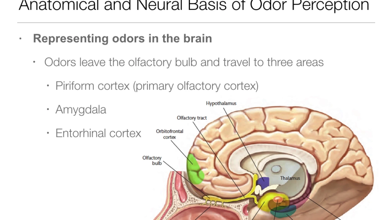
Taste and smell are the primary chemical senses that detect chemical stimuli in the environment; taste involves five basic tastes (saltiness, sweetness, sourness, bitterness, and umami) detected by taste buds on the tongue through specific receptor cells using different molecular mechanisms (simple ionic changes for salt/sour, G-protein coupled receptors for sweet/bitter/umami), with information transmitted via cranial nerves VII, IX, and X to the nucleus of the solitary tract and then to the thalamus and insular/frontal taste cortex; smell involves olfactory receptor neurons in the olfactory epithelium expressing single odorant receptor proteins, with axons converging onto glomeruli in the olfactory bulb before projecting to multiple cortical areas including the piriform cortex, amygdala, and entorhinal cortex, uniquely bypassing the thalamus in a direct pathway to cortex.
Taste and Smell Explained: Chemical Senses | Neuroscience Lecture
Added:we were now transitioning to another set of of the sensory system so we spent two lectures talking about the visual system it's a very complex system the sensory organ the eye is very complex and there's a lot of very interesting processing that happens within those systems we're now going to discuss two other sensory systems all in one lecture they share some similarities uh but obviously they're also very different and um the we will be talking about these systems uh all in one lecture because they're buying relatively more simple and there's also a little bit less than we understand about these systems than we do about the visual system so today we're talking about the chemical senses that is the main commonality that it is that these two systems have and we're going to be talking about the sense of taste and the sense of smell but before we get into that i wanted to discuss here in this lecture the facial nerves so this is a very important concept within understanding the nervous system and uh and and so we're going to cover this um in some detail right here and we won't be going over this uh thoroughly but it is something that is critical that you are familiar with and something that you you understand there are 12 major nerves that leave the central nervous system in the head and innervate various parts of the face these nerves uh can be they use roman numerals to describe them and it goes from one to twelve we won't be talking about all of them and i don't necessarily expect you to memorize all of them you do need to know the nerves that we talk about and you should know which number those nerves are okay so i do expect you to have memorized the number for the the nerves that are going to be most important that we're going to be talking about now the nerves nerves can be broken into two main categories there are sensory nerves and then there are motor nerves the sensory nerves obviously sense the world and then there are motor nerves that control the muscles the there are some nerves that are actually are mixed as well but primarily you're going to find that many of the nerves are specialized into one or the other we have talked so far about the visual system and the main nerve there is the optic nerve which goes here into the optic chiasm the optic nerve is connected to the eye and this is known as the secondary the second facial nerve okay so this is facial nerve number two the optic nerve we will be talking about three nerves that are critical for taste the facial nerve which is nerve seven the glossopharyngeal nerve which is nerve nine and the vagus nerve which is nerve 10.
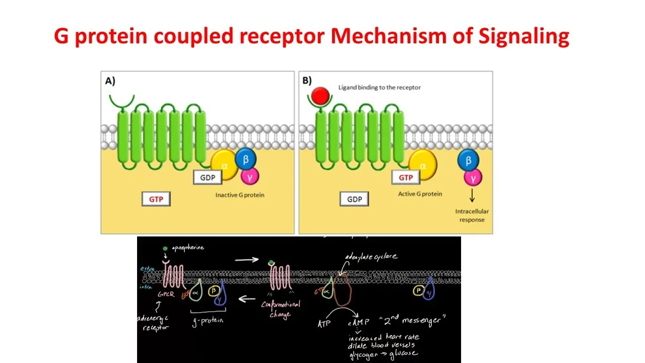
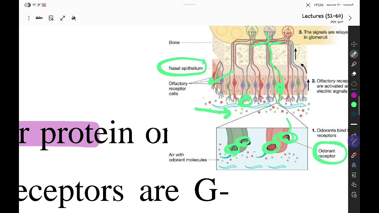
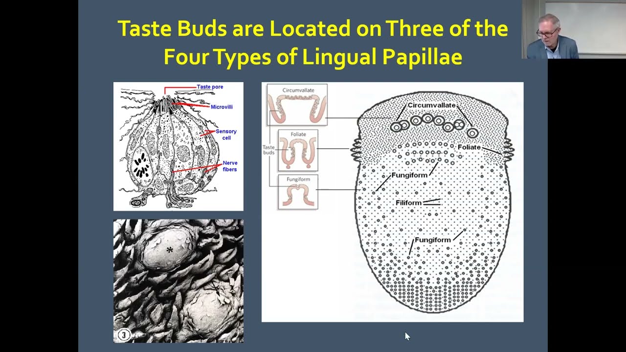
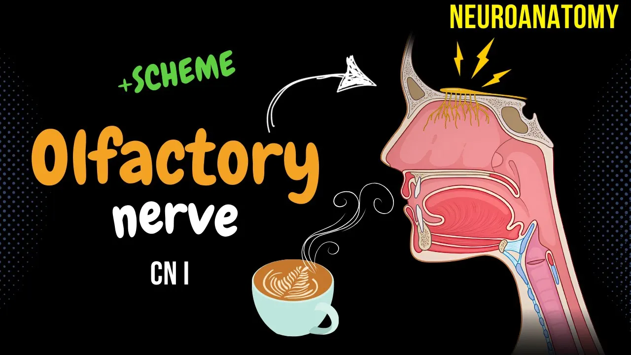
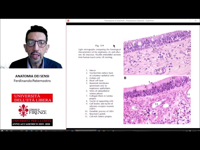
all of these innervate the tongue and then the second half of the lecture we're going to talk about the olfactory system and that is nerve number one the olfactory nerve going from the olfactory bulb to the brain so those are the three nerves that we uh sorry these are the three categories of nerves okay so let's talk about taste humans recognize five basic tastes we have saltiness sweetness sourness bitterness and umami these are self-explanatory but umami is a relatively new taste everyone for centuries has known that we could have these four basic tastes but umami was one that was recognized as a very specific category um it was originally identified by by the japanese and they're the ones who named it so um so it has this japanese name umami which means delicious in japanese and it provides um there are there's a specific subset of taste buds that have umami receptors on them that help us to detect the savory taste of amino acids and in particular it detects glutamate and i'm sure you all have heard about msg and how bad it is but msg is just monosodium glutamate it is essentially a glutamate salt and so the debate about how bad msg is is relat is complex and since it is basically just an amino acid but the reason why that that amino acid is added to foods is because it gives a very savory meaty kind of taste okay so we got five basic tastes but there are all kinds of different tastes out there when you eat pizza or lemon ice cream or bubble tea any of these things um you know there's a lot more than just five of them right so with this limited number of basic tastes how do we perceive the countless flavors in food and drink well that's because foods activate distinct combinations of taste receptors so for instance lemon ice cream you're going to have a very strong combination of sweetness plus sourness that gives it its unique set of tastes the flavors are also combined with the smells as well and smell helps uh to give us the um the combination of sensory inputs that gives us the distinct experience of particular tastes lemon also has its own unique smell as does pizza and so those things also help color how what we describe as taste texture and temperature also contribute so the sensations of uh pain which we will talk about in a bit uh in in the next lecture i think yes next lecture um so having spicy flavors such as uh hot sauces those that actually are not acting directly on to the um taste receptors at least not exclusively there are also pain receptors that help carry the information of of of being spicy or minty or other things as well which contributes to this process as well we're not going to talk about those today we'll talk about those in the next lecture so taste is primarily a function of the tongue there are other regions within our mouth that also have taste sensitivity but the tongue has the vast majority of them taste sensing cells are found in what are called taste buds that are distributed on the surface of the tongue and these other regions so most taste stimuli they're called tastings are non-bilateral uh and they're hydrophilic so these are things that like to be diluted into water and they don't just float around like odorants do that we smell so these are going to be soluble in saliva that allow us to process what we're actually tasting so it has to be able to be put into liquid format the perceived intensity of taste is usually directly proportional to the taste and concentration so the more salt that you have on something you're going to increase the concentration of salt which is going to make us perceive things to be particularly salty for instance okay many of us have this impression or have grown up with the idea that we have very distinct regions of our tongue that are specifically directed towards um a particular taste so in the back we uh we have this assumption that that's where bitter taste is sours on the back side salty's on the front side and then sweet right up at the front now there is it is true in general that there is sort of regional um increased sensitivity as you can see here so that it is true that sour is generally going to be better detected on the sides of the tongue and then in the middle bitter towards more in the back but it is not true that that's where these receptors are exclusively found in fact there are the receptors for these four different tastes five tastes are found all across the tongue it's just that their density tends to be a bit higher in certain parts of the tongue so if you touch something that's bitter towards the tip of your tongue you will taste it there and it's not because it's slipping towards the back of the tongue it's because there are bitter interceptors located towards the front of the tongue same for salt and put that towards the back of the tongue there are salt receptors all the way in the back of the tongue it just so happens that the higher density of those salt receptors are located towards the front so there aren't distinct regions uh where you're only going to find that sensitivity and in fact you'll find sensitivity across the whole tongue it's just that the density tends to differ depending upon where you're talking about on the tongue tastings are detected in receptive specializations called papillae which contain taste buds and taste cells so we're going to talk about these papillae a little bit each so papilla is the plural form papilla singular so a papilla has one to several hundred taste buds that contain neuroepithelial taste cells let's see this is what a papilla looks like right here it's these kind of big bumps on the tongue that's a papilla there that's a papilla there but if you zoom in and look what you'll see is yes there's this bump but then there's this ridge that actually is like a deep canyon that goes down into the tongue and then if you get closer and zoom in even more you can zoom in on what then is an individual taste bud so this is what the taste bud looks like in cross section taste bud will have a taste pour uh several different taste cells and then there are additional cells called basal cells and then gustar gustatory afferent axons we will talk about uh the organization of these different taste cells across the different tastes in a little bit more detail across the next several slides now each person has around two thousand to five thousand different taste buds but there's actually a huge range of variability across individuals there's some people who have really poor senses of taste those individuals tend to have fewer taste buds as few as many as 500 taste buds across their entire mouth but there are some people that are super tasters they can have as many as 20 000 taste buds that's a lot so there's a huge range but on average it's somewhere between two to five thousand teaspoons so these um taste buds have an apical end which is located here and that has the uh taste pour and that is what interacts with the saliva so saliva comes down into these channels and then the chemicals that are within the saliva will interact uh there at the taste pore where the taste cells have little microbially that attach that are interacting with the chemicals there basal cells are the stem cells that are continually dividing to replace taste cells your taste cells actually have a life cycle they're present for some time and then eventually they just die so the taste cells are not neurons you can just look at how they look you can see they're definitely not neurons sort of like the photoreceptor cells so taste cells in fact are epithelial cells sort of deriving from the same uh parts of the embryo that skin does so is the nervous system the taste receptor cells they form synapses with the endings of gustatory afferents near the bottom of the taste bud so they have this kind of bulb like shape and then they have the synapses down near the bottom the type of that you can find at any given synapse is going to be dependent on the specific stimulus type that that particular taste cell is sensitive to so sour and salty taste cells they use serotonin as its neurotransmitter to interact with the primary gustatory afferent uh neurons so it's serotonin that is released from salty or sour neurons the other three taste cells sweet bitter and umami they use atp as a neurotransmitter that acts on to those gustatory neurotransmitters released onto the uh within the synapse onto the gust gustatory afferent and that causes that's uh that that sensory neuron to to fire an action potential so that's saltiness it's a pretty straightforward process it's simply driven by changes in sodium concentration sourness uses a somewhat similar mechanism so sour tasting food is due to high acidity it's very acidic acids have low ph and ph is determined by the imbalance of free hydrogen ions so with more free hydrogen ions in a solution the more acidic it's going to be so let's say you eat some of that lemon ice cream you there's a relatively acidic citric acid it's going to cause that's going to include a relatively high level of free hydrogen ions that gets into your saliva and so then from there hydrogen can affect taste receptors by binding or blocking potassium channels so that'll block the potassium channels but then it also includes um going through high uh proton channels so then you can have this depolarization that happens by the potassium channels being blocked and hydrogen flowing through sort of like sodium being positive so you have this cation flowing through this leads to membrane depolarization which from there then causes the same basic mechanism that's found in salt cells with an increase in intracellular calcium leading to the vesicles that are filled with serotonin or transmitter to bind to the cell membrane of the sour taste cells leading to serotonin release causing the postsynaptic uh gustatory axon to have an action potential pretty sim similar and simple mechanism all right from here uh now we're going to talk about the three other categories of taste cells and they have somewhat more complex mechanisms isn't simply just differences in iron concentrations so bitterness sweet and umami they are sensed via dimers of g protein coupled receptors we refer to them as ti1 uh t sorry t1r and t2 are proteins that's the so they have these dimers that will give you a complex set of of more specific receptor types so with bitter there's actually 25 different types of bitter receptors in humans and and so one of their major functions is to act as poison detectors poisons um have certain compounds in them we have evolved a wide variety of these t2r proteins to allow us to detect a range of different things that we refer to as bitter bitter a lot of people don't care for bitter at least bitter is sort of a an acquired taste in many cases such as like coffee kids will kids will tend to not like coffee and you have to kind of grow to appreciate coffee as an adult that's because there's all kinds of bitterness that are in there and in fact like raw coffee has all kinds of poisons and toxins for other animals for humans it's not so bad so the process then is that bitter tasting foods uh there'll be some kind of protein or compound that'll bind to a particular t2r g protein coupled receptor this diamond this induces um stimulates the enzyme of phospholipid c plc right here so this g protein coupled mechanism you get to a secondary messenger system that increases the production of ip3 ip3 triggers the release of intracellular calcium stores so we've got calcium stores endoplasmic reticulum typically um induces release of calcium into the cytoplasm the calcium uh uh opens up sodium channels and then this calcium so then leads to further membrane depolarization the calcium also then will trigger then the release of atp and then atp acts as a neurotransmitter at these synapses where it acts on atp specific receptors that then causes an action potential in the accurate sweetness sweet tastings are natural and the artificial forms they're detected by the same taste receptor protein so with sweetness we have a much more simpler family of taste receptors they are also dimers of g protein couple receptors but we only have two t1 receptor types we have t1 r2 and t1r3 if either of these are present um then these are going to be sweetness receptors and if there's a mutation uh in in one of these then um or if it's missing then sweetness may not be perceived you have to have one of these two in order to detect sweet sweet tastings such as glucose for instance or sucralose or sucrose fructose they will bind to this g protein coupled receptor to the dimer and subject like i said if you have a mutation in one of these or one of them is missing you can't detect it because you can't move the dimer this leads to the activation of the plc increases ip3 production triggers the release of intracellular calcium stores calcium stimulates atp release to activate the postsynaptic pyrogenic receptors which is sensitive to the atp so it's very very similar processing um and obviously we have similar proteins that enable us to detect bitter and sweet tastes so how do we then not confuse bitter tastings from sweet ones it has to do with the specific receptor protein so the receptor protein that is taking the sensory perception um uh this this reception and the transduction side is allowing for the um taking chemical information in the environment perceived by these particular receptors and then those particular receptors are found in different taste cells we have a sweet taste cell and a bitter taste cell you won't have a cell a taste cell that expresses both bitter and sweet so the the the the differences that we can see between bitter and sweet is that they are different in different act they're in actually different taste cells okay and then they connect to different gustatory axons so the transduction of this information into the brain then goes down very separate pathways you will have a sweet pathway and a bitter pathway and it keeps the umami so let's walk through mommy a little bit these receptors also involve um a g protein coupled receptor mechanism requires t1 r1 and t1 r3 so you need to have this g protein coupled receptor dimerization and it has a very again a very similar mechanism so the amino acids such as glutamate will bind to this monomial receptor activating a plc mechanism leading to increase in ip3 production this triggers the release of calcium stores and calcium stimulates the atp to act on the postsynaptic cell so how do we not confuse umami bitter and sweet again the receptor proteins for each type are expressed in different taste cells so you have umami taste cells bitter taste cells sweet taste cells they all have a very similar internal molecular mechanism but the difference is these g protein coupled dimers we have two different kinds of dimers it allows for the specificity of the kind of tasting that it's going to perceive and each taste cell has its own only one particular combination of g protein coupled receptors okay that's the difference all right so then how do we get the uh um uh the how do we get transfer of this information up into the brain as i mentioned before we have three facial nerves that innervate the tasting areas of the mouth we have the facial nerve which innervates the first two thirds of the tongue so the facial nerve is the seventh uh um facial nerve um the uh um the glossy pharyngeal nerve which is nerve cranial nerve nine cranial nerve nine innervates the back third of the tongue and then we have the vagus nerve which innervates the epiglottis but there are all kinds of taste receptors back there as well so the the vagus nerve the glossopharyngeal nerve and the facial nerve these are all carrying that the the five different uh taste information or uh up into the central nervous system each of these nerves synapse the ones that are carrying the sensory information they synapse onto the nucleus of the solitary tract this is also known as the gustatory nucleus so that this nucleus is really the first main station within the central nervous system that's receiving this taste of information and then that then ascends up into the brain uh synapsing onto the ventral posterior medial nucleus of the thalamus so it's now carrying this information to the thalamus which we've described before as being this relay station that's critical for carrying sensory information to the cortex so and then this is showing again the synapses so this part is shown here so these are the ascending pathway from the um nucleus of the solitary tract synapsing onto the ventral posterior medial nucleus of the thalamus thalamus okay so the ventral posterior nucleus of the thalamus then uh these neurons synapsed onto the insular taste cortex and the ossible synapse onto the frontal taste cortex so two different brain areas the insulin and in the the frontal cortex as well and those are going to be the main initial parts of olfactory cortex okay so this is a very simplified schematic of what this looks like um there are parts of this that we are not going to talk about in detail now instead we will talk about the when we talk about these brain areas such as the hypothalamus and the amygdala but i but this includes some of these important parts right so the first four stations that we have the taste buds on the uh on the tongue sensing the taste information it is carried on these three cranial nerves the facial facial glossopharyngeal and vagus um though that information is carried up to the nucleus of the solitary tract also known as the uh gustatory nucleus up to the the ventral uh posterior portion of the medial thalamus or vpn of the thalamus um and then that connects to the insula in the temporal lobe and the frontal cortex cortical areas that are olfactory cortex okay then from there that car that taste information is then processed by other brain areas especially the amygdala and the hypothalamus but like i said we will cover those when we get to that gustatory cells they also project to other brain stem regions involved with swallowing gagging and vomiting this is in more instinctual circuits that allow you to throw up so if you have something in your mouth that's particularly gross uh these circuits will be activated and cause you to throw up so i will let you discuss uh try to figure out what are the four basic steps for sensory pathways and taste again sensory reception transduction transmission and perception and i want you to be able to do and describe all these four basic steps and the sensory pathways for all the sensory pathways that we're going to be talking about so if we talk about what sensory interception is for vision taste smell uh touch or hearing i want you to be able to to give some detail about what each of these different steps are and know what are the similarities differences between each of these the sensory systems all right that is taste now we're going to switch over to smell smell your sniffer your nose um it's actually a very interesting and um i think a relatively complex uh system it's actually quite elegant quite beautiful it's one of my favorite um sensory modalities um and i hope that you'll grow to appreciate it just as i do um you know it's the only way that we can you when you smell the roses there's actually a lot of beauty happening inside your head to make that happen that's very poetic okay so we have the basic function of smell is you inhale air it's going to be filled with all kinds of chemicals called odorants and then it's going to touch upon what's called the olfactory epithelium so our nose itself the part right here that uh that you can kind of see and just on the inside that isn't what is doing any of the smelling instead the air actually has to go up quite up inside the nasal cavity and it interacts with the olfactory epithelium which in humans is located on the top part of the nasal cavity so the olfactory epithelium is a very thin sheet of cells and it's comprised of three different cell types olfactory receptor cells neuron and these are these are actually neurons so unlike taste cells and photoreceptor cells olfactory receptor cells in fact are neurons and they transduce the olfactory signal and send that information into the central nervous system there are also supporting cells these supporting cells act very similar to glia um they um but they also produce mucus to keep which to create this mucous layer at the surface of the epithelium and then there are basal cells and these are basal cells that are very similar to the kinds of basal cells you find in uh in the taste system in that they uh they are stem cells that also produce new receptor cells so these basal cells will divide and give rise to new receptor cells so there's turnover receptor cells as well uh just like the taste cells that connects to the olfactory bulb sorry too fast so you have membrane depolarization which can which then as long as there's enough odor receptors that are activated uh leading to an action potential and these olfactory receptor cells slash neuron okay so we talked about the taste receptors that like sweet there's basically one combination of the dimer g protein coupled receptors same for umami bitterness there's a little bit more variety you have 25 different kinds giving you a little bit more complexity to the bitter tasting right so there's uh you can imagine the there's a wider variety of chemicals that we can pick up and and and perceive as bitter olfactory receptor proteins blow all those out of the water there's a huge range of olfactory receptor proteins and each individual neuron will express a single olfactory receptor protein each olfactory receptor neuron is going to have a particular odor receptor that it expresses which is why we have these different colors here to represent a particular receptor that each neuron is expressing um the number of olfactory receptor proteins varies enormously across species animals that are particularly olfactory sensitive such as rodents they have more than a thousand different receptors that they express uh in fact um you know if you think about each receptor it has to be expressed uh has to that information has to be contained in the genome nearly something like i know in rodents it's something between a thousand and two thousand of the genes that make up the rodent genome which is thirty thousand genes those are just odor receptors they have a huge range of voter receptors that they express humans we are not particularly olfactory uh um centered so we have a lot less than rodents we have around 350 odor receptors in our genome so because each olfactory receptor neuron expresses a single factor receptor protein um that gives us then um a specificity of that particular neuron being fired but we have all kinds of different smells that we can pick up right and again that has to do with the combination of the different kinds of receptors that uh neurons that have been activated so the olfactory receptor neurons will fire they don't necessarily have super high specificity to a single chemical some of them do but many of them will have will pick up different kinds of chemicals and they'll have differing levels of sensitivity depending upon the chemical and how much of the chemical is there so for instance let's say we're looking at this particular green olfactory receptor neuron you can see that this neuron particularly particularly like citrus the the citrus chemicals it kind of likes floral chemicals it has a few action potentials but not a lot it also seems to like peppermint but it doesn't respond to almond at all uh the red neuron though doesn't like citrus but it also but it's particularly sensitive to almond and it also happens to be sensitive to peppermint so you can see that okay we don't have super high specificity but instead because different chemicals are going to act on different olfactory receptor neurons in different ways it is the combination of the response that'll tell you which odor that you're smelling and this is what's referred to as a population coating scheme so if we are smelling citrus you're gonna have these green neurons that are firing an awful lot a little bit of the blue and none of the red and that will tell you hey there's some citrus in here if we have almond there we're not gonna have any green tiny little bit of the blue and but the red responding quite a bit that will tell you that that's almond and it'll be this combination this population coding scheme that tells us hey this is almond or citrus or both right so a little bit of the history of this and how this was discovered richard axel and linda buck they were both winners of the nobel prize in 2004 and it was linda buck she was working as a postdoc in richard axel's lab um she discovered she and him as well as other people but they discovered the the family of odor receptors and they worked out a lot of these details of that different olfactory receptor neurons express just a single uh odorant receptor but that there are hundreds and hundreds and hundreds of different odorant receptors that are found and they also worked out some of the details of that g protein coupled mechanism as well that got them the nobel prize in 2004.
central olfactory pathways then um so this is another part of the system and i find particularly elegant and it's actually quite beautiful to look at in uh brain slices as well so uh so let's walk over it the um so again we have the olfactory epithelium right here at the the top of the nasal cavity and you've got all your olfactory receptor cells and the olfactory epithelium there's the little cilia there so let's say an odor uh receptor neuron starts to fire um and here's a group of them that then send their axons uh up to the olfactory bulb so now this is where we have our first uh part of the central nervous system getting involved so these axons connect to the olfactory bulb that's our first order neuron and then from there we have second order neurons there are neurons on the other side of that that then send their axons up into the cortex in fact so this interaction is happening at what's called a glomerulus and we're going to look at a little bit more detail of what's happening in a glomerulus and learn a little bit more about the secondary order olfactory neuron fly there is specific mapping of olfactory cells and receptors onto specific glomeruly within the olfactory bulb so the olfactory bulb on the outer surface it'll be covered with all these little circles called glomeruline glomeruly is plural glomerulus is singular each glomerulus is receiving input from uh recep olfactory receptor cells that express only a single receptor protein so these blue cells they have one particular olfactory receptor protein that they're expressing and all that information is connected into a single glomerulus so you can see we're now sort of gathering the sensory information and organizing it into us an olfactory receptor specified way so the blue uh receptor receptor neurons are connected to this glomerulus red is over here and green is over here and this is referring to the different kinds of receptors that are expressed another note on the olfactory bulb so just as the rat olfactory epithelium has a very large representation in its nose relative to its brain compared to humans this is also reflected in the olfactory system itself and the olfactory bulb in rats is huge it makes up something on the order of about twenty percent of their entire brain whereas in humans it's less than one percent so that's this is the old factory involved in humans it's super duper tiny and um yeah so very tiny compared to uh rodents so here is a missile stain section of a cross-section of a mouse olfactory bulb okay so this is remember nissl labels cell bodies and so what you're seeing are the different cell bodies and a cross-section of the olfactory bulb has uh it reveals that there are different layers so you have the glomerular layer and that's where all the glomeruli are located and you can see these empty spaces here that's this is a glomerulus here and that's where those um axons from the olfactory receptor cells are connecting to dendrites of the second order neurons then we have a layer called the outer plexiform layer then we have a layer called the mitral cell layer and so the mitral cells are really the secondary order axons uh neurons and then you have the inner plexiform layer and then you have granule cells down below and so there's a ton of these granule cells located here here's a schematic of how this is organized okay um so you've got your olfactory epithelium the olfactory receptor cells going up through the bone and connecting to the different glomeruli and then the glomeruli are connected to primarily mitral cells mitral cells are the main olfactory uh receptor output from uh that's carrying the olfactory receptor information to the brain these are the important parts that you need to know the olfactory receptor cells obviously you need to know those the glomerulus the mitral cells and the granule cells granule cells are also important to this process right so just so that you're familiar like what what we're looking at here this this was the dorsal side of the olfactory bulb right here we were looking over here and the uh um and so it's reversed compared to what this is describing here right so the glomerulus is down here and the mitral cells are right here well here we're looking at the glomerulus over here in the mitral cells here because they're describing it from the ventral side of the olfactory bulb but this side is showing the dorsal side so you need to know that like depending upon what side of a structure that you're looking at you'll have to know that order okay so if you're looking at the dorsal side you'll need to know that the glomeruly are on the outside or if you're talking about the ventral side a glomerular located on the bottom okay top box you got to know the order but the order may be reversed depending upon whether you're talking about the top or the bottom dorsal potential all right so these mitral cells these are the critical outputs uh going through the olfactory neuron which is the first cranial nerve they connect up into multiple areas of the cortex so they project to a wide variety of cortical areas so this is showing a couple in one cross section a bunch of different areas of cortex we've got the piriform cortex the olfactory turbical accessory olfactory nucleus the amygdala is located here the intel rhino cortex i'm listing all these names but these are just different cortical areas that receive old factory input including the amygdala and then this is showing in that same section so this is the same section but now it's showing in in red the axons coming from mitral cells and these mitral cells they will split off their axons and they'll connect to all kinds of different olfactory areas throughout the brain so these the mitral cells we learned in the previous slide they project very widely throughout the central nervous system um they and this figure is showing a in a little bit more schematized version of what particular areas receive input from the mitral cells so this is the olfactory bulb here in this part so we're looking at a human brain here are the olfactory receptor cells and so this would be this is a very schematized version the synapse would be normally in a glomerulus but it's just showing it projecting to an olfact a mitral output neuron these neurons they project to a bunch of different areas an area called the olfactory tubercle they project to a bunch of other olfactory cortices including interrinal cortex the piriform cortex and then as well as the amygdala which some of you probably have heard of we have an entire lecture dedicated to the amygdala later when we talk about fear and stress and it will come up over and over again but it also receives input directly from the old faction one important point here that i hope that you can see is that there is a direct parallel projection to the olfactory cortex that bypasses the thalamus so there is a connection that kind of makes its way through the thalamus which is through the olfactory tubercle technically this is actually part cortex so it goes from the sensory system cortex goes through thalamus and then up to the orbital frontal cortex but there's a ton of other information that goes straight from the olfactory system directly to cortex this is the only sensory system that has a substantial bypass of the thalamus and connecting directly to cortex itself all the other systems don't they don't have a direct connection to cortex and it is thought that this is part of the reason why smell has kind of a different salience when we have when we talk about memory that in some respects you get this strong sense of deja vu when you smell something it'll bring up some kind of experience and it'll be very different than if you were seeing or hearing something but then there are other times when it's really hard to place that memory uh that that you know that you're experiencing some kind of emotion or some sort of feeling but it's not something that you can place on a very specific event and recall the event that often happens with smell it is thought that part of the reason why that might be is because so much of the olfaction bypasses the thalamus and goes to cortical areas so it's not being organized by the thalamus and gating its way through certain memory centers and instead is dedicating itself to olfactory specific parts of cortex really interesting stuff a lot of sort of mystery around it as well so to review this again we've got the olfactory receptors connected to the olfactory bulb to a variety of olfactory bulb targets sorry yeah targets of the olfactory bulb including the internal cortex and then from there we have a number of other secondary structures and again we will discuss amygdala and hippocampus later so we'll discuss like the role of smell in partner preference and how it can affect the hippocampus and that kind of thing we'll talk about orbital frontal cortex and smells much later in the semester when we talk about these brain areas but because i haven't described these brain areas we can't quite get into it too much um instead you just kind of really need to know this pathway that the olfactory bulb connects to these areas okay so that's going to be the important part and then we'll get to the to the other parts later all right so there are a bunch of different ways that neuroscientists can measure neural activity in live animals and so far in just in the last lecture you've seen two major important ways to look at neural activity in live intact animals one that we saw was doing electrophysiological recordings electric electrophysiological recordings of individual neurons or groups of neurons using a glass pipette or say an electrical wire this gets lowered into a particular brain area and then you can record from a particular group of neurons or even an individual neuron we saw uh what people in weasel did recording in the visual primary visual cortex and cats this is a very common technique it is widely used and it's been used for decades and we will be revisiting it over and over again the readout of this information is spikes right so we'll have raster plot for instance like we saw with the uh facial recognition um in uh in monkeys and also the halle berry cells in humans uh you you see individual spikes and you have to summarize sum up what that activity looks like over time across multiple trials in order to see patterns and change that is how electrophysiological recordings are are analyzed we also have functional magnetic resonance imaging or fmri there's a couple of other different kinds of imaging which we will see across the semester but last time we talked about that in humans you can see changes in blood flow in uh in this brain area called nt which is critical for looking at and detecting movement within the visual field and mt is particularly sensitive to movement and so for this you can put a human inside a machine show them something so this human would be seeing something and it's got like a little readout here pushing some buttons or something some kind of task is going on but they're in this giant fmri machine and it can detect changes in blood flow across the entire brain in real time and it'll look at changes activity happening on the order of seconds so it doesn't provide quite the resolution that the electrophysiological recording does of of an individual neuron or like a handful of neurons but it looks at big brain areas across time but it's a very non-invasive way to do this and it's oftentimes the only way that we can get any reporting of activity in humans and sometimes this is also done in animals as well so those are two ways of looking at measuring measuring neural activity in live animals i just want to emphasize these things because it's really critical that you know uh what these different kinds of techniques are so we're going to talk about another way of measuring neural activity in live animals and this is becoming a more and more common way of doing this kind of work and it's really kind of exciting and cutting edge almost science fiction-like and it uses um it takes advantage of the fact that there's a change in intracellular calcium that occurs when a neuron fires so uh a a neuron at rest will have a certain relatively low level of intracellular calcium there's intracellular stores of calcium like in the endoplasmic reticulum and then you have calcium channels that are closed and pumps that are continuing to push out calcium out of the cell so intracellularly it's relatively low calcium concentration is low at rest as soon as there's an action potential the calcium channels open and there's also there can be release of calcium stores happening from the endoplasmic reticulum there will be an increase of calcium concentrations within the cytoplasm of the cell so that will go up alongside an action potential so we're looking at membrane voltage here potential goes up it's depolarized after hyperpolarization but over this whole time course we have this sharp increase in calcium and then it slowly goes down over time and by slowly i mean on the order of 100 to 200 milliseconds compared to an x potential so we have all this calcium within the cell and over the last 15 years or so scientists have been using genetically modified green fluorescent protein to detect changes in intracellular calcium so what uh the most commonly used um genetic tool that's that's done for calcium uh doing calcium imaging is called uh g-can and it can detect an increase in calcium so if calcium increases what will happen is that uh one domain of this will then interact with calmodulin calmodulin is is a calcium sensitive protein it will take on the excess calcium causes this protein to translocate here which allows this green fluorescent protein to glow when you shine a light on it but if there's no calcium this protein is is now dissociated from the calmodulin and you can't get it to glow it doesn't glow very well at all so you can detect calcium calcium will be present by looking at just how much the cells are glowing and the change can happen very very quickly so that's shown here in fact so calcium imaging can be used to monitor activity in neural populations in whole animals so you can make a genetic mouse or transfect a mouse using viruses you can inject it into a particular brain area causing the neurons in that area then to express uh this g-camp uh calcium indicator so the cells will start expressing this protein and then you can make a little window on the surface of the mouse's head and lower a uh a microscope onto its skull and then uh record um using a microscope looking at the the fluorescence of cells within that area and so at any one time you might see a little bit of fluorescence but as a population of cells gets activated you will see this increase in fluorescence which will be driven by the influx of calcium happening at that time so what you're seeing here is just a repeated image over and over again of neurons that are have relatively low calcium and then some changes happen say the animal has smelled something and then a subset of all those neurons are now having an increase of intracellular calcium and you can see the sharp increase in fluorescence and so you can see that they fired and you can see which one's fired so this has been done over and over again in the olfactory system across a bunch of different labs in mice and what we've discovered is that if you're looking at a particular part of cortex um piriform cortex has been the most heavily studied but you can see similar things happening in the torment rhinocortex what happens is that when you're imaging in that area and you put on say uh one odor you will see a subset of the neurons fire so that's green here so we're this is looking at alpha piney which gives a kind of piney smell uh as far as we perceive those green neurons are firing here so and you can see them all firing all at the same time using this calcium indicator technique so all at once you can see a big population a subset of the population firing then in the next 10 seconds later you can then puff on octanol which gives a fruity or citrus smell and then you'll have a different subset of neurons that fire so then these neurons are frying they're showing the red ones fire you can see some of them are yellow and that's because those that subset of neurons fired for the alpha pine and the octanol so it's the again more population coding that's happening in these areas of the olfactory cortex that's giving you the ability to detect piney or octanol or both so let's say then we do the next one so in the next phase comparing these two different odors we have hexanol so this is the smell that gives you cut green grass and that's going to give us a green color here so we're using green for that so that's a different subset compared to the ones here but in this same animal we do octanol again you're going to see red and it's going to be the same red ones if you go from here to here you can see it's the same red ones here here here there same red ones because it's the same odor but the green one is different and so you see different neurons responding to this odor than this because it's a different set of olfactory receptor neurons that have been activated but again there is some overlap some of these neurons are receiving octanol information and hexanol information but they're not receiving necessarily the same uh neuron information from the alpha pining so here we're starting to get some convergence of this information and it's the combination of this information that allows us to tell whether we're talking about hexanol or octanol and it's that population coding activity and it's that constellation of neurons that fire upon odor exposure it gives us the salience of which odor is is it being expressed which and allows us to tell and create memories of the odorant context which odors were present at a particular time feeding forward into the hippocampus and the amygdala and allowing us to connect particular events to odorant experiences and that's how that's done from a population coding perspective so in this slide i'm just going to briefly go over the four basic steps that all sensory pathways have with respect to olfaction we have sensory reception we have odorants coming into the nose and they are sensed by the olfactory receptors that are located on the cilia of olfactory receptor neurons in the olfactory epithelium that induces a change in the membrane potential so there's transduction of that chemical signal into the kind of information that the nervous system can then read out so that there'll be changes at the membrane potential those cells depolarize and then start to fire and then there's transmission of that signal where the information is gathered into specific glomeruli that will have odor information inducing firing in mitral cells that then is transmitted up to the cortex where there is processing of that information so that it can be perceived and we talked about what happens on the population level within the cortex with populations of neurons firing with specific odorants and then a different odorant might have a different population of neurons that will fire but there may even be some overlap depending upon the odorant so to wrap up um what are the five major tastes how are taste cells organized anatomically you know what how are they connected in the papillae what does the taste bud look like um and then you also should know the key molecular events that underlie taste perception in each kind of taste cell which cranial nerves are involved in relaying taste information to the brain what are uh what are the ascending pathways as well um what are the olfactory receptor neurons and how are they organized what are glomerularly and how are they organized what is the basic circuits and how are they connected up to the olfactory bulb and then what brain areas are targets of mitral cells and what particular areas are not targets and that there's one more thing that i would add to that i suppose which is have some familiarity with these concepts of population coding and have some understanding of how uh how we record and and understand neural activity in live animals and because we're going to build upon these concepts as well all right so that's it for this lecture we'll discuss this more at the appropriate discussion section the one last thing that i want to just add to this is the this is just a helpful summary of some of these uh different the different taste receptors so it's a lot of detail here but this can be a useful summary for studying so um so i included this as well has a little bit more detail than you might need to know so everything that i discussed before um in the lecture is going to be the key parts that you'll need to know for say exams quizzes that kind of thing but this covers almost all of it so um i still think it's a useful table all right thank you very much
Up Next

Neuroscience of Taste and Smell: Gustation and Olfaction
@TheBarkerLab
292 views•2023-05-28

Deciphering Neural Circuits Controlling Anorexia in Mice | Richard Palmiter
@AllenInstitute
2.3K views•2011-10-10

Neuroscience of Vision: The Visual Pathway Explained
@Dr.ChrisThompson
3.6K views•2020-09-01

How Exercise Benefits Your Brain: Science Explained
@TED
11.4M views•2018-03-21
Related Study Plans & Knowledge Roadmaps
Structured learning paths in Neuroscience