















![[LIVE] เรียนรู้จักพื้นฐานเครื่อง Pacemaker CRT ICD](https://i.ytimg.com/vi/K21r4kv-Zdc/sddefault.jpg?v=6a620343)
Bundle branch block is a cardiac condition where the electrical signal from the AV node is completely blocked or delayed along one of the bundle branches in the ventricles, typically caused by fibrosis or scarring from acute conditions like heart attack or chronic conditions like hypertension and cardiomyopathies; it manifests on ECG as a widened QRS complex (>120ms) and can be differentiated between right and left bundle branch block using chest leads (V1 and V6), with treatment ranging from observation for asymptomatic cases to cardiac resynchronization therapy for severe cases with heart failure.
Bundle Branch Block: Causes, ECG Signs & Treatment | Pathology Review
Added:Anatomy and physiology of the cardiac conduction system, specifically the pathway from the Bundle of His to the Purkinje fibers.
Fundamentals of a normal 12-lead ECG, including the physiological meaning of the P wave, QRS complex, and T wave.
The concept of vector cardiography and how electrical depolarization vectors project onto precordial leads (V1 through V6).
Basic cardiac pathology, particularly myocardial ischemia, infarction, and hypertrophy, which can disrupt conduction pathways.
Advanced ECG interpretation to differentiate Left Bundle Branch Block (LBBB) from Right Bundle Branch Block (RBBB) using diagnostic criteria.
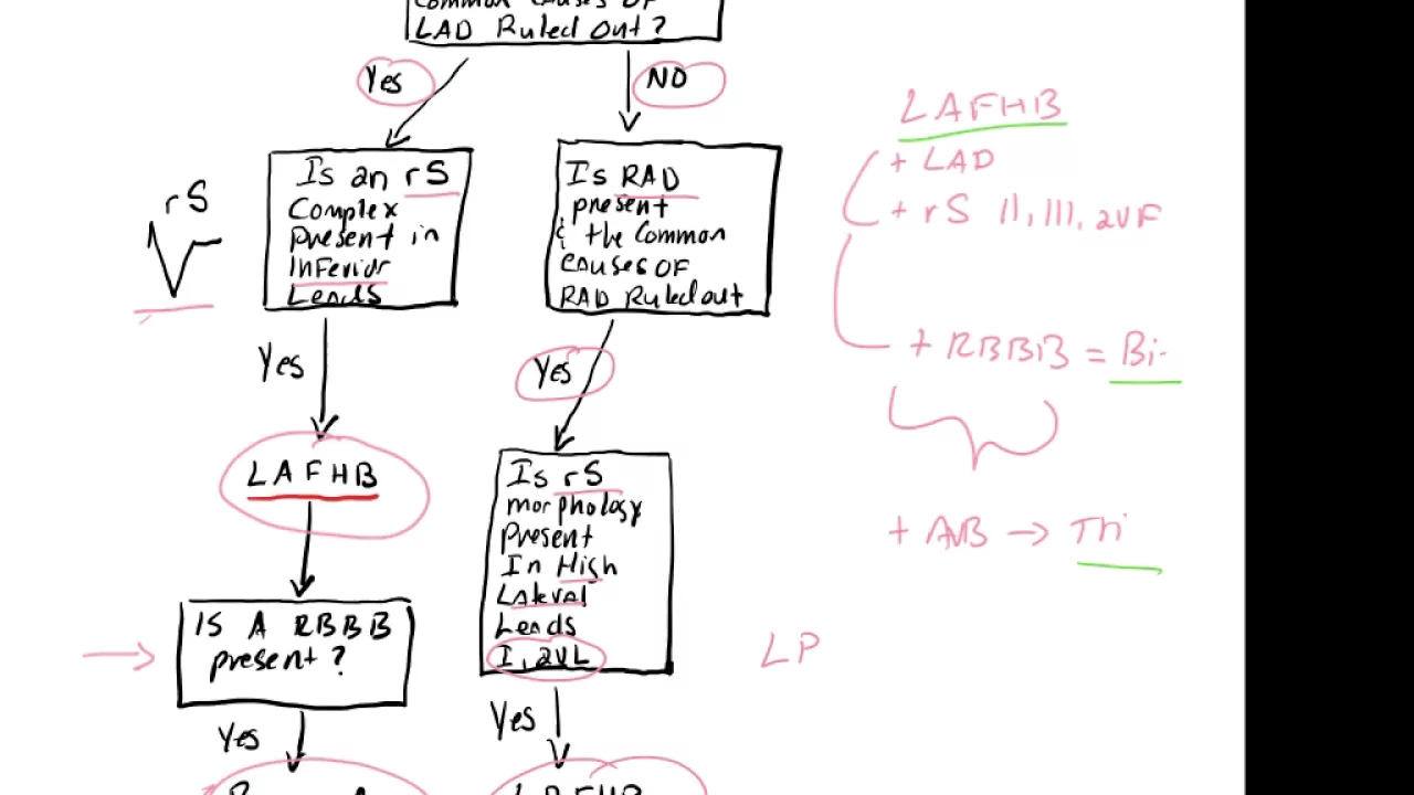
Understanding fascicular blocks (hemiblocks) and the clinical significance of bifascicular and trifascicular blocks.
Evaluating new-onset LBBB as a potential indicator of acute myocardial infarction (including the use of Sgarbossa criteria).
Clinical management of bundle branch blocks, including the indications for artificial pacemakers and Cardiac Resynchronization Therapy (CRT).
324.6K views5.4Klikes6:34Original Release: 2022-08-15
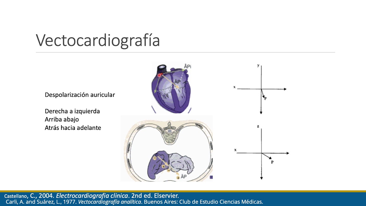
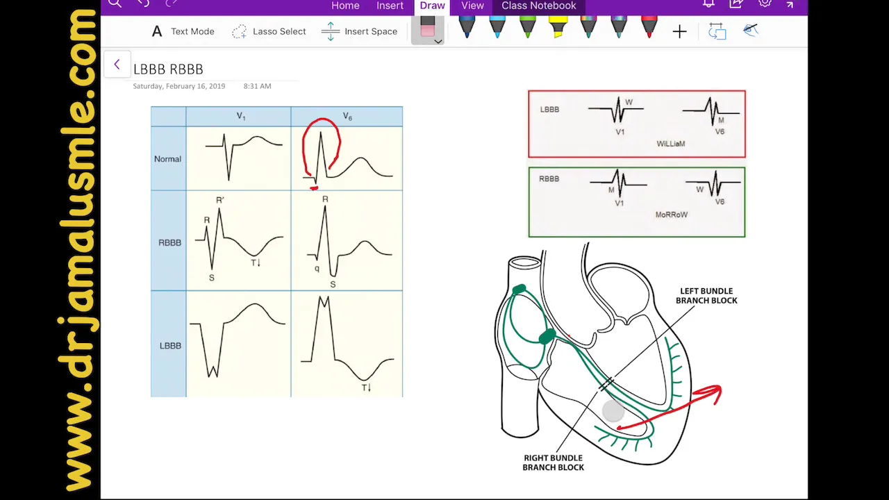
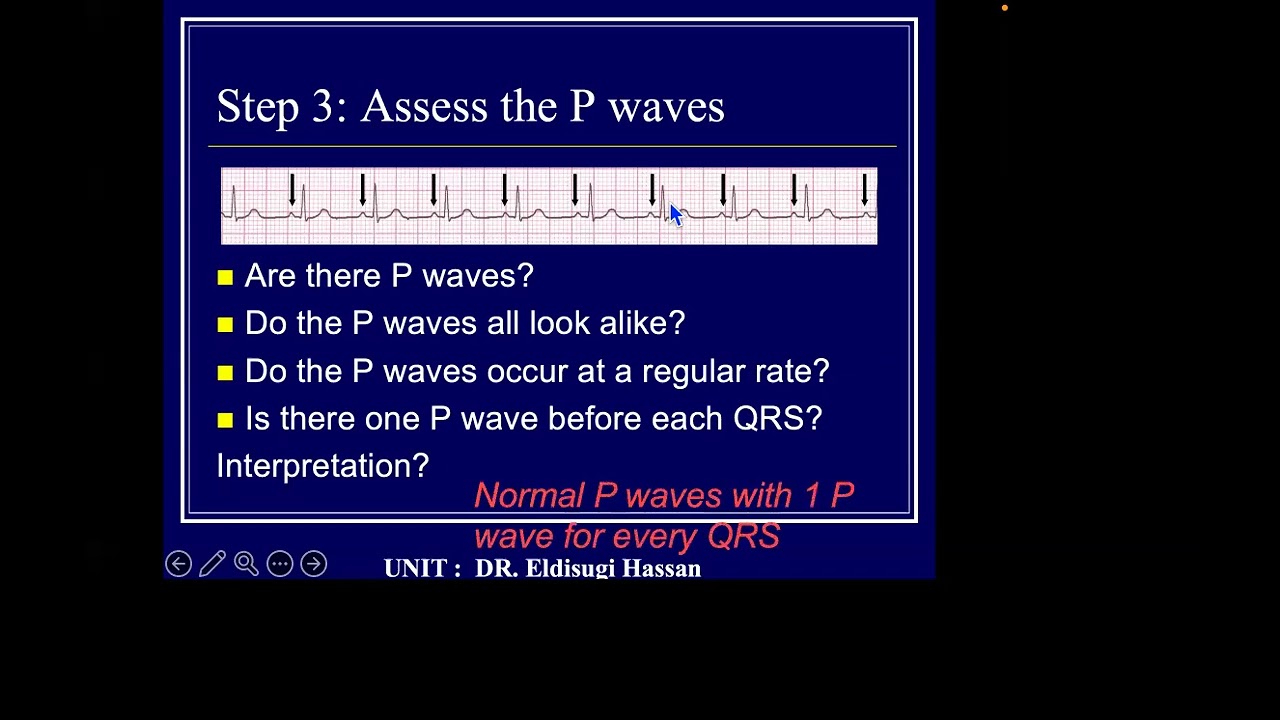
each heartbeat starts with the heart's pacemaker cells in the sinoatrial node or sometimes sa node in the right atrium the sa node sends out an electrical signal that propagates out and contracts both upper chambers then moves through the atrioventricular or av node down into the lower chambers where it reaches the bundle of hiss and then splits into the left and right bundle branches serving the left and the right ventricles respectively the signal then goes onto each ventricle's purkinje fibers which leads to ventricular contraction now a bundle branch block describes when the electrical signal gets completely blocked or held up along one of the bundle branches in most cases this block or delay is caused by fibrosis or scarring that either occurs acutely or chronically acute causes can be things like ischemia and heart attack or myocarditis inflammation of the heart tissue chronic conditions that might lead to fibrosis of the heart tissue include hypertension coronary artery disease and cardiomyopathies because they can all cause slow and steady remodeling of the heart muscle so if the block happens on the right side it's referred to as a right bundle branch block so with this type the electrical signal starts in the sa node contracts the atria moves through the av node splits at the bundle of hiss and then moves down the left bundled branch but is blocked on the right bundle branch and so the left ventricle contracts first the signal then spreads from the purkinje fibers to the left ventricle over to the right ventricle which contracts but this happens after the left contracts so the right bundle branch block the right ventricle contracts late if the block happened to be on the left side instead a left bundle branch block the signal would be delayed on that side and so the right ventricle would contract first and then the left ventricle would contract late now on an electrocardiogram or ecg normally you have a characteristic p wave which corresponds to the atria contracting then the qrs complex which corresponds to both ventricles contracting and is usually between 80 and 100 milliseconds long if there's a delay in depolarization of one of the ventricles the qrs complex gets wider because depolarization starts on time but ends later than usual since now one of the ventricles is contracting late a qrs complex longer than 120 milliseconds is common to see in a bundle branch block okay so far we've been looking at a relatively common view of the heart called lead 2 which is a type of limb lead since we get it by attaching leads to the right arm and left leg both of which are limbs even though this lead can give us some solid information it's often helpful to use other leads as well especially when differentiating between left and right bundle branch block the chest or precordial leads are really useful for doing just that chest leads look at the heart's electrical activity when viewed from a slice parallel with the ground so it's a bit different from the limb leads which look at the heart in the frontal plane which is perpendicular to the ground for chest leads they go from v1 coming straight out the front of the chest to v6 going out the left side of the chest each of these is like looking at the heart from a different perspective right v1 and v6 are particularly helpful because they're the farthest apart from each other and therefore have the most contrast between views normally the first thing to depolarize is the septum and the wave of depolarization moves from the left bundle branch toward the right ventricle so toward v1 but away from v6 meaning you'll get a positive deflection on v1 and a negative deflection on v6 then that wave moves to the ventricles themselves and since the left ventricle is usually more massive you'll get a large depolarization directed away from v1 but toward v6 so negative on v1 and positive on v6 now let's say that there's a block along the left bundle branch so now the overall direction of depolarization comes from the right side and moves toward the left so away from v1 and toward v6 meaning negative in v1 and positive in v6 v6 will also often be notched from the left ventricle depolarizing late so when it's all said and done v1 usually has a qs or a little rs complex qs meaning an absent r wave and little r s meaning a small r wave either of which give it a w shape on v6 the large notched r wave gives it an m shape all right now let's say that there's a block in the right bundle branch the first part is actually similar to the normal situation right so the septum depolarizes from the left bundle branch toward the right ventricle so toward in positive in v1 and away a negative in v6 next the left ventricle depolarizes and you get a negative deflection in v1 and a positive deflection in v6 finally as the right ventricle depolarizes in a roundabout sort of way you get a positive deflection in v1 and a negative in v6 so v1 has a large terminal r wave meaning it ends with a large r wave and this gives it an m shape on v6 there should be a slurred s wave giving it a w shape now if we look at all of these you've got a w shape in v1 and m shape and v6 for left bundle branch block but then an m shape in v1 and a w shape in v6 in right bundle branch block which sets us up for this awesome mnemonic william morrow nailed it some cases of bundle branch block are present since birth and don't cause symptoms and therefore probably don't need any treatment in a lot of cases though the block is acquired through some other heart disease in severe cases where the block is accompanied by heart failure a special cardiac resynchronization pacemaker can be implanted that has electrical leads going to both the left and the right ventricles and can paste them at the same time causing them both to contract at the same time which helps improve cardiac function all right as a quick recap a bundle branch block is when the electrical signal from the av node gets blocked or delayed along one of the bundle branches in the ventricles it's usually caused by fibrosis of heart tissue from acute or chronic heart disease it shows up as a wider qrs complex on an ecg and using chest leads on the ecg can help confirm the bundle blocks location current and future clinicians focus learn retain and thrive learn more
Up Next

14:36
ECG Diagnosis of Cardiac Ischemia: STEMI and NSTEMI Explained
@ECGSadhana
3.2K views•2022-01-25

48:47
Graphic Medicine: Comics for Collaborative Healthcare Communication
@nationalpatientadvocate
189 views•2023-12-04
11:10
Understanding Systemic Lupus Erythematosus: Causes, Symptoms, and Diagnosis
@osmosis
2.7M views•2016-05-23

3:05
Stages of Labor and Vaginal Birth | Childbirth Animation
@nucleusmedicalmedia
52.1M views•2017-08-18
Related Study Plans & Knowledge Roadmaps
Structured learning paths in Medicine
medicine
Abnormal LFTs: Diagnostic Workup & Pathology
01Liver Anatomy and Basic Physiology
02Introduction to Liver Function Tests (LFTs)
03Hepatocellular vs. Cholestatic Patterns
+ 3 more learning modules
12h6 modules
Explore Roadmap
medicine
Analyzing Blood Smears: RBC, WBC & Platelets
01Introduction to Blood Components & Smear Preparation
02Normal Morphology of Blood Cells
03Erythrocyte Pathologies & Morphological Variants
+ 2 more learning modules
12h5 modules
Explore Roadmap
medicine
Mechanical Ventilation: Waveforms & Alarms
01Anatomy and Physiology of Breathing
02Introduction to Mechanical Ventilation & Core Modes
03Ventilator Settings and Waveform Interpretation
+ 2 more learning modules
12h5 modules
Explore Roadmap
medicine
Neuro Exam: Localization & Stroke Syndromes
01Foundations of Neuroanatomy and Vascular Supply
02The Neurological Examination: Step-by-Step
03Principles of Neurological Localization
+ 3 more learning modules
18h6 modules
Explore Roadmap