The MRC (Medical Research Council) Scale rates muscle strength on a 0-5 scale: Grade 0 indicates no muscle contraction; Grade 1 shows only a trace or flicker of movement without limb movement; Grade 2 allows full range of motion only when gravity is eliminated (tested in side-lying position); Grade 3 enables movement against gravity without resistance; Grade 4 demonstrates weakness with moderate resistance; and Grade 5 represents full strength against maximum resistance. For accurate assessment, clinicians must compare both limbs and use appropriate patient positioning for each grade.
MRC Muscle Strength Grading: A Comprehensive Guide
Added:Deep Dive
Basic anatomy of the musculoskeletal system, specifically the structure and function of the knee joint and the quadriceps femoris muscle group.
무릎에 제일 좋은 근력 운동
4.4M
The knee joint consists of two joints: where the femur meets the tibia, and where the femur meets the patella. The patella is embedded within the quadriceps tendon, making it essential for knee function. The quadriceps femoris muscle performs critical functions: absorbing impact during walking, generating propulsion during running, supporting body weight during stair climbing, enabling the patella's gear-like rotational movement, and managing synovial membrane movement within the knee joint. As people age, the quadriceps becomes increasingly important for knee health. Strengthening this muscle is crucial for maintaining knee function and preventing age-related decline.
Click to watch →
【膝×筋肉】膝関節の動き&主要な筋肉がすべてわかる動画
39.5K
The knee joint is a hinge joint (ginglymus) located between the femur and tibia, composed of three bones (femur, tibia, and patella) that allows primarily flexion and extension movements. The knee's main muscle groups include the quadriceps femoris (the largest muscle group in the body, consisting of rectus femoris, vastus medialis, vastus intermedius, and vastus lateralis) responsible for knee extension, and the hamstrings (semimembranosus, semitendinosus, and biceps femoris) responsible for knee flexion. Additional muscles include the gastrocnemius (calf muscle) for knee flexion and plantar flexion, the popliteus for knee stability, and the sartorius and gracilis for knee internal rotation. The quadriceps femoris has the largest muscle volume (approximately 1,900cc), followed by the gastrocnemius (3,202cc), and the hamstrings collectively (approximately 900cc).
Click to watch →

Knee Muscle Anatomy | Expert Physio Guides you through 3D Anatomy Tutorial!
12.9K
The quadriceps femoris consists of four muscles (vastus lateralis, vastus intermedius, vastus medialis, and rectus femoris) that originate from the femur and insert via the quadriceps tendon into the patella, then via the patellar tendon into the tibial tuberosity. All four muscles function primarily as knee extensors. The rectus femoris uniquely originates from the anterior inferior iliac spine and has a role in hip flexion in addition to knee extension.
Click to watch →

Knee muscles
106.2K
The quadriceps femoris is a four-headed muscle group (rectus femoris, vastus lateralis, vastus medialis, and vastus intermedius) that extends the knee joint. All four heads originate from the femur and converge to insert via the patellar tendon into the tibial tuberosity. The patella (kneecap) acts as a sesamoid bone that protects the knee joint and provides mechanical advantage to the quadriceps tendon.
Click to watch →
Anatomy and Functions of the Quadriceps Femorii and Hamstrings
97.1K
The quadriceps femoris is a four-headed muscle group on the anterior thigh, consisting of the rectus femoris, vastus lateralis, vastus medialis, and vastus intermedius. The term 'quadriceps' derives from 'quad-' meaning four and '-iceps' meaning heads. Three muscles originate from the femur while the rectus femoris originates from the iliac crest. All four muscles insert via the patellar tendon onto the tibial tuberosity, enabling knee extension by pulling the tibia anteriorly. The rectus femoris uniquely crosses two joints (hip and knee), allowing it to contribute to both hip flexion and knee extension.
Click to watch →
Deep Dive
The physical concept of gravity and how 'gravity-eliminated' versus 'against-gravity' positioning is utilized during physical examinations.

Stages of Motor Recovery: What are the best arm and leg exercises?
42.9K
Active range of motion has two levels based on gravitational demands: (1) Gravity eliminated - movements performed parallel to the ground, making them easier to execute, and (2) Against gravity - movements performed perpendicular to the ground, requiring significantly more effort. To achieve gravity-eliminated movement, position the body so the movement occurs parallel to the ground, such as lying on your side for arm or leg exercises.
Click to watch →

ROM and MMT --Meagan
123
Gravity elimination position assessment involves lying on the back with head on the side, lifting the arm to touch the examiner's hand, lifting the arm to touch a specific point, rolling over to the stomach, lifting the hand up, moving the hand back toward the examiner, lying on the side, moving the arm toward the head, turning to the other side, moving the arm toward the knee, moving the arm straight up, moving the arm out toward the examiner, moving the arm back to the middle, crossing the body, moving the arm toward the body, moving the arm out toward the examiner with palm facing forward, bending the elbow as far as possible, extending the arm toward the examiner with palm face down, turning the hand up toward the examiner, turning the head down, and turning the palm face up. This systematic assessment evaluates arm mobility and function without gravitational influence.
Click to watch →

Faradism Under Pressure
1.5K
Electrical stimulation should be performed in an anti-gravity position rather than gravity-assisted positioning. Gravity-assisted positioning would work against the natural pumping action of the treatment. Anti-gravity positioning allows the body's own mechanisms to assist in fluid drainage and lymphatic return.
Click to watch →

Unintentional ASMR Patient Medical Exam
40K
To grade strength at level 2 (movement with gravity removed), the examiner positions the limb so gravity assists movement. For example, the arm is positioned so the patient can move it toward their body against gravity. If the patient can move the limb but cannot lift it against gravity, this indicates strength of at least 2 out of 5.
Click to watch →
🎯👩⚕️AIIMS physiotherapy exam PYQ's/MCQ's 2025✅
8.8K
Grade 2 in manual muscle testing represents a gravity-eliminated position. For quadriceps testing, this means the patient must be positioned so that the affected leg can move without gravity's influence, requiring side-lying position on the sound side.
Click to watch →
Deep Dive
Fundamental concepts of neurological assessment, including how motor pathways (upper and lower motor neurons) control voluntary muscle contraction.

Sir Ashutosh Technological Research Institute is live! physiotherapy class join all
121
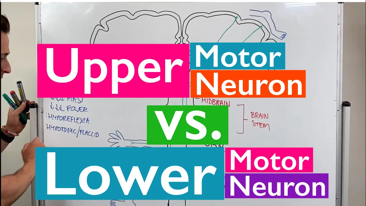
Motor neurons control muscle movement and are classified into upper and lower motor neurons. Upper motor neurons originate in the brain and send signals down the spinal cord. Lower motor neurons originate in the spinal cord and directly control muscle contraction. Motor signals travel from the brain through the spinal cord to reach muscles, causing them to contract and produce movement. This motor pathway works in coordination with sensory pathways to produce coordinated body movements.
Click to watch →

General Anatomy | b d chaurasia Handbook- Chapter 4 ( PART- 5) | Hindi Lectures
13.7K
Muscle contraction is controlled through a two-neuron pathway: upper motor neurons originate in the brain (cerebral cortex) and send signals down the spinal cord, while lower motor neurons in the spinal cord directly innervate muscle fibers. Both types are involved in muscle contraction. Damage to lower motor neurons causes flaccid paralysis with muscle weakness and loss of reflexes, while upper motor neuron damage causes spastic paralysis. This pathway ensures coordinated muscle activation for voluntary movement.
Click to watch →
Spinal Cord Injury, Motor Neurons, and Reflexes
183.7K
The motor pathway consists of two neurons working together. The first neuron (upper motor neuron) originates in the brain's motor cortex and travels down through the brainstem to the spinal cord, where it synapses with a second neuron (lower motor neuron). The lower motor neuron then exits the spinal cord and directly innervates the target muscle to cause contraction. This two-neuron system allows voluntary movement control.
Click to watch →

Medicine - Introductory Class by Dr. Sakthivel | MECEE-PG 2027 Classes | Prepolic
268
Motor pathways involve upper motor neurons (brain to spinal cord) and lower motor neurons (spinal cord to periphery). Primary motor cortex (precentral gyrus, Brodmann areas 4,5) sends commands via upper motor neurons. Upper motor neuron defects cause weakness in whole muscle groups, hypertonia, clasp-knife spasticity, minimal wasting, and extensor plantar reflex. Lower motor neuron defects cause paralysis of individual muscles, hypotonia, severe wasting, and flexor plantar reflex.
Click to watch →

Topic : Extrapyramidal Tracts - Part II
1.1K
The motor system consists of two neuron types: upper and lower motor neurons. Lower motor neurons arise from spinal cord anterior horn cells or cranial nerve nuclei and serve as the 'final common pathway' since all motor impulses must pass through them to reach muscles. Upper motor neurons terminate in these lower motor neurons and include pyramidal tracts (cortico-spinal and cortico-nuclear) and extra-pyramidal tracts (reticulo-spinal and vestibulo-spinal). Pyramidal tracts control voluntary movement, while extra-pyramidal tracts regulate muscle tone, posture, and equilibrium. Understanding this hierarchical organization is essential for neurological examination and diagnosis.
Click to watch →
Deep Dive
The general purpose and clinical indications for performing a manual muscle test (MMT) in healthcare settings.

Manual Muscle Testing (Lecture 2) | Nitin Kumar Arora | PhyX Pain Solutions
2.4K
Manual muscle testing serves as a quantifiable measure for assessing muscle strength in various conditions. It is indicated for any condition involving reduction of muscle strength, including upper and lower motor neurone diseases (such as poliomyelitis, stroke, traumatic brain injury, paralysis, hemiparesis, cerebral palsy), arthritis (particularly osteoarthritis where muscle strength typically decreases), and back or neck pain where nerve root involvement may cause muscle weakness and atrophy. MMT provides objective data to track patient progress and rehabilitation effectiveness.
Click to watch →
Intro Evaluation and Measurement | شرح
731
MMT is contraindicated in patients with acute inflammation, unhealed fractures, dislocations, severe cardiac or respiratory conditions, hemophilia, osteoporosis, and myositis. Precautions include careful monitoring for cardiovascular patients, special positioning for abdominal surgery patients, and avoiding testing patients with severe weakness from malnutrition or chronic conditions. MMT limitations include unsuitability for neurological disorders, muscle tone abnormalities, patients with strength below Grade 4, and children under 5-6 years old. Successful MMT requires proper setup: patient must be comfortable and pain-free, environment must be quiet with comfortable temperature, examination table must be stable, patient positioned correctly, and all equipment (goniometer, chart, paper) must be within reach. A helper may be needed for positioning and stabilization. Documentation includes patient history, test results, and assessment findings. These considerations ensure patient safety and test validity.
Click to watch →

Pemeriksaan MMT (Manual Muscle Testing) UE - Nisrina Virnanda (2006480433) - OT UI 2020
319
Manual Muscle Testing (MMT) is a clinical examination procedure used to assess voluntary muscle contraction ability. The Medical Research Council (MRC) grading scale ranges from 0 (no contraction) to 5 (full ROM with maximal resistance). MMT for shoulder horizontal abduction tests the posterior deltoid, while shoulder adduction assesses pectoralis major, latissimus dorsi, and teres major. Procedures vary by grade: Grade 1 involves palpation, Grade 2 uses supported movement, and Grades 3-5 require gravitational and resistive challenges. Only qualified medical professionals such as orthopedic doctors, physiotherapists, and occupational therapists can perform MMT examinations.
Click to watch →
Manual Muscle Testing
29.5K
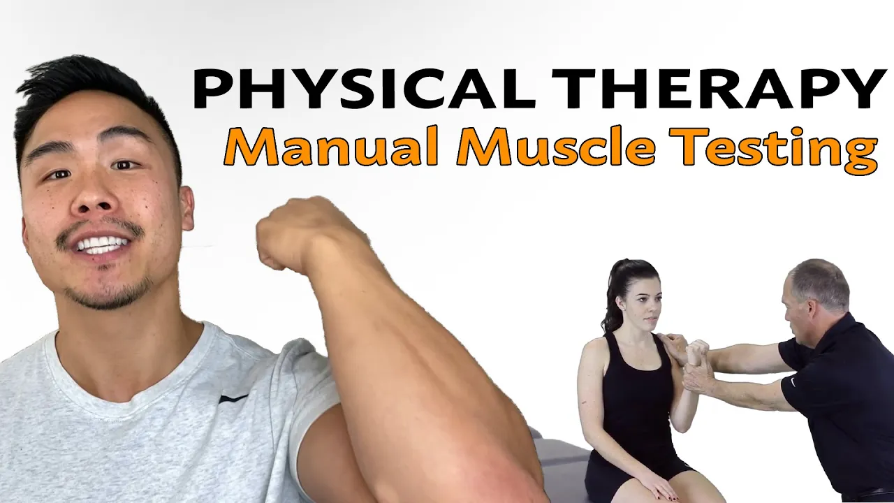
Manual Muscle Testing (MMT) is used to objectively measure the strength of a specific muscle in a patient. Physical therapists use MMT to assess whether muscles are weak or strong, which helps identify potential causes of injuries or diagnoses. For example, if someone has a hamstring strain, it may be due to weakness in the hamstring itself or synergistic muscles like the glutes. MMT provides baseline information that can be used to track progress over time, such as moving from a grade of 3/5 to 4+/5.
Click to watch →
Manual Muscle Testing (MMT) | Pengujian Otot Manual
376
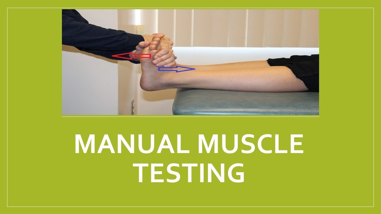
Manual Muscle Testing (MMT) is a clinical examination method used to assess muscle strength. The primary purpose of MMT is to determine or evaluate an individual's ability to contract their muscles. During the examination, the patient is asked to perform specific movements while the healthcare provider applies resistance to measure the strength of the muscle group being tested.
Click to watch →
Prerequisite Knowledge
- Concept 01Basic anatomy of the musculoskeletal system, specifically the structure and function of the knee joint and the quadriceps femoris muscle group.
- Concept 02The physical concept of gravity and how 'gravity-eliminated' versus 'against-gravity' positioning is utilized during physical examinations.
- Concept 03Fundamental concepts of neurological assessment, including how motor pathways (upper and lower motor neurons) control voluntary muscle contraction.
- Concept 04The general purpose and clinical indications for performing a manual muscle test (MMT) in healthcare settings.
Subsequent Learning
- Step 01Applying the MRC scale to other major muscle groups of the upper and lower extremities (e.g., deltoids, biceps, hamstrings, and tibialis anterior).
- Step 02Understanding the clinical significance of MRC grades in determining a patient's functional independence and designing physical rehabilitation protocols.
- Step 03Learning advanced grading modifications, such as the use of '+' and '-' modifiers (e.g., 4- or 4+) to capture subtle changes in muscle strength.
- Step 04Recognizing the limitations of the MRC scale, including subjectivity in grade 4, and exploring objective alternatives like hand-held dynamometry.
- Step 05Correlating muscle weakness patterns (identified via MRC scoring) with specific neurological or neuromuscular conditions, such as stroke, nerve root compression, or Guillain-Barré syndrome.
Grades 0-2
Grades 3-5
Grades 0-2
0:00Playing Section
- 1
MRC scale defines muscle strength from 0 to 5.
- 2
Grade 0: no contraction; grade 1: trace flicker.
- 3
Grade 2: full movement without gravity.
Limitations of the MRC Scale and the Shift to Quantitative Muscle Testing (QMT)
While the MRC Muscle Strength Grading system is a clinical standard due to its simplicity, it faces significant criticism for its subjectivity, poor inter-rater reliability, and "ceiling effect" (particularly in distinguishing between grades 4 and 5, which span a vast range of actual physical strength). Opponents and modern clinicians advocate for Quantitative Muscle Testing (QMT) using Handheld Dynamometry (HHD) as a superior alternative. QMT provides objective, continuous, and precise force measurements (in kilograms or Newtons) rather than subjective ordinal categories. This objective approach is more sensitive to minor changes in muscle strength, making it more effective for tracking patient recovery, detecting early neuromuscular decline, and ensuring consistent measurements across different examiners.
Deep Dive
Applying the MRC scale to other major muscle groups of the upper and lower extremities (e.g., deltoids, biceps, hamstrings, and tibialis anterior).
Taller. Exploración neurológica básica
54K
Meningeal signs indicate meningeal irritation and include neck stiffness, photophobia, headache, Kernig's sign (painful leg extension in flexed hip/knee position), and Brudzinski's sign (involuntary hip/knee flexion with neck flexion). These signs are positive in meningitis, subarachnoid hemorrhage, and other inflammatory conditions. Motor strength is graded on a scale from 0 to 5 using the Medical Research Council scale: 0=no contraction, 1=visible contraction without movement, 2=movement against gravity without resistance, 3=movement against gravity and some resistance, 4=movement against full resistance, 5=normal strength. Upper limb examination tests deltoid, biceps, triceps, carpal muscles, and finger movements. Lower limb examination tests psoas iliacus, quadriceps, hamstrings, tibialis anterior, gastrocnemius, and peroneals.
Click to watch →
MRC Muscle Strength (Updated Version in description)
54.3K
The MRC (Medical Research Council) scale is a standardized system for assessing muscle strength on a scale from 0 to 5, where Grade 0 indicates no movement, Grade 1 shows only a trace or flicker of movement, Grade 2 allows full range of motion only when gravity is eliminated, Grade 3 enables movement against gravity without resistance, Grade 4 permits movement with moderate resistance, and Grade 5 represents full strength with maximum resistance; this assessment method requires comparing the tested limb to the contralateral side and can be applied to various muscles and joints by positioning the patient appropriately.
Click to watch →

Examen Físico del Sistema Nervioso (Semiología)
38K
Muscle strength is graded using the Medical Research Council (MRC) scale from 0 to 5: 0 = no contraction, 1 = flicker, 2 = movement with gravity eliminated, 3 = movement against gravity, 4 = movement against some resistance, 5 = normal strength. Assessment involves comparing both sides and documenting asymmetry. Upper limb assessment includes shoulder, elbow, wrist, and finger movements. Lower limb assessment includes hip, knee, and ankle movements.
Click to watch →

AULÃO DE VÉSPERA - FAMEMA (VUNESP) - FISIOTERAPIA - PROF MARA RIBEIRO
1.2K
The MRC scale assesses muscle strength in six muscle groups (three upper, three lower extremities): 0=complete paralysis, 1=trace contraction, 2=active movement with gravity eliminated, 3=active movement against gravity, 4=active movement against some resistance, 5=normal strength. Maximum score is 60 (5 in all groups), minimum is 0. A score of 48 or below indicates significant weakness, possibly suggesting critical illness polyneuropathy.
Click to watch →

Como avaliar a força muscular em pacientes internados usando a escala MRC
1.5K
The six major muscle groups to assess are: shoulder abduction, elbow flexion, wrist extension, hip flexion, knee extension, and dorsiflexion of the foot. Each muscle group is scored from 0-5, with 6 muscle groups tested bilaterally, resulting in a maximum total score of 60 points. For shoulder abduction: Grade 2 involves therapist assistance; Grade 3 is independent movement against gravity; Grades 4-5 involve resistance testing. For elbow flexion: Grade 2 involves therapist assistance; Grade 3 is independent movement against gravity; Grades 4-5 involve resistance testing. For wrist extension: Grade 2 involves therapist assistance; Grade 3 is independent movement against gravity; Grades 4-5 involve resistance testing. For hip flexion: Grade 2 involves therapist assistance with knee support; Grade 3 is independent movement against gravity; Grades 4-5 involve resistance testing. For knee extension: Grade 2 involves therapist assistance; Grade 3 is independent movement against gravity; Grades 4-5 involve resistance testing. For dorsiflexion: Grade 2 involves therapist assistance; Grade 3 is independent movement against gravity; Grades 4-5 involve resistance testing. For all lower limb assessments, the patient should be positioned supine with the limb supported appropriately. The therapist should apply resistance while the joint is in a static position, not during active movement.
Click to watch →
Deep Dive
Understanding the clinical significance of MRC grades in determining a patient's functional independence and designing physical rehabilitation protocols.
Como avaliar a força muscular em pacientes internados usando a escala MRC
1.5K
MRC assessment has limitations that must be considered in clinical interpretation. A patient with MRC score of 60 may still have functional limitations that are not apparent from the MRC score alone. MRC assessment should be complemented with other functional assessments such as Barthel Index and grip strength testing. A patient with MRC score of 60 but poor grip strength may have different rehabilitation needs. MRC assessment should be interpreted in the context of other clinical findings and functional assessments.
Click to watch →

Terapia Intensiva - Parte 1
7.8K
The Medical Research Council (MRC) scale grades muscle strength from 0 to 5: 0 = no visible contraction, 1 = flicker of contraction, 2 = movement with gravity eliminated, 3 = movement against gravity, 4 = movement against gravity and some resistance, 5 = normal strength. In ICU settings, three muscle groups are assessed bilaterally in each limb: upper limb (shoulder abduction, elbow flexion, wrist extension) and lower limb (hip flexion, knee extension, ankle dorsiflexion). Maximum score per limb is 15, total maximum is 60. A score below 48 indicates ICU-acquired weakness. The scale is simple, reliable, requires no special equipment, and has excellent inter-examiner reliability. It is essential for monitoring neurological recovery and predicting functional outcomes in critically ill patients.
Click to watch →
Neurological Examination - Power, Reflexes, Sensation, and Coordination - Dr Gill
177.5K
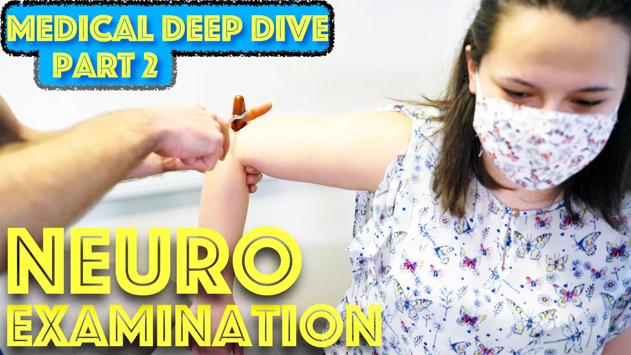
The MRC scale grades muscle power from 0 to 5: Grade 5 indicates normal power where the patient can push and pull against the examiner with reasonable strength; Grade 4 shows weakness but the patient can still move against resistance; Grade 3 allows movement against gravity without resistance; Grade 2 permits movement only when gravity is eliminated (e.g., arm can be moved side-to-side if held); Grade 1 indicates no visible movement but muscle contraction can be felt; Grade 0 represents complete absence of movement. Upper motor neuron lesions typically cause weakness in extensors of the upper limbs and flexors of the lower limbs.
Click to watch →

Stefan Nessizius: Assessments zur Frühmobilisation
251
The MRC Sum Score assesses muscle strength across six groups (dorsal extension, wrist flexion, elbow flexion, shoulder abduction, dorsiflexion, knee extension, hip flexion) on a 0-5 scale. Scores below 48 indicate ICU-acquired weakness. The score correlates with one-year survival, ICU length of stay, and 6-minute walk test performance.
Click to watch →

PROVA DE TITULO DE ESPECIALISTA EM FISIOTERAPIA EM UTI
447
This segment covers ICU rehabilitation barriers and assessment tools. Barriers to ICU rehabilitation include: patient not responding to commands at sedation level (strategy: discuss with multidisciplinary team about lighter sedation goals with analgesia), high number of ICU professionals with musculoskeletal lesions and stress (strategy: acquire auxiliary mobilization equipment, specific training, adequate formation), and patients starting rehabilitation too late (strategy: daily triage to identify patients for early rehabilitation program implementation). The MRC muscle strength assessment evaluates upper limb muscles (wrist extensors, wrist flexors, elbow flexors, shoulder abductors) and lower limb muscles (plantar flexors, knee extensors, hip flexors) with a cutoff score of 48 out of 60. Dependence on invasive mechanical ventilation can indicate acquired muscle weakness. ICU discharge is defined as a process for timely and responsible hospital discharge that should be part of routine care and management. Its main advantage is treating patients in a more appropriate environment for their clinical condition and more comfortable, avoiding hospital complications and infections.
Click to watch →
Deep Dive
Learning advanced grading modifications, such as the use of '+' and '-' modifiers (e.g., 4- or 4+) to capture subtle changes in muscle strength.

Manual Muscle Testing simplified | Nitin Kumar Arora | Physical Therapy | Ishant Arora
20K
Advanced MMT incorporates plus (+) and minus (-) modifiers to enhance diagnostic precision, particularly for grades 2, 3, and 4. A single grade reduction represents approximately 40-60% loss of active motor units, making fine-grained assessment clinically significant. Grade 3+ indicates completion against gravity with minimal end-range resistance. Grade 2- indicates inability to complete full range in gravity-minimal plane, while grade 2+ indicates partial completion against gravity. These modifiers enable clinicians to track subtle functional changes and rehabilitation progress more accurately, distinguishing between patients with very limited versus slightly better function.
Click to watch →

Round 1 | PT Traumatology ( Ortho )
1.1K
The plus and minus grading system modifies the standard 0-5 scale. Plus grades (1+, 2+, 3+, 4+) indicate strength above the standard grade but below the next grade. Minus grades (1-, 2-, 3-, 4-) indicate strength below the standard grade but above the previous grade. This provides more precise assessment.
Click to watch →
Manual Muscle Testing- grades, testing procedure detailed explanation with notes.
8.6K
The MMT grading scale ranges from 0 to 5 with plus and minus modifiers. Grade 0 shows no muscle contraction. Grade 1 shows flicker without movement. Grade 2 allows full range only in gravity-eliminated positions. Grade 3 allows full range against gravity. Grade 4 allows full range against gravity with moderate resistance. Grade 5 allows full range against gravity with strong resistance. Plus and minus modifiers indicate resistance levels: 1+ is minimal resistance, 2+ is slight resistance, 3- is 50% range against gravity, 4- is full range with near minimal resistance, 4+ is full range with near strong resistance, 5- is full range with strong resistance, 5+ is full range with very strong resistance. Testing procedures include the Break Test (resistance at end-range) and Make Test (resistance throughout movement). Proper stabilization of the tested muscle is essential to prevent compensation by other muscle groups.
Click to watch →
Síndromes Neurológicas Parte I - Professora Karina Lebeis
17.6K
Muscle strength is graded on a scale of 0-5: (0) No contraction, (1) Minimal contraction, (2) Movement against gravity but not resistance, (3) Movement against gravity and some resistance, (4) Movement against moderate resistance, (5) Normal strength. The scale can be modified with 4- and 4+ for more precise assessment.
Click to watch →

Неврологический осмотр: есть ли разница в подходах между отечественной и западной школами?!
3K
The muscle strength grading system (0-5 scale) should include intermediate grades (4+ and 4-) to detect subtle weakness. Many clinicians skip these intermediate grades, which can lead to missing important findings. The physician should use the full range of the grading system for accurate assessment.
Click to watch →
Deep Dive
Recognizing the limitations of the MRC scale, including subjectivity in grade 4, and exploring objective alternatives like hand-held dynamometry.

DEBILIDAD ADQUIRIDA EN LA UCI
857
The Medical Research Council (MRC) scale establishes standards for muscle strength testing in bedridden patients. Originally developed for Guillain-Barré syndrome, it evaluates six muscle groups bilaterally: shoulder flexion, forearm flexion, wrist extension, hip flexion, knee extension, and dorsiflexion. Scoring system: 0 = no contraction, 1 = visible contraction without movement, 2 = movement against gravity, 3 = normal movement against gravity, 4 = movement against moderate resistance (3 fingers), 5 = movement against maximum resistance (full hand). ICU weakness is diagnosed when sum of scores is <48, with <36 indicating severe weakness. Manual dynamometry is preferred for simplicity: reduced grip strength is <11 kg in men or <7 kg in women. Proper technique requires patients to be alert, cooperative, and motivated, with three consecutive measurements and 30-60 second intervals. Patients should be positioned with elbow at 90 degrees if seated, or arm supported against bed if supine.
Click to watch →

Rede Sarah: tópicos essenciais de Fisioterapia II
1.1K
Muscle strength is assessed using the Medical Research Council (MRC) scale (0-5): Grade 0 (no contraction), Grade 1 (contraction without movement), Grade 2 (movement without gravity resistance), Grade 3 (movement against gravity), Grade 4 (movement against moderate resistance), Grade 5 (normal strength). Hand dynamometry measures grip strength. Deficit maneuvers (Mingauzinho and triple flexion) assess sustained strength by having patients maintain postures for two minutes. Muscle tone assessment includes palpation, percussion, and passive movement. The Ashworth scale grades spasticity from 0 (normal) to 4 (very high rigidity).
Click to watch →

Manual Muscle testing my Ton Schreuders
3.9K
This section covers the foundational principles of hand muscle strength testing. The MRC grading scale (0-5) is explained, with emphasis that these are ordinal variables where intervals between numbers are unequal. Dynamometry provides quantitative data showing Grade 3 ≈ 11-12N, Grade 4 ≈ 20N, and Grade 5 ≈ 33-34N. A critical insight is that M4 represents only 40-50% of actual maximum strength. The step-by-step testing approach is introduced: first assess passive range of motion, then apply resistance only after confirming the patient can maintain position. This systematic method prevents misgrading and ensures reliable assessment before resistance is applied.
Click to watch →

Respiratory System History Taking - Common Symptoms for OSCE Revision - Dr James Gill
227.7K
The Medical Research Council (MRC) dyspnea scale grades breathlessness severity: Grade 1 - troubled only by breathlessness except on strenuous exercise; Grade 2 - short of breath when hurrying on level or slight hill; Grade 3 - walks slower than most people on level or stops after 1 mile or 15 minutes; Grade 4 - stops for breath after walking only 100 yards or few minutes on level ground; Grade 5 - too breathless to leave house or even undress. This scale helps quantify subjective dyspnea and assess impact on daily activities.
Click to watch →

Escala de fuerza modificada o MRC
2.5K
The Modified Medical Research Council (MRC) Scale is a standardized 0-5 grading system used to assess muscle strength, where 0 indicates no muscle contraction, 1 shows trace contraction, 2 demonstrates movement against gravity without resistance, 3 allows movement against gravity with resistance, 4 enables movement against moderate resistance, and 5 represents normal strength against full resistance; this scale is considered a reference standard for concurrent validity in clinical studies and is commonly used alongside isokinetic dynamometers for objective muscle strength measurement, though it is not suitable for patients with spasticity due to altered muscle tone affecting force generation.
Click to watch →
Deep Dive
Correlating muscle weakness patterns (identified via MRC scoring) with specific neurological or neuromuscular conditions, such as stroke, nerve root compression, or Guillain-Barré syndrome.

examen de la fuerza
244
Muscle strength evaluation is a fundamental component of neurological examination that helps differentiate between central and peripheral causes of weakness; the Medical Research Council (MRC) scale grades muscle strength from 0 (no movement) to 5 (normal), and systematic testing of at least 18 muscle groups—including axial, proximal, and distal muscles—allows clinicians to identify specific patterns of weakness that indicate particular neurological pathologies.
Click to watch →
Neurological Examination - Power, Reflexes, Sensation, and Coordination - Dr Gill
177.5K
The MRC scale grades muscle power from 0 to 5: Grade 5 indicates normal power where the patient can push and pull against the examiner with reasonable strength; Grade 4 shows weakness but the patient can still move against resistance; Grade 3 allows movement against gravity without resistance; Grade 2 permits movement only when gravity is eliminated (e.g., arm can be moved side-to-side if held); Grade 1 indicates no visible movement but muscle contraction can be felt; Grade 0 represents complete absence of movement. Upper motor neuron lesions typically cause weakness in extensors of the upper limbs and flexors of the lower limbs.
Click to watch →

Infecções do sistema nervoso central e neuropatias | Cortes de Aulas: Clínica Médica
1.9K
Muscle weakness patterns differ based on the anatomical location of the lesion: upper motor neuron lesions (first motor neuron) cause hyperreflexia, spasticity, and Babinski sign with mild atrophy, while lower motor neuron lesions (second motor neuron) cause hyporeflexia, hypotonia, and significant atrophy; myopathies typically present with proximal weakness affecting the shoulder and pelvic girdles.
Click to watch →

Saiba tudo sobre síndrome de Guillain Barré
154.7K
Guillain-Barré Syndrome causes progressive motor weakness that follows a characteristic pattern: it typically begins in the lower extremities and ascends upward toward the head. Patients experience difficulty walking, may trip, and have trouble climbing stairs. The weakness is voluntary and progressive, with patients becoming increasingly aware of their declining mobility. Sensory function is generally preserved, though some patients may experience pain initially.
Click to watch →

Fraqueza Muscular é Só Cansaço ou Algo Mais Sério
1.5K
Neurological diseases represent the primary cause of muscle weakness, requiring systematic differential diagnosis. Stroke causes abrupt, unilateral weakness affecting brain motor regions, often without pain, and may be subtle enough that patients dismiss symptoms as disuse. Multiple sclerosis produces characteristic cyclical flare-ups and remissions where strength gradually diminishes, stabilizes, then slowly returns. Amyotrophic lateral sclerosis causes progressive bilateral weakness through degeneration of both upper and lower motor neurons, leading to muscle atrophy. Neuropathies involve peripheral nerve damage through mononeuropathies (single nerve injury, e.g., peroneal nerve causing foot drop) or radiculopathies (nerve root compression affecting specific muscle groups). These conditions require detailed neurological examination, electromyography, and sometimes surgical intervention. Early recognition of these patterns enables appropriate specialist referral and treatment initiation.
Click to watch →
Grades 0-2
Grades 3-5
Grades 0-2
0:00- 1
MRC scale defines muscle strength from 0 to 5.
- 2
Grade 0: no contraction; grade 1: trace flicker.
- 3
Grade 2: full movement without gravity.
Limitations of the MRC Scale and the Shift to Quantitative Muscle Testing (QMT)
While the MRC Muscle Strength Grading system is a clinical standard due to its simplicity, it faces significant criticism for its subjectivity, poor inter-rater reliability, and "ceiling effect" (particularly in distinguishing between grades 4 and 5, which span a vast range of actual physical strength). Opponents and modern clinicians advocate for Quantitative Muscle Testing (QMT) using Handheld Dynamometry (HHD) as a superior alternative. QMT provides objective, continuous, and precise force measurements (in kilograms or Newtons) rather than subjective ordinal categories. This objective approach is more sensitive to minor changes in muscle strength, making it more effective for tracking patient recovery, detecting early neuromuscular decline, and ensuring consistent measurements across different examiners.
Basic anatomy of the musculoskeletal system, specifically the structure and function of the knee joint and the quadriceps femoris muscle group.
The physical concept of gravity and how 'gravity-eliminated' versus 'against-gravity' positioning is utilized during physical examinations.
Fundamental concepts of neurological assessment, including how motor pathways (upper and lower motor neurons) control voluntary muscle contraction.
The general purpose and clinical indications for performing a manual muscle test (MMT) in healthcare settings.
Applying the MRC scale to other major muscle groups of the upper and lower extremities (e.g., deltoids, biceps, hamstrings, and tibialis anterior).
Understanding the clinical significance of MRC grades in determining a patient's functional independence and designing physical rehabilitation protocols.
Learning advanced grading modifications, such as the use of '+' and '-' modifiers (e.g., 4- or 4+) to capture subtle changes in muscle strength.
Recognizing the limitations of the MRC scale, including subjectivity in grade 4, and exploring objective alternatives like hand-held dynamometry.
Correlating muscle weakness patterns (identified via MRC scoring) with specific neurological or neuromuscular conditions, such as stroke, nerve root compression, or Guillain-Barré syndrome.
578K views9.8Klikes3:45@PhysiotutorsOriginal Release: 2015-11-30
This is going to be a video on the MRC Scale to rate muscle strength.
MRC stands for Medical Research Council which is the institution that set up the standard for muscle strength testing.
The strength can be categorized on a level from zero to five.
In our example, we will use the extension of the knee joint.
So, the muscle which we are going to test is the Quadriceps.
The levels are as follows: Grade 0: The patient cannot activate the muscle, so no movement is observed.
For grade 0, ask the patient to contract his quadriceps.
He can do this by pushing the back of his knee into the bench.
For grade 0, I will not see or feel a flicker or trace of contraction or movement.
Grade 1: the patient can activate the muscle, without moving the limb.
So only a trace or flicker of movement is seen or felt during palpation of the muscle.
For grade 1, ask the patient to do the exact same thing, and this time, you will see or feel a muscle flicker or trace of movement.
Grade 2: movement over full range of motion can only occur if gravity is eliminated.
In order to distinguish between grade 1 and 2, we have to bring our patient in side-laying position to eliminate gravity.
Then, I will support the leg of my patient, bring it into full flexion and ask my patient to move into extension.
If my patient is able to move through the full range of motion, this is a grade 2.
If no movement is possible at all, we are talking about grade 1.
Grade 3: the patient can overcome gravity and move through the full range of motion without resistance coming from the examiner.
For grade 3, I’ll ask my patient to extend his knee against gravity.
Grade 4: weakness with resistance.
So your patient can move through the full range of motion with moderate resistance coming from the examiner.
For grade 4, I will give moderate resistance against the extension of my patient’s knee.
And at last, grade 5: full strength.
So your patient can move through the whole range of motion against full resistance coming from the examiner.
And for grade 5, I’ll give full resistance against the extension of my patient’s knee.
In order to distinguish between a grade 4 and 5, make sure to compare both legs.
Now that you’ve seen the basics of how to test according to the MRC scale, make sure to practice this with different joints and muscles and figure out a way of how to position your patient.
Okay, this was our video on the MRC Scale, I hope this video was helpful to you. If it was, give it a thumbs up and subscribe to our channel if you haven’t and if you want a quick information about your patient’s strength of his hip and knee muscles, check out our resisted isometric videos right here and see you next time, bye.
Up Next

146:49
Regenerative Agriculture with Will Harris | White Oak Pastures
@joerogan
73.2K views•2024-06-27

12:54
Triumph of Orthodoxy Icon: Byzantine Art & History Explained
@BenCallan
2.1K views•2024-08-06

9:49
FastAPI vs Flask vs Django: Choosing the Right Python Web Framework
@TechWithTim
302.5K views•2024-05-26

1:42
Game of Thrones Opening Credits: A Cinematic Analysis
@gameofthrones
46.3M views•2011-04-18
Related Study Plans & Knowledge Roadmaps
Structured learning paths in General & Interdisciplinary Studies
general-studies
Elevator Engineering: History, Physics & Cities
01From Hoists to Safety Brakes: The Dawn of Vertical Lift
02Hydraulic vs. Traction: The Engineering Physics of Elevators
03Modern Speed, Safety, and Algorithms
+ 2 more learning modules
12h5 modules
Explore Roadmap
general-studies
Rubber: Material Science, History & Geopolitics
01Origins of Natural Rubber & Mesoamerican History
02The Chemistry of Natural Rubber & Vulcanization
03The Geopolitics of the Rubber Boom & Colonization
+ 2 more learning modules
12h5 modules
Explore Roadmap
general-studies
Containerization: Evolution, Logistics & Trade
01Ancient Trade & Bulk Cargo: Amphorae to Barrels
02The Industrial Revolution & Cardboard Packaging
03The Containerization Revolution: Malcom McLean
+ 2 more learning modules
12h5 modules
Explore Roadmap
general-studies
Textile Tech: Evolution, Labor & Global Impact
01Pre-Industrial Textiles: Handloom & Cottage Industry
02Mechanization: The Spinning Jenny to the Power Loom
03The Factory System & Global Labor Transformation
+ 2 more learning modules
10h5 modules
Explore Roadmap