






![👑الوحدة 1: تركيب البروتين✍🏻الدرس1: مقر تركيب البروتين🍖[علوم تجريبية+رياضيات] بسم الله سنبدأ رحلتنا](https://i.ytimg.com/vi_webp/bPgBHZzXxsM/maxresdefault.webp)


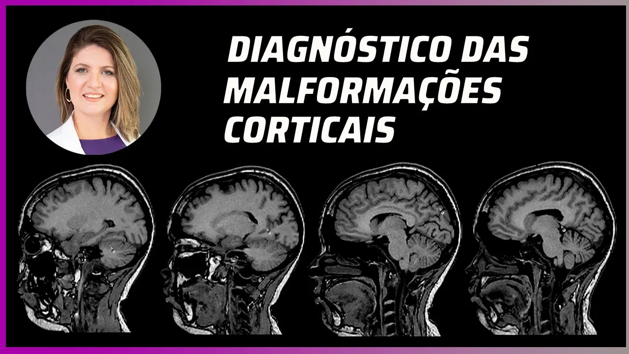




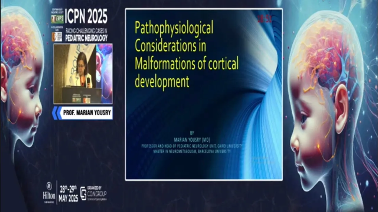






Neural migration disorders arise from defects in the molecular machinery that orchestrates neuronal movement from the ventricular zone to their final cortical positions during early fetal development, resulting in conditions such as periventricular nodular heterotopia (no migration), lissencephaly (impaired migration), and polymicrogyria (disrupted migration), each characterized by distinct pathological features including abnormal cortical lamination, disorganized neuronal placement, and disrupted gyral patterns.
Neuronal Migration Disorders: Embryology & Pathology Explained
Added:Fundamentals of early neuroembryology, including neurulation and the proliferation of neural progenitor cells within the ventricular zone.
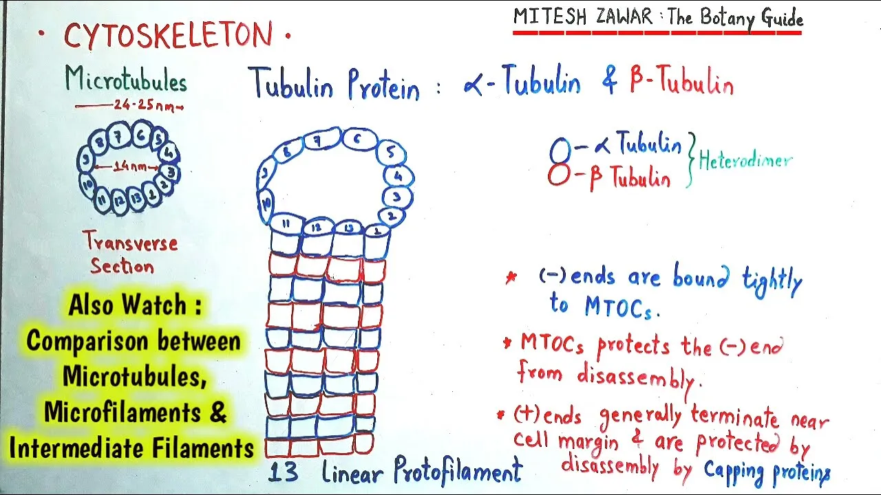
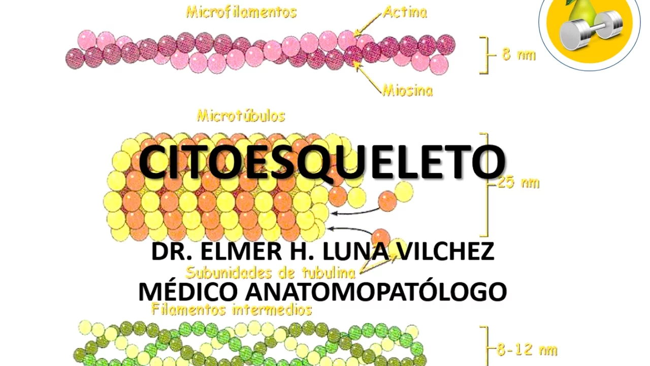
The structure and function of the eukaryotic cytoskeleton, specifically the roles of microtubules, actin filaments, and motor proteins in cell motility.
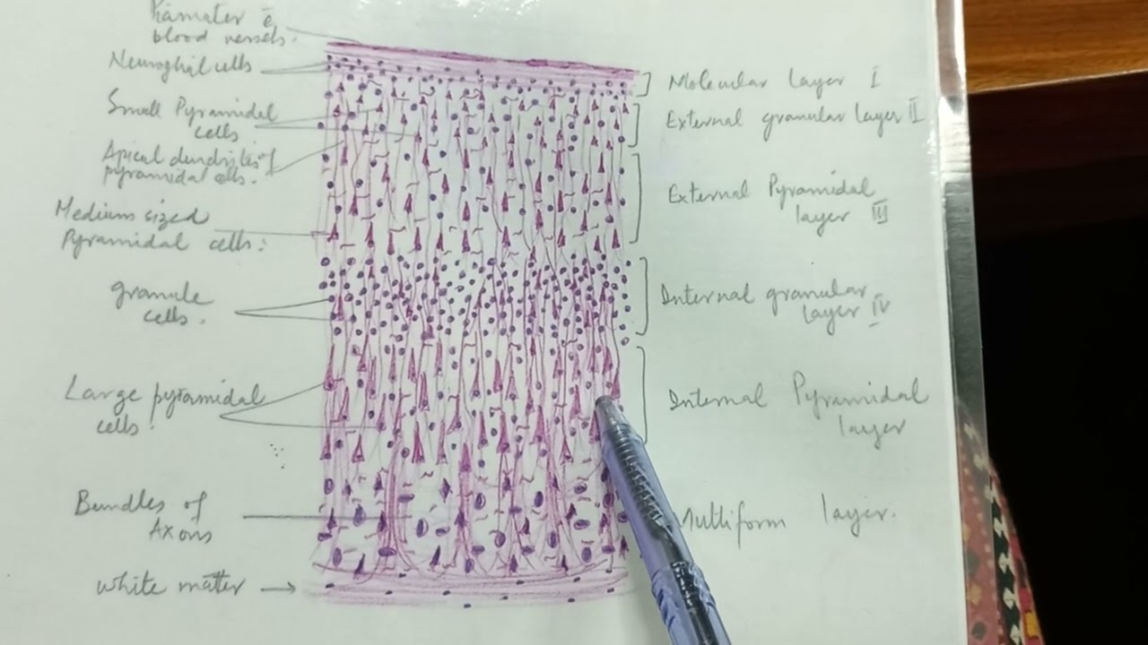
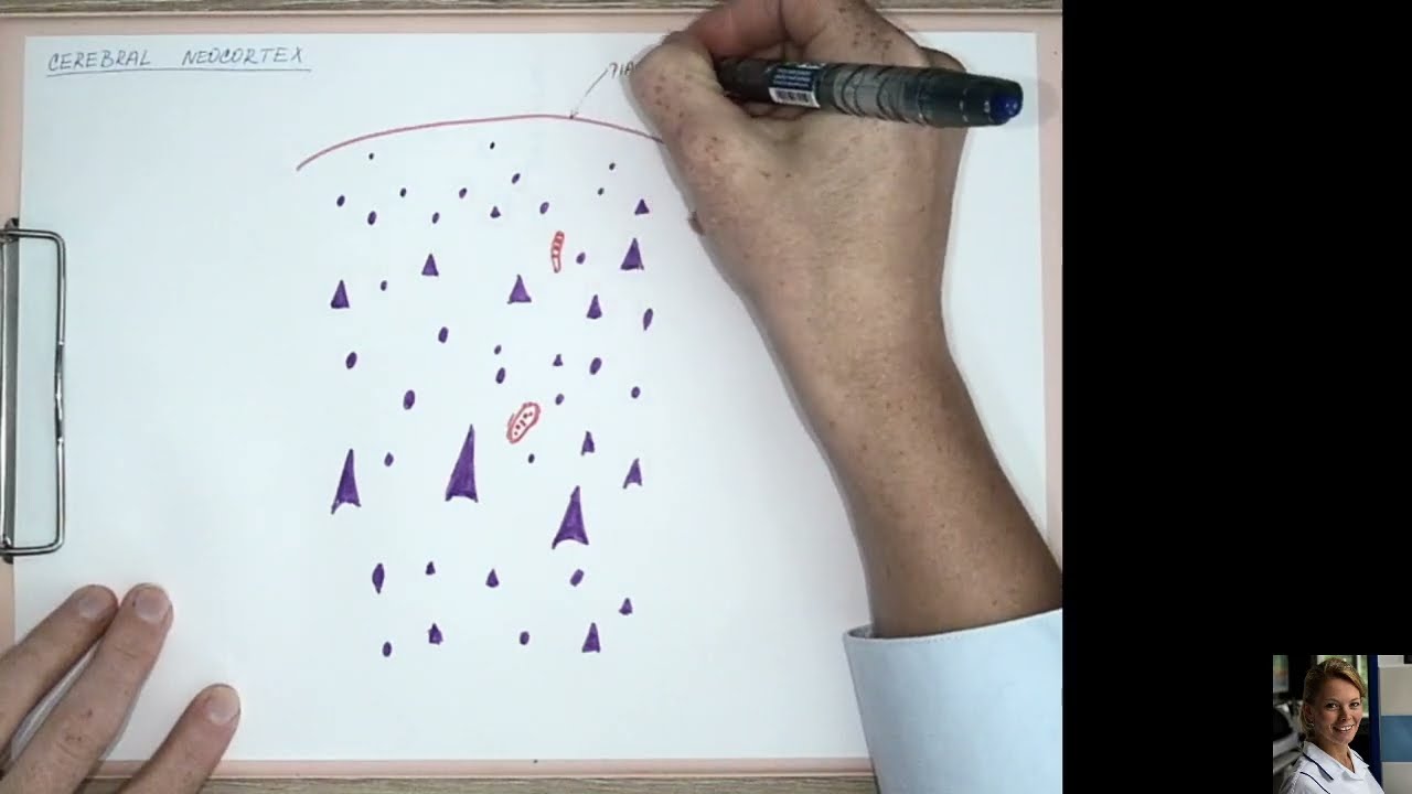
The normal histological architecture of the cerebral cortex, particularly its inside-out, six-layered development pattern.
Basic concepts of molecular genetics, including gene expression, mutation types, and how genotype influences protein structure and cellular function.
Clinical identification and diagnostic neuroimaging (MRI) features of cortical malformations such as lissencephaly, subcortical band heterotopia, and polymicrogyria.
The physiological consequences of migration defects on neural circuitry, including the pathophysiology of pharmacoresistant epilepsy and cognitive impairment.
In-depth analysis of specific signaling cascades, such as the Reelin-DAB1 pathway, and genes like LIS1 and DCX in developmental neurobiology.
Current research into therapeutic interventions, including gene replacement therapies, molecular chaperones, and prenatal diagnostic screening.
6.2K views141likes1:22:58Original Release: 2020-02-22
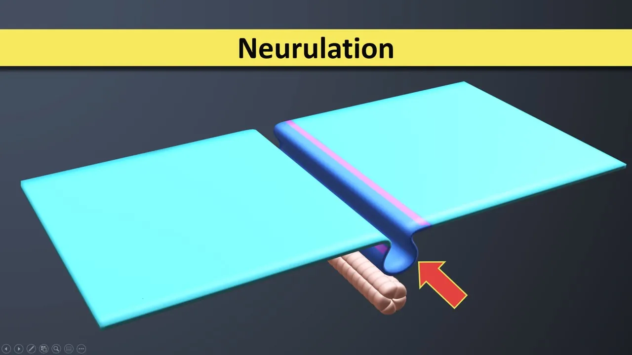
the human brain is one of the most complex pieces of matter known in this universe and many questions that can be asked how is this complexity so reproducibly programmed in each and every one of us what are the molecular machinery that coordinated this process to give us features like personality creativity and emotions one of those mechanisms is the ability of our brain to program the production and the organization of our nerve cells in the correct locations in the brain this all occurs in the uterus in a highly coordinated fashion that we now understand thanks to many genetic and neuro pathological studies the goal of today's episode is to review some of the molecular machinery that drive neural migration from the area where they're created all the way to the ends of the brain the cortex where the neurons reside permanently till drought our whole life in addition to reviewing the normal physiology of this process we're gonna see what happens when there are genetic mutations that perturb this process and lead to a number of diseases known as neural migrational disorders these are very complex and devastating disorders that can occur in newborns so hope you enjoy this lecture let's get to it [Music] my name is Phidias Diamandis I'm a neuropathologist at the University Health Network and a assistant professor at the University of Toronto today we're gonna be talking about one of my favorite topics neural migration providing an overview of the theory and of course the disorders that arise when when things go wrong I'm supposed to be giving this presentation to some of the residents in our neuropathology program but I thought I'd also share it here on the channel so of course disclaimer this is for educational purposes only it's not a diagnostic tool by any means I'm a adult neuropathologist with really more of a hobby in in neurodevelopmental disorders so please keep that in mind so today we're gonna go and cover an overview of normal neural development with a focus on some of the molecular players of neural migration and then kind of use that theory to try to understand the the wide array of neural migrational disorders that have been characterized so let's begin with again just an overview of neuro development this is a little cartoon one of my former master students jennifer cow created and basically as we know neural development occurs almost entirely in utero and really our brain begins to be formed after the fourth week of in utero life during a process called neural ation where you have this neuro ectoderm this one of the three germ layers fold up upon itself to form this neural tube that will go on to form the spinal cord and and cerebral cortex and cerebellum and so on and so forth so very important structure and once it's formed it's at the fourth week stage it's a it immediately begins to form the brain through progenitor proliferation differentiation and migration of the neurons to correct location so once this Union layer structure is formed it will go a massive proliferation potential to form these germinal centers or ganglionic eminence at menses that really house a large amount of of neural precursors and while they're being produced they undergo neurogenesis to form neurons and shortly after and it kind of coordinated process these these elements will migrate from this ventricular zone which is the hollow aspect of the tube to the most distal parts of the of the brain to form the cortical plate and as you'll see there's two types of migration there's radial migration from this dorsal part of the subventricular zone and there's also a tangential migration that occurs from these medial and lateral ganglionic eminences and really that that occurs in the first 20 weeks of gestational life and it's a profound statement but at that point most of our neurons are not only formed but they're in their correct location for almost the rest of our lives so what happens then the brain continues to grow by changing its developmental program in these germinal zones and producing glia so glia are astrocytes and oligodendrocytes that are needed to support the circuitry of the neurons and of course there's additional maturation steps in the cortex where were things undergoing neural maturation synapses are form there's dendritic maturation and synaptic pruning and of course myelination to form the kind of the adult human brain that we're all familiar with so obviously a very complicated process and many things can obviously go wrong and when they do there's a number of diseases that have been attributed to defects in one of these complex processes and of course the one that we're going to talk about today is neural migration and really the diseases that are hallmarks of neural migrational disorders are Liz encephalo Polly microg area heterotopia z-- epilepsy and four cortical dysplasia these will be as safe for a different lecture just just to kind of keep it a reasonable length so there's some key points that I want to emphasize that are important to understand is that at four weeks you have this ventricular zone and a relatively fin pre plate as it's called which has a very small number of neurons the pre plate neurons and the Kahala red cells either kind of the the founder neurons that are produced in the ventricular zone and the first neurons that kind of exit this the this key structure this progenitor region and and as we know the the neurons lie in the cerebral cortex at the surface of the of the brain and thus it is there traveling quite some time so errors that occur can occur at any point during their migration or process so as I mentioned you have your pre plate in Kahala red cells once these are formed these proliferative zones will generate what are called radial glial cells these are your kind of definitive neural stem cells that really extend a process to the glial limit and here at the at the peel brain surface and also extend the process to the to the ependymal zone to form this kind of Reed real scaffold which is provides a nice route for newly formed neurons to migrate this large distance in a kind of very coordinated fashion towards the sub plate splitting the sub plate and the marginal zone which is where your Kahala red seed cells are to populate the the six layered cortex now interestingly the cortex has a characteristic pattern known as an inside-out pattern of generation in which these migrating neurons will first populate the the the inside layer or this or layer six and five of this cortical plate and then newly formed neurons will reside at a superficial level to these original neurons and thus you can actually date the neurons by understanding where they are located in the cortical plate as to their age relative to one another now how do they know where to stop one of the key ways is that these caja these Kahala red seed cells secrete a protein called reelin which binds to a receptor known as the very low density lipoprotein receptor on migrating neurons and and tells it that you've reached the top and it's time to jump off so the next neuron can go and because of that phenomenon new neurons really have to go to the edge of the of the cortical plate to be subjected to this reelin secretion to tell them to stop migrating and that's really why you have this inside-out pattern and we'll see what happens when when that becomes defective so some key points here to note that will hopefully help make this process more understandable as we go through the notes is that again neurons are slide away from their site of birth so there in the ventricular zone and really you can see they they have to travel a long distance to reach their final destination and as the brain is maturing new neurons have to travel even further than the original neurons due to the brain growing but also having to travel that extra mile for this inside out pattern now to do this again they need a lot of complex migratory machinery there's extracellular matrix proteins there's adhesion molecules cell set of skeletal proteins that help maintain the shape of the migrating neuron receptors and of course secreted molecules as we talked about things to note again there's both a radial and tangential migration the the the excitatory neurons really arise from this dorsal subventricular zone and and radially migrate to form your glutamatergic neurons or your are your pyramidal cells that really form these long connections and then you're kind of GABAergic neurons arise in this medial ganglionic eminence and carry out this tangential migratory pattern into the cortex and then will undergo radial migration to find their correct location so again these orange ones are undergoing radial migration and then these green inter neurons or inhibitory neurons are undergoing ten tangential migration and you'll see why that's important as we cover some of the many diseases of neural migration one thing to note is although it's not clearly figured out yet of how the brain forms gyri it's understood that some of the theories suggest that it's the connections that neurons form that actually drive gerra fication or the or the gyri that we see in normal brains and thus if you have abnormal neurons these types of Jiraiya may not form due to this you know insufficient environment to to foster these connections that lead to cortical folding so one of the hallmark features as we'll see of neural migrational disorders is in fact also the lack of folding that occurs after neural migration and has completed um so it's always important to have a framework to remember these different diverse diseases so I created this summary slide that hopefully you can ingrained in your brain of how I memorize these or try to think of them in an illogical way and basically you can think of neural migrational disorders as a kind of spectrum of defects in migration you can have no migration so so cells do not migrate out of the ventricular zone and when that happens we get a disease called peer ventricular nodular heterotopia you can have impaired migration where the neurons haven't well will migrate towards the cortical plate but will do so another in a very slow or uncoordinated fashion and lead to defects in this inside-out pattern but also in this nice organized lamination pattern you can also have partial migration where some neurons migrate normally but others do not and as we'll see that leads to a disease called subcortical band heterotopia through some interesting mechanisms so if you can have under migration well why can't you have over migration and that's really what happens in cobblestone type tool is encephalo as opposed to type 1 in an impaired migration these are different diseases as we'll see and then a kind of more subtle version of this is it's called leptin and in joke little neuronal heterotopia and then and then you could have disrupted migration where it's still unclear what the exact mechanism is but perhaps there's a an insult that occurs during the process of migration that leads to this disease called polymicrobial or focal cortical dysplasia so let's look at those separately let's start with what happens when you have no no migration and really this is the disease called periventricular nodular heterotopia so here's an image from this very nice website that i recommend you all use for your studies neuropathology web org that really highlights the disease very very nicely so clinically this can occur unilateral or bilateral so on one side of the brain or both it can be focal or diffuse and really patients present with seizures and really the the clinical picture as you'll see from many of these is quite it's quite wide depending on the severity of the mutations and the defect it can be an incidental finding patients can be actually quite normal it usually only occurs in females as we'll see why and usually when it occurs in males it's incompatible with life but there are cases where males can have this pattern as we'll see due to very mild mutations or mutations and non-canonical genes involved in this process so have you've never seen this before this is a brain scan and as you'll see you have your grey matter here at the periphery and and then you have your white matter which is white now what you what you may appreciate is that in the normal brain the ventricular zone is lined by a very thin layer of ependymal that's not very visible on brain scans but here instead it looks like it's lined by a a grey matter like substance that looks like has the characteristics of the gray matter so this is this is your subcortical this is your periventricular nodular heterotopia it's periventricular as the descriptive name suggests it's nodular and it's heterotopia this is brain tissue not in the correct location and you can see that again no migration these neurons are exactly where they were born in the ventricular zone and didn't migrate up now why did some migrate out and some didn't we'll see that when we talk about the pathobiology so this is either a familial or sporadic loss of function mutation in a gene called filament a which is on the X chromosome the protein filament a is an active binding protein involved in cellular structure and migration so when it's defective cells are unable to migrate properly and affects things like neuro migration unless you get this failure of migration in this heterotopic differentiation of neurons so it's excellent dominant so males only have one X chromosome so when they have a filament a mutation all the cells in their brain are unable to migrate out and thus they cannot form a cortical ribbon that would be required for a basic function interestingly females do the process called lyanna's a lie on lie on ization where you have this random X inactivation of one of their two x chromosomes if this was random half the neurons would actually have a normal filament a protein while half of them would not and as a result half of them are able to migrate to their correct location while while the other half remain in the ventricular zone and form these modular modular structures and the ventricular zone usually this would be smooth and that's why this is a disease found primarily in females just because the male the male phenotype is incompatible with life there are however hypo morphs or other mutations as we'll see that can can can foster this type of sufficient migration to to make males also compatible with life so so again the rules keep changing as we see more and more of these cases there's a lot of as you can imagine set a set of skew the structure is an important it's important for all cells and thus many of these disorders can also be seen as part of a constellation of multi organ syndromes in patients with with these mutations so histologically we saw some gross images this is luck so fast blue hne stain where the luck so fast blue will stain white matter and the H&E will primarily stain the white and gray matter but here you can only see the grain the gray matter because of the luck so fast blue staining of the white matter and you can see you have your cortical ribbon here which is what you would expect you have your gray matter again what you would expect but again here in the normal human brain this is completely a very thin structure and you have these kind of heterotopic nodules of grey matter so this is very very in keeping with parent tricular nodular heterotopia these islands are disorganized they have neurons they lie under kind of ependymal lining obviously which is the lining of the ventricles and because again these are forming in a somewhat coordinated fashion you get these kind of rudimentary laminations where you have different types of neurons being generated with respect to their developmental age so the overline cortex can be dysplastic or intial poly my courage area but it can also be normal and females just because of this process where half of the cells are able to on average are able to migrate in a kind of normal fashion here's some more pictures from another great book developmental neuropathology also a book I use during my residency again we have this gray matter here we have white matter this is a kind of formalin-fixed brain it appear you have your periventricular nodules here one and you can see that they're very bumpy and the in the ventricular in the ventricles now one teaching point that you don't want to over call this by normal brain structures so keep in mind where things like the basal ganglia should be just to make sure that you don't get overexcited and call a normal subventricular gray matter structures nodular header Tobias and and you can see here on histology you get these nodules they're very disorganized and again they will line the ventricular zone this is I include this this is a more recent paper in this journal called development that really highlights some of of the kind of current knowledge of different mutations that can cause a ventricular nodule heterotopia x' and other migrational disorders as we'll see as we cover the other ones here's our filament a the the kind of classic gene involved in this disorder but of course there's many many genes that have now been implicated in this disorder so the literature is always changing reassuringly when you look at the main function of these proteins which is a common theme here is that it's not random proteins involved in different functions it's really the migratory machinery and the set of skeletal structure that are the major kind of biological pathways implicit implicated in these diseases so let's talk about kind of a a milder form as you may milder in terms of biology obviously these are much more severe diseases from a disease like pyramid tricular nodular heterotopia where where females can actually have very subtle findings so let's look at some of those disorders so classic we'll begin with kind of classical type one Liz encephalo this is a pretty rare disorder Liz encephalo is is Greek for a smooth brain and as you can see here we have this very smooth brain lacking convolutions except for this Sylvian fissure which is highly abnormal depending on what stage of development you're in so it's quite rare one one in a million but has very very distinctive genetic and in pathological findings so considered exclusively a genetic disease as we'll see some of the diseases that we talk about can have both a genetic and acquired pattern it's it's the kind of reported incidences are equal in males and females but there are two x-linked forms as we'll see that occur or a more severe in male so you know probably because of the of the of how rare these disorders are they just the the the the the additional burden that may occur in males it does not come out well in studies so severe psychomotor and intellectual impairment intractable seizures dysmorphic faces we'll see especially with this Miller Dieker syndrome or you get the bitemporal hollowing hyper Taylorism frontal bossing many many other facial dis morphisms and so on and so forth and even congenital heart defects and defects outside the brain so multiple fingers so this this is a very special disorder and we'll see the kind of genetics behind it a rx is is another form of this disorder that's good to know it's one of the x-linked disorders and usually manifests with ambiguous genitalia and small adrenal glands there's you can have little encephalo with really mutations where you can get what's called congenital lymphedema where the child is completely full with lymph tissue that's not draining properly eating - this kind of kind of swollen look so these are really more historic to help you determine what type of mutation and thus what type of genetics the family has to help them with family planning but but now these are perhaps less relevant in an area where you can sequence new cases and understand exactly what mutation exists life expectancy is highly variable some say 10 years but really this depends as all these diseases depend on the severity of the mutation so more kind of hypo morphic mutations that have less severe phenotypes patients may have severe intellectual disability but actually live longer and of course if if there's very severe mutations may be more challenging to the outlook so um what brings these all these diseases together um so again there's impaired migrational machinery resulting in a disorganized thick thicker cortex and and really their defects in and radial migration and I put this asterisks here because one of them particularly ARX is actually thought to be a defect in a tangential migration but for the most part they're thought to be defects and in radial migration and you can see the the list of mutations that are being discovered continue to grow and they have different types of inheritance patterns from autosomal dominant recessive x-linked and some de novo mutations and again there's some syndromes that are associated with with each one of these mutations that help you kind of localized based on the clinical findings what the presumed mutation is now what are all these genes have in common they're all involved in the migratory machinery so imagine this is the imagine this is the the molecular machinery on these migrating neurons and really the way these cytoskeletal proteins work is by trying to pull not only extending the the the leading process but also pulling the nucleus as the the neuron migrates and to do this in a coordinated fashion you need to have very strong set of skeletal proteins like your microtubule ins and then you have to have these motor proteins these dying's to really drive the pulling of the nucleus and in migration and and when you have defects and proteins involved in this complex like DC X which is a kind of a classic one list 114 EE 3 epsilon which is the the protein implicated in Miller Dieker syndrome tubulin appa these that have mutations and tubulin you you you get less encephalo type 1 if it's severe mutation now interestingly there's this other machinery as we talked about called the v LD r d l r receptor which is the very low-density lipoprotein which is really the receptor that binds real in and tells the cell to stop migrating when it reaches the top here at the Kahala wretched cells in the marginal zone and and and so mutations in this will obviously also lead to migrational defects because this ability to jump off and move out of the way to allow new neurons to come in is significantly impaired when when you don't have a stop signal these these will keep my greeting in and an informed disorganized kind of migratory kind of a car crash at the top of the of this readable scaffold now interestingly when you look at some proteins close together some of the molecular phenotype some of the clinical phenotypes are very similar so when you look at your VDRL you get cerebellar hypoplasia reelin which is the the ligand also has cerebellar hypoplasia and and cdk v which is just immediately downstream also has cerebellar hypoplasia so although this can be seen as one unit there are that there do seems to be some intricacies that lead to additional things in addition to with encephalo when particular regions of this molecular machinery are are impaired so let's look at some of the kind of defining pathology so again you have Liz encephalo or or very smooth cortex that that lacks cortical sulci and usually the there is one sulcus the Sylvian fissure that remains in some of these brains just for comparison this is a normal brain you can see this is what normal gyri look like for for those newer viewers and this is the kind of this is what if a smooth brain looks like so quite dramatic now there's different spectrums so Liz encephalo is reserved when you have no gyri and in fact is often synonymous that used with age area whereas the term package area really really is reserved when there are fewer gyri packaged IRA actually means thick fig gyri but like I said it's um there's a spectrum where you can have zero to some depending on the kind of severity of the mutations for whatever reason medial so um so this is a good example of what a thick cortex looks like so this is a normal brain this is again another great book Allison loved reference text and Atlas a must for for all neuropathology trainees you see the the thicker cortex compared to the normal the normal adult brain with less white matter so this is typical again not only is it thicker but you don't get any of these general patterns because you really need normal cortex to 200 gojira fication for whatever reason most medial and ventral gyri seem to be spared so you can see here there's a hint of a cingulate gyrus here which is interesting so definitely something you can look for so thick and Syria but cortex one or two centimeters compared to what's normal 0.0 0.3 to 0.5 these are I think MRI kind of measures and then you have this intervening white matter band which will see nicely on on additional images you have enlarged ventricles because of the decrease in the white matter and again you can have these cerebellar hypoplasia that that should make you suspicious for real in c DK v and v VL DRL mutations so here's another nice picture really highlighting the thickened cortex and again the the kind of classic appearance is this thick cortex interrupted by this white matter my length in Milan strip I'll give you my theory of why I think it's there and in just a little bit but then you have this reduced white matter and enlarged ventricles here's some more nice pictures to really highlight the point this seems against smooth cortex you have a nice Sylvian fissure here this is a Miller Dieker syndrome which as we'll see is just a variant of the classic list1 autosomal dominant mutations that occur and then when you when you cut this brain the vast majority of the of the diameter here is of the radius from the ventricle it is this thickened cortex with with very few convolutions and again here perhaps there's some convolutions occurring again medially so so interesting so um microscopy this is a nerve filament stain really highlighting the the normal abnormal architecture of this brain usually usually we have a six layered cortex I've shown here you have your molecular layer which has your Kahala red cells early in life these eventually vanish you have your external granular cell layer your your external pyramidal cell layer your internal granular cell layer your internal pyramidal cell layer and then your your polymorphic layer here this is the normal cortex and again this occurs in the inside-out fashion so these neurons are younger than these ones but they probably were brother and sister because they're derived from the same radial glial cell that formed their scaffold and then in inless encephalo you have this classic four layered structure where you have this preservation of the of the molecular layer that contains the col red seed cells you have instead of a granular cell layer you have your this disorganized pyramidal cell layer layer three is this posse cellular white matter layer and and then you have this fourth layer that's really broad and made up of partially my grid neurons now uh so there's some interesting things to I think really appreciate here my suspicion is again this is just the theory or how I kind of try to understand it is that the majority of white matter that that we have is made by these projection neurons and the cortex are layer five and so um when so because layer five is now displaced here it's disorganized you have a thin not not fully thick white matter layer I just immediately beneath it that are probably axons from these pyramidal cells that are kind of aberrant and and trying to at least carry out their the northern normal function now the other thing to note is this is it completely inverted right so that the neurons that are supposed to be newly formed and in in the in the outside position of this inside-out pattern it's in fact inverted and and why this is the case in my opinion is it's because as the brain is growing the distance to the cahal red seed cells gets further and further away so actually the newer neurons are given the tact to travel the furthest distance compared to their to their older counterparts so obviously if there's a defect in migration it's going to more negatively affect the the newer neurons because of this extra distance that they must travel and and thus this this creates this kind of very broad and inverted cortex other things to note is that there's simplification of other structures like inferior Oliver a nuclei in the hippocampus this is probably not entirely unexpected given that these are highly intricate and and and complex ly organized structures so if you have defects in in the migratory machinery you would expect that such structures would be difficult to form again there's cerebellar dysplasias in some cases and again cortical spinal tracts are very going to be difficult to form when you don't have very nicely oriented pyramidal cell layers here's some hne images showing the microscopy again you have this medially looks actually quite normal this is the corpus callosum this is the cingulate gyrus and then immediately it's quite normal and then as you go laterally it gets thicker you have this abnormal layer 3-band here and you get these kind of these these islands of inappropriately migrated neurons here's just another high-power view of this from Allison and love really showing the the the white matter in layer three so this is a nice picture that really highlights the different morphologies based on genotype-phenotype correlation so again the common theme here is that the more we learn about them the more genes we implicate but I think the key ones are really list one which is your autosomal dominance you have your DC X and your AR X which is your x-linked and then you have your tubulin mutations that are also you know tube you know tubulin appa the-- is that that causal is essentially I would add reeling into this just because of its unique biology I think that's important to know but this I think cartoon really highlights some key features so this is the normal cortex you have a very tight and and and thin normally thin cortex and you have an abundance of white matter because of these nicely formed pyramidal neurons and then as you unless one you have four layer cortex again we said it's inverted it's upside-down it's four layers and then for whatever reason one of the clinical correlations is that there's a posterior predominance so in DC x you get anterior predominance and and similarly appearing cortex and then you don't know this is actually accurate but you maybe get these nodules as we saw in the previous histological image in the white matter air X is special in the sense that it has a three layered cortex and really as we'll see it's hard to understand what it's hard to really I think draw these that are disorganized but the key feature is has a very cellular molecular layer and that's that's interesting as well as well note because tangentially migrating neurons may be using this molecular layer to to actually get to their correct location so this may this may have a nice kind of biological portlet as well and and then and then for whatever tubulin mutations seem to be very very disorganized so let's see what I wrote here just to make sure we didn't leave anything out so list one again is autosomal dominant form and can give you isolated let encephalo sequence so just listen thoughtfully you can also have the Miller Dieker syndrome which is basically a deletion of list one but the deletion is actually much larger and encompasses genes telomeric so closer to the end of the chromosome 17 and include a number of genes including 14 - 3 - 3 epsilon which is part of that migratory machinery that we saw but seems to create a syndrome where multi organs and structures are affected and again this is most more severe posterior lis than anteriorly we'll see why that's important when we talked about X list so X list is the excellent version of what encephalo found on chromosome execute 22.3 and really DC X encodes another micro tubular associative protein as we as we saw in that picture and in males you get Liz encephalo and females you get this very kind of similar biological mechanism that we saw in pyramid tricular nodule or heterotopia where females have a kind of more subtle finding as we'll see due to ionization and this seems to occur more severely anteriorly allowing you to again make these genotype-phenotype correlations in the previous era where you know genome genomic testing may have not been ubiquitous readlyn mutations again these are autosomal recessive they're associated with cerebellar hypoplasia lymphedema right from birth and neuromuscular problems ARX is the other x-linked disorder this is unique in a number of ways it's a transcription factor probably involved in migration but needless to say it's not a kind of set of skeletal protein so that's one of the reasons why it's unique in males it has it leads to ambiguous genitalia because of their small size making them hard to identify at birth and again it's one of these features it's why it's this kind of disorder that's thought to affect tangential migration rather than solely affect our radial migration so that's another reason why it's unique and interesting so let's just go quickly through these again listen sefa lee lists one this is this is this protein here interacts with both tubulin and and dining and encodes this protein called beta subunit of platelet activating factor acetal hydroxylase PAF a h-1b one and again this really interacts with with dining that's really required to pull the nucleus during migration along these tubular tubulin fibers and you can see that you get a similar picture with mutations in in in some of the dining jeans again Miller dicker syndrome is just kind of list one plus where you have loss of contiguous telomere genes such as 14-3 Destry epsilon and leads to this sequence of of dismorphism that we'll see so this is a case of Miller dicker syndrome from Allison and love this is the kind of stereotypical face you have this frontal bossing you have these by temporal hollowing x' you have facial dismorphism x' hyperthyroidism polydactyly and so on and so forth and then and then at autopsy you'll see a very smooth brain preserved Sylvian fissure thick cortex when you cut the brain in half and then you get this histology that has a very thick cortical plate with some obvious migrational defects as well these are pre ventricular nodules as they would appear again with these listen cephalic pathways you get defects in these very intricate nuclei such as the inferior Oliver a nucleus as shown here the hippocampus so look out for that too when you're looking at the histology so X list is again one of these interesting cases where it's another a microtubule associated protein required for neural migration in males that have one X chromosome you get Liz encephalo very similar to list one mutations in females because half the the cells are able to make up for that defect using the other normal X chromosome you get half cells with with a normal DC X and thus you get kind of a normal pistol G in the cortex and then this subcortical ban is shown here on the imaging that's gray matter within juxtapose between the the ventricle and in the gray matter so this is beautiful here on this on this section as well from Allison and love you get a ventricle here's your white matter here's your normal-appearing cortex and then you get this second band of a subcortical band of heterotopia so again more severe anteriorly usually kind of associated with females but as we learn more and more and see more and more cases it's now being increasingly seen in in males when there is a hypo morph mutation allowing DC x to function somewhat but not perfectly so keep that in mind that these are classic associations that are not by any means definitive and the histology of in females at least as a normal cortex clinical phenotype is variable you know anytime you have a topic heterotopic grey matter that's electrically active seizures is always a risk these are in fact act active areas of neural tissue they respond on pet on imaging that allows you to see what parts of the brain are active so these are in fact electro electro physiologically active areas of the brain and really the it's it's it's sometimes hard to predict the clinical severity just based on the imaging so um there's of course other mutations that we talked about ARX mutations this is an x-linked Liz encephalo with ambiguous genitalia it's a transcription factor but obviously the phenotype would suggest that this transcription factor is involved in neural migration it's associated with a genesis of the corpus callosum again the kind of the trivia question is that this this genius is involved in tangential migration and not just radial migration as seen in this one or Excellus and thus produces different cortical pattern interestingly again because of this random X inactivation even though we think of this as a male disease it can occur in females but in females it doesn't seem to cause those and safley because of this ionization process and instead they get epilepsy so again histologically you have this three layer structure where you have a hypercellular layer one which is unique I guess compared to the other lives in cephalus you have this second layer which is composed of these larger pyramidal neurons and then you have a kind of more heterogeneous layer three underlying this I think this is from Allison and loved apologize for that letter so again there are a growing number of genes that are being implicated again more than most of them are involved in neural migration but just just to keep it current I added this is this nice reference here so moving on to over migration so as we mentioned you can have a partial migration or impaired migration in type 1 wasn't sefa lee subcortical band heterotopia spectrum but you can have also over migration where we're neurons migrate beyond this marginal zone which is where your cahal red seed cells secrete reeling to tell them to stop and in that case you get two types of diseases maybe a spectrum of the same disease you have your cobblestone type tool of encephalo and then you have more focal leptin injeel heterotopia x' so cobblestone type tool is encephalo this is genetically and pathophysiological ii distinct from classic type one less encephalo patients have severe psychomotor impairment they have seizures but what's very distinct clinically from type one is that they also have ocular abnormalities and congenital muscular dystrophy which is not a feature of type one so if you're a clinician again in the past this would kind of help you determine the type of list encephalo that you would be most suspicious about patients have hydrocephalus we'll see y equal male female all of these are autosomal recessive as we'll see and really the life expectancy is highly variable depending on the type of mutation and the severity of the mutation so what's the what's the issue in type 2 Liz indefinitely we said it's passive physiologically distinct and and and really it's the disorder of defects in glycosylation or oh man oscillation specific proteins so glycosylase is a very important process about 1% of our proteins are thought to be involved in black oscillation and like oscillation of proteins is really thought to enhance protein stability mediate that ligand receptor interactions and help shuttle proteins for secretion so when these are defective you have you can have aberrations in normal biology there's two types of glycosylation there's n linked like oscillation of asparagine and then there's Oh link like oscillation of serine and threonine and really it's the old link that's the problem in this disease and specifically Oh link is a very kind of a very specific modification affecting a select number of proteins and one of those proteins is this alpha District like an protein that's that's important in forming the glial P limit ends that outer boundary of where the PM meets the edge edge of the edge of the cortex and so when that happens you have these defects in your glial peel limit hands which leads to improper alignment of the radial glial cells and as we'll see over migration into the subarachnoid space of the migrating neurons and glia cells off of this drug like an is is very very important in muscle biology it helps keep it helps keep membrane integrity by connecting muscle set of skeletons with external ECM protein such as lamin ins and that's when there's defects in in this dystrophic or district like can you get muscular dystrophy so this is why these these two clinical manifestations co-occur in these patients and really to do this as we'll see in the next in the next slide you need a number of different enzymes to add these different alcohol moieties or sugar moieties to to these serine threonine residues and so when that happens when you have defects in those proteins you you get you get defects in the Alpha District like in pathology so this is a nice kind of picture showing showing what the defect is so normally you have your ventricular zone you have your radio glial cells we're neurons migrate and then they reach the top here where they interact with your red cells and then they jump off to allow a new neurons to migrate further in normal development of course this leads to this inside-out cortex that we we talked about and type tool is encephalo you have these breaks in the glial peel limit and this leads to your carotid cells extending into the subarachnoid space it leads to these radial glia fibers extending in the subarachnoid space and thus migration is also as a result of these abnormally a place radial glial kind of scaffolds will also migrate into the subarachnoid space and lead to this over migration phenomenon so there's a different genes that can affect this glycosylation process and these are associate with different syndrome is about 16 total genes identified we're just going to go over the classic ones which is really Walker Warburg mutt muscle I brain disease Fukuyama congenital muscular dystrophy are the real main ones and you can see these are the enzymes that we saw in the previous cartoon that are required for this almond acyl transferase or o-o type glycosylation so the classic diseases are Walker Warburg syndrome this is a mutation classically in this protein Omen also transferase protein or gene but now it's I think realized that it can occur in other mutations as well the key thing here is this occurs worldwide and that's important because the other two have very specific geographic location so if you're practicing outside of Finland or or Japan and you see these types of diseases this is the most likely cause that you're going to be experiencing this is the most severe as well patients survive less than two years with with this with this disease on average so muscle I brain disease again it's a it's due to this mutation in p OMG NT one this is another type oh-oh-oh Manos transferees and really the the key thing here is that it's found mostly in Finland for whatever reason so Fukuyama congenital muscular dystrophy is probably the kind of most common and the most interesting of these because of its biology so this is due to mutations in this protein called foo Curtin and interestingly the kind of common mutation is shown here this is the normal kind of mRNA sequence with this with these ten exons in this untranslated region and really the defect in in these in these cases is this retro transposon insertion into the non-coding region that leads to this aberrant splice site that that causes defect in in in the final exon and destabilization of the kind of mRNA protein leading to degradation this is the the most common type it's found almost exclusively in Japan incidence is three and a hundred thousand some some some references we've seen suggest that it's one in ninety Japanese carriers are carriers for this specific mutation and interestingly 87 percent of of cases in Japan appear to come from one ancestral founder given this unique biologist it's it's unlikely that this has occurred multiple times and so it's thought that this is actually quite incredible just how common this this mechanism is in Japan this is this is a little bit milder it occurs patients are bedridden about less than ten years and died after twenty years again bedridden because of the muscular dystrophy most likely but again they have severe psycho psycho motor impairments and seizures due to their listen safley these are other forms of this disease again related to proteins that are important manos glycosylation so again nice to see this this ability for the mutations to cluster in a specific in a specific family of proteins so again Olga like oscillation is all the all the proteins that have been found related to this disease really affect this the specific pathway so microscopy you have very you have small brains you have this irregular cobblestone pattern and where you have again this very thick and white leptomeninges covering which is your over migrated neural tissue now one of the key air one of the key functions of the subarachnoid space is to help divert CSF into the into the veins to drain out of the brain when this is clogged up by by these neural elements these ectopic neuro elements obviously this draining system doesn't work so you get what's called communicating hydrocephalus where all your ventra contributor system kind of backs up it and it's filled with extra CSF one of the nice mnemonics ways to remember Walker Warburg syndrome is this hard e syndrome where you have hydrocephalus a diarrhea retinal dysplasias occipital lobe and syphilis seals in in five to ten percent of case and that's why it's plus or minus e you can also have a Vermeil hyperplasia and dandy water dandy Walker malformations in 20% of cases now interestingly deep nuclei are preserved again because this is not a necessarily a defect of neural migration but rather defects in the boundaries of we're neurons will migrate to the cortex so these deep nuclei perhaps because they're not reliant on that reelin signal to stop in their correct location actually form nicely which is a very distinctive from from type one again you have this thick gray matter layer and then again whenever you have problems with your with your neurons you're gonna have a very very small amount of white matter and you're not going to have these nice Jireh that you see in the normal brain so you have a very smooth cortex you have big enlarged ventricles and again I think a thick ill-defined gray matter so um microscopy wise again it's very hard to identify cortical layers you have this disorganized you have disorganized neurons and and really you have these intermingled bundles of white matter that can that can also be seen in in these different sections the PIA is interrupted if you stain with collagen you'll see fragments of pia and again you'll see these over migrated these neurons now how do you know that these are over migrated well if you look closely in this picture here you have this large thick vessel this is way too large to be in the cortex this is a leptomeninges vessel on the surface of the brain so anything above this you can interpret as being over migrated the cerebellum again is disorganized muscle if you take a muscle biopsy or an autopsy you have congenital muscular dystrophy with with regenerating and degenerating fibers and fibrosis and again the I show retinal dysplasias and cataracts here are some more pictures showing the difference between normal and type tool is encephalo here you're ventricular zone with some precursors still present you have this very nice tight cortical ribbon here in the normal whereas you get this very disorganized and thicker cortical structure here in in type tools encephalo and again this error really is highlighting these these large vessels that have no business being in deep within the cortex here's just a picture of the cerebellum again highlighting that this disorganization is found diffusely throughout the brain again because wherever you're going to have pia you're gonna have this this problem some more pictures from ellison and love highlighting the disorganized brain tissue and again this here you have these large vessels these appear to be disorganized migration of neurons before the wheel peel limit and whereas everything up here is in the subarachnoid space so very very profound and an obvious pathology shown here so leptin meningioma gelareh topia this is a kind of milder form of this disease where you have just these kind of focal protrusions of glial neural tissue that appeared to have migrated beyond the the the the boundary of the molecular layer and and and and and in the leptin meningeal space and and and it's unclear these are poorly characterized in my opinion can be caused by focal hypoxic or toxic injuries their significance is unclear because they're hard to see on MRI you can't even see them at pathology you really can only see them under the microscope and they kind of commonly occur with other pathologies when you do see them and thus their significance is in my opinion unclear so again you get focal protrusions of tissue in the leptin maninjau space there's usually some sort of central vessel or some radial structure showing suggesting some normal organization but but but other than that I just leave it here as a kind of milder form of disruptions in this glia glial peel limit ends so two to finish things up we're gonna end the discussion of neural my creational disorders by talking what are often referred to as disruptions in migration and ironically these are in my opinion some of the more poorly understood disorders at least poly micros area but it's perhaps the most common neural my creational disorder that we see we're gonna say focal cortical dysplasia for another time in epilepsy and really just end with poly micro G area so this usually occurs once migration has finished or during the kind of final steps of migration and as we'll see you get very specific patterns because of that so what is poly micro Jharia this name suggests it's um an excessive folding of the cortical ribbon into form miniaturized gyre gyre I like structures fused together giving the cerebral cortex this Morocco leather type of appearance again it's it's quite common compared to some of the other disorders that we've discussed today one in 2000 and clinically the findings are going to really depend on the extent and the location of this defect you can have hemiparesis quadruped rhesus if it's diffuse seizures or common developmental delay psychomotor impairments again really depending on the location and and then the function that that area of the brain contributes to so the the risk factors include both genetic and acquired causes there's a number of regions of the genome that have been implicated in kind of familial disorders including autosomal dominant recessive and x-linked diseases they're often seen in other types of disorders such as metabolic disorders Tonetta fork dysplasias with fgf mutations and other kind of congenital anomaly syndromes probably the more common causes that you may encounter are usually due to either intrauterine torch infections CMV toxoplasmosis syphilis and any kind of inter inter uterine hypoperfusion so any kind of hypoxic ischemic insult where there's blood loss that compromises blood flow during the the period of neuro migration is thought to cause poly micro area including competitive syndromes conditions where you have twin or triplet pregnancies and blood supply to each fetus may be compromised so pathi physiologically again more commonly it's the most commonly acquired [Music] malformation and occurs during month three to five again that's weeks 12 to 20 really around the time when neuronal migration is at its peak that's why it's considered a neural migrational disorder and it's unclear what the mechanism is but it's possible that the the kind of hypoxic insult or the viral insult could cause damage to the deeper cortical layers and the overlying Pia giving it the as we see the appearance that we see so what may happen is that the the newly formed neurons will pass through these damaged layers the deeper layers and become superficially disorganized and can and if there's breaks in the PR breached in the PIA they can connect by nearby gyri forming these kind of fusions between gyri and and you can see here how these miniaturize fused Jarrell patterns form one of the characteristic features that you can tell that this is occurring is that there again Lee's leptomeninges vessels that get entrapped while these gyri are being fused kind of helping us understand the sequence of events that are occurring to form these miniaturized gyri again we know that the hypoxia is a big driver of this process because they're often seen in vascular territories and watershed areas where the the blood flow can be easily compromised they're all they also border poor encephalic lesions which are much more severe hypoxic ischemic injuries that occur in utero and and again the there's some patterns where the first four layers appear to really represent the the normal four layers of the cortex suggesting that there's kind of post migrational destruction of the deeper layers other theories from CMV suggest that because CMV has a predilection for the ventricular zone where your stem cells are destroying the stem cells in radial glia could lead to impaired migration and neurogenesis that could lead to this aberrant disorganized process again these these are less well characterized than some of the other disorders based on the current available literature microscopically though you're gonna see an irregular bumpy surface again usually in the see territory perhaps there's some micro gyri here this area suspicious as well are really composed of mini fuse gyre I giving this again this more rock and leather appearance it can be diffuse or focal depending on where the insult is bilateral and unilateral symmetric or asymmetric and again in two thirds there in this parasya sylveon distribution with the mca distribution here's another picture just again highlighting the the Moroccan leather appearance on external exam and again when you cut the brain you can see these miniaturized IRI forming again which gives it it's a very appropriate name histologically when you cut and stained these sections you get this very very convoluted cortical pattern as shown here there's um there's entrapped leptomeninges vessels as we'll see but again really the the kind of hallmark feature at low power is this fusion of the molecular layer layer to form these irregular patterns which is basically nearby Jarrah being fused together so that's very very characteristic it's almost like a low power diagnosis and once you see it it's very clear what's going on there are two histological patterns that have been described there's this two layer pattern that really is made up of the fuse molecular layers with the central up the meningeal vessel and then the kind of disorganized cortical layers beneath it and then there is a four layered structure where you have your outer molecular layer you have two neuronal layers separated by this posse cellular band of myelinated fibers so again [Music] this is what you see and again it's a it's a disorder that's common with metabolic disorders things like funada for dysplasia so it's often seen concurrent with other other findings and of course even in acquired conditions like CMV look for your viral inclusions look for other areas of hypoxic injury to suggest a kind of more diffuse process here's another H&E lfb really showing this complex pattern of molecular layer fusing and forming this kind of advanced a very complex pattern that is very abnormal and again if you zoom in you're gonna see these large vessels encased within the this molecular layer that's fused which is very characteristic that gives you evidence that there's a fusion of these Jason gyri so there you have it we've kind of covered the neural migrational disorders of early neural development we didn't cover the first two this is a nice table that really summarizes I think many of these disorders we discussed when there's process affecting progenitor cell proliferation apoptosis you can get microcephaly which is obviously hypo proliferation and microcephaly which is abnormally increased proliferation and this gives you abnormally small and big heads respectively hey Meg encephalo is when it occurs only on one side focal cortical dysplasia we'll see you later are these kind of disrupted lamination patterns with these very atypical cells such as balloon cells that are known to cause epilepsy the ones that we really focused on that you should kind of ingrained in your head is Liz encephalic type one eye these are really the defects in the migrational machinery involved in neural migration you have your periventricular heterotopia x' with the classic filament a mutations where cells alright are unable to leave the ventricular zone and then you have your subcortical band heterotopia x' do 2dc X mutations where you get that second band of gray matter in the in the white matter cobblestone was encephalo we discussed this is a problem not with migration but rather a defect in the grille P element ins which leads to over migration into the subarachnoid space and again we have Polly my courage area which is really the the the excess amounts of convolutions due to this fusion of nearby moleculars layers with intervening leptomeninges vessels excuse and thoughtfully again these are more kind of dramatic lesions that can occur with kind of destructive destructive lesions during development whether it be a hypoxic ischemic injury or some sort of torch infection but you know we didn't cover those too closely but perhaps we'll do that in another session one thing to take away as again these were at one point very discrete and and well-defined entities the more and more we understand them and the more and more we study them we see that they're in fact somewhat heterogeneous and overlapping I don't want to say emphasize that too much because again there is unique biology as we as we as we saw but of course these diseases can co-occur and depending on the severity of the mutations can lead to very confusing phenotypes so just be aware that these nice little compartments that we've categorized the diseases as undergoing evolution so I want to I want to provide the references and acknowledgments for the amazing photos that took many many years to accumulate these are great books that I use during my training and where the the photos have come from another great resource here in our pathology web org Ellison and love nerd developmental neuropathology and the review paper that we discussed I also want to mention that we want to acknowledge the University of Toronto for providing funding through an instructional Technology Innovation Fund award to really help produce these videos and neuropsych you in general so thank you and really we're going to be using these funds to understand the impact of providing these educational resources in a kind of free and scalable manner to the community so again your feedback is very much appreciated as we try to understand the value of making these free and available for everyone so thank you again for your attention and see you at the next one so there you have it I hope that was informative those were an overview of neural migration and the disorders associated with defects in the normal physiology and biology of these complex mechanisms I hope you learned a lot and we hope to see you at the next one and don't forget to share with your friends like and subscribe so you never miss an episode [Music] you
Up Next

9:59
Axonal Growth Cones & Neural Circuit Formation | Neurobiology
@DrLetour
17.4K views•2010-03-09

50:12
Bacterial Communication: Quorum Sensing and Biofilm Formation Explained
@JHUAAP
12.6K views•2011-06-28

5:35
Enteric Nervous System Explained: The Gut's Brain | Neurobiology Lecture
@alumniu6029
438 views•2018-09-12

7:08
Bacteriophages: Earth's Deadliest Killers and Future Antibiotics
@kurzgesagt
34.6M views•2018-05-13
Related Study Plans & Knowledge Roadmaps
Structured learning paths in Biology
biology
RNAi: Biogenesis, RISC & Gene Silencing
01Molecular Biology Foundations: DNA to Protein
02Introduction to RNA Interference (RNAi)
03Molecular Biogenesis of miRNA and siRNA
+ 2 more learning modules
10h5 modules
Explore Roadmap
biology
Mycorrhizae: Signaling, Biophysics & Ecology
01Foundations of Mycorrhizal Symbiosis
02Molecular Signaling and Root Colonization
03Biophysics and Bioenergetics of Nutrient Exchange
+ 1 more learning modules
11h4 modules
Explore Roadmap
biology
Circadian Clocks: TTFL, SCN & Periphery
01Foundations of Molecular Biology & Gene Expression
02Introduction to Chronobiology and Actograms
03The Core TTFL: Molecular Architecture of the Clock
+ 3 more learning modules
14h6 modules
Explore Roadmap
biology
Population Genetics: Math, Drift & Selection
01Genetic & Probability Foundations
02The Hardy-Weinberg Principle and Baseline Models
03Mathematical Modeling of Natural Selection
+ 3 more learning modules
18h6 modules
Explore Roadmap