





![Биология поведения человека: Лекция #5. Молекулярная генетика, II [Роберт Сапольски, 2010. Стэнфорд]](https://i.ytimg.com/vi/E5eMH0HLEno/maxresdefault.jpg)









The nitrogen-fixing symbiosis between legumes and rhizobia bacteria involves complex signal exchange where bacterial nod factors trigger a cascade of plant responses including membrane depolarization, calcium spiking, and gene expression changes, with specific plant genes like NFP, DOI1/DOI2, NSP1/NSP2, and later-stage genes like DNF1 (a signal peptidase) and DNF2 (a phospholipase C) being essential for successful nodule formation and bacterial differentiation.
Nitrogen-Fixing Symbiosis: Plant Genetics & Cell Response (Part 3) | Sharon Long, Stanford
Added:Basic principles of biological nitrogen fixation and the ecological role of the legume-rhizobium mutualism.
Fundamentals of plant cell biology, including signal transduction pathways, receptor-ligand interactions, and second messengers like calcium.
Concepts in genetics and molecular biology, specifically how transcription factors regulate gene expression in response to external stimuli.
Basic plant root anatomy and developmental biology, including root hair development and cortical cell division.
The Common Symbiosis Pathway (CSP) and how evolutionarily older mycorrhizal signaling pathways relate to nodulation genetics.
Genetic engineering strategies to transfer nitrogen-fixing capabilities to non-leguminous staple crops like wheat and maize.
The biochemistry of nitrogenase, energy costs (ATP usage), and the regulation of oxygen levels via leghemoglobin within the nodule.
Advanced molecular characterization of specific host-symbiont recognition molecules, such as Nod factors and their corresponding plant receptors.
12K views122likes1:02:57Original Release: 2011-04-23
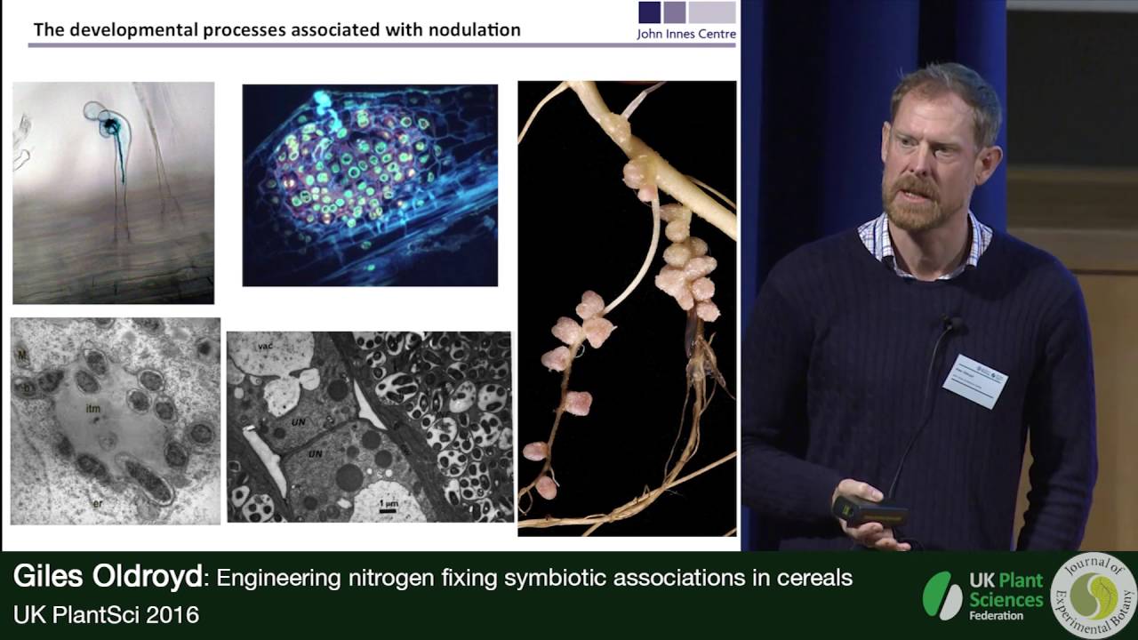
hi I'm Sharon long from Stanford University and I'm here today to talk about our recent work on the plant side of the nitrogen fixing symbiosis specifically about genes and cell responses by way of review I'll mention again that the rium symbiosis happens in organs called root nodules and Within These nodules bacteria are able to fix nitrogen and provide that to the plant for nutrition this is a complex developmental uh process it goes through a number of stages and you'll see some of those today it is species specific which I've discussed in some of my earlier talks and U importantly for today's lecture bacterium and plant each respond to signals from the other and outline of my talk today is shown here uh at the beginning I'll give you some introduction reviewing some of the Dynamics of how bacteria and plants interact then I'd like to talk uh about some recent work in our lab first about infection and about a group of proteins called flotillin and also about a nod factor receptor from the plant then I'd like to talk about two plant mutants dnf1 and dnf2 which we've identified um as being defective in late stages of nodulation and by describing the genes that we've now cloned from those mutants uh I can uh share with you some of our ideas on how the symbiosis is working so here I'm showing a review of the basic idea of signal Exchange in the early uh phases of nodulation the plant secretes a flavonoid signal this flavonoid acts as a trigger in the bacterium and that causes the transcription of genes called nod genes which encode enzymes those enzymes are able to synthesize this second signal the so-called nod Factor down here sometimes abbreviated as NF in some of the slides to come this is a modified um kiten fragment you can see there's four residues of in aetel glucosamine these are modified at the reducing end with a sulfate and at the non-reducing end with a both an acetel and an nasil group and in one of the uh slides yet to come I'll be talking about the differences between the plant response to kiten and to a true nod Factor so nod factors are very powerful and this gives you an idea of just how powerful they are on the top we have an alfalfa route and on this route Cobian melodi the symbiot has established a nodule and this is an intact nodule it's been cleared and you can see the uh deep brown color which is from the metals that are in the various cytochromes and enzymes of the bacteria while they're fixing nitrogen now at the bottom we see a form that looks very similar and yet this has no bacteria in it this entire structure on an alala route was uh caused just by a small droplet of nod Factor so this one chemical is able to produce an entire organ on the plant here is another one of the characteristic early events in rium uh uh legum interactions this shows uh root hairs on the root of a plant and here on the right you can see a normal root hair which grows straight and here you see a root hair that has been provoked by Ryobi uh to grow in such a way that it top over and forms a curl and here in the uh crook is uh where the bacteria are trapped and these deformed root hairs of various kinds are very characteristic of the effect of uh the correct rium on its compatible host now the bacteria are then able to uh uh travel by proliferating and burrowing their way into the plant cell itself and this white Arrow shows an infection thread this is formed by the plant in response to the bacteria within the infection thread bacteria are proliferating and they are uh invading through the root hair cell and as you'll see eventually down into further cell layers so there's the infection thread and you'll see more about that in a little while I'd now like to show you something about the Dynamics of how Ryobi uh affect plants let's start by looking at normal root hair so on a root the youngest part of the root is over here and you can see that they don't have any root hairs yet and then the root hairs get longer and over here they're full sized so root hairs get to a particular size and then they stop now let's watch in this movie to see how root hairs elongate normally that's without Ryobi so here you see root hairs they start they grow and then at some point they reach a mature length and they just stop like that but they grow fairly straight during this entire process now we're going to look at root hairs on a plant that has been treated with its rium um symbiant so here the root hairs are in the process of growing and if you watch what happens to them as they grow you'll see how different it is from unperturbed growth so here for example we see root hairs that are curling over on this side if you look at this root hair you can see that it's growing and then it pauses see that pause and then it emerges and now it pauses in branches again so there's two things going on one is this characteristic pause so right here you can see there's a pause right before it starts to top over and the other is that you're getting branching or curling other kinds of deformation so rium has a profound effect on the morphogenesis of this particular plant cell now we've seen a little bit now about root hair deoration and I'll mention along the way that uh rium can also cause roots to engage in specific transcription which we've studied with microarrays and they provoke cell division but for the moment I just want to look a little bit more about what happens to these root hairs because if you uh think about it root hairs are on the surface of the plant they're single cells they have a high surface to volume ratio it makes sense that these are going to be exposed to the signals from the outside and they might be the place where signal transduction begins so we might ask um by cell biology and by genetics are there receptors and what is the signal transduction pathway and ask whether we can study root hairs as a way of capturing those now signal transduction is often a complex and fairly rapid multi-protein process so we set out to look for events that occur in root hairs that um are cell autonomous and fairly rapid one of the earliest uh uh studies that we did in this area was simply to look at the um electrochemical potential across the plant membrane so in this we're taking a single root hair here and by putting in a micro electrode and measuring the difference in potential between the interior of the cell and the exterior of the cell we were able to uh find that untreated root hairs have a stable uh very negative potential at about 130 to 140 uh molts however if we treated a root hair with Ryobi what we found was that the Ryobi and its nod Factor are able to cause a depolarization of that plasma membrane so that the the potential change across the membrane here diminishes and that happens actually within a minute uh in our studies and others this has been shown to be uh accompanied by ion fluxes ion currents near the tip now one ion in particular is very interesting to us and that is calcium uh we've used different techniques but here's one of them that we have used on the uh left you can see um a root hair and into this root hair we're delivering a mixture of two different fluorescent proteins one is fluorescent irrespective of the calcium concentration the other increases its fluoresence when calcium is high using those together we can take a ratio and get uh the value of calcium corrected for the concentration of the cytoplasm and then we contract that over time so now we're going to follow calcium in this slide now this is a pseudocolor representation of uh the fluorescence corrected for cytoplasmic concentration cool colors mean low calcium warm colors mean high calcium if you take a look at the root hairs here they're all blue that means low calcium As you move in time and these are 10-second intervals you see the warmer color yellow appearing from the tip of the root here so calcium is uh getting high at the tip as you continue the time series up here you can now get to a point where you see this extremely high calcium shown by red and this happens to be in the part of the cell represented by the nucleus so what can we say and I'll quantify that with the following graphs first if you compare an untreated cell that's here and here's the Baseline of the calcium with uh the presentation of one nanomolar of Nod Factor what you find is that about 10 minutes after the presentation of the nod Factor you get these sharp upswings in calcium and that's what's going on here this is the calcium spiking represented there now number two if you put in a higher amount of Nod Factor 10 nanomolar now you get something more complex you get a faster response and this turns out to be the tip flux of calcium coming in and then calcium spiking happens at the same time now the the other data that I'll show you here is that if you take kiten um alone now remember uh I pointed out that nod factor is like a an olive poer of kiten and aetl glucosamine residues with some modifications but what if you take the modifications off it's not aactor anymore it's just kiten if you add very very large amounts of kiten up at um uh let's see we've got one microa here so a thousandfold higher you get some spiking but you need much more uh kiten than nod factor and it's not completely normal so for that we can say that the uh plant is exquisitly tuned to the nod Factor finally we do know that the uh calcium flux and calcium spiking are not just um uh separated uh in uh in space but they're really independent in that you can add nod Factor after uh High nod Factor after calcium SP spiking has started and you'll get the big influx so they really do appear to be two separate events now a third uh response that we were able to document uh very early in nodulation is this one using a whole seedling assay and uh with um an indicator that shows the presence of hydrogen peroxide we can follow normal uh Roots over time and we can assess how much peroxide are they producing and in a normal untreated root hair that's shown here they don't produce zero they produce a modest amount in here here's what it looks like over a period of about 1.5 hours now what if we treat those with nod Factor what we find very intriguingly is that nod factor causes the rate of evolution to be diminished so a lower level of Nod a lower level of hydrogen peroxide is produced um in the presence of Nod Factor now what if we add an elicitor pathogenic elicitor we would expect an elicitor to cause the plant to mount a defense response and sure enough if you look at the amount of hydrogen peroxide coming in the plant after the treatment with the liter it's accelerated so it seems that uh root roots are able to elaborate uh hydrogen peroxide an example of reactive oxygen species that nod Factor diminishes that which would suggest it's lowering its defenses and H and elicitor increases it consistent with an increase in defense now this is only over the couple of hours we're interested here only in very early events uh in fact uh the rea the uh defense reactions appear to have a very interesting and complex role later on in nodulation but that's uh this is not related to those this is just uh um in the first two hours or so so now we can fill in a bit what we know about the early responses of the plants to the Ryobi nod Factor we know that it causes um nodules to form cell divisions in the plant we now can fill in that in addition to the overall root hair curling which we could see um uh in the microscope other kinds of assays demonstrate that there's a rapid depolarization across the plant plasma membrane in response to nod Factor this is accompanied by calcium flux slightly later there's calcium spiking in the cytoplasm and a suppression of the rate of reactive oxygen uh uh production now one other topic that I'll just mention briefly is transcription we've been able to assess transcription uh during nodulation uh at various stages uh with a specialized uh approach which is uh shown on the next slide we following our work on determining the uh complete genome sequence of the bacterium we then then constructed an aphim metrics chip in which we had the complete bacterial genome plus about 10,000 uh sequences representing um probable genes from EST libraries in the plant and we put the two genomes on the same apim metric a chip so we call this our Symbio chip and uh through analysis of RNA species from the uh nodules we can actually get a readout of both bacteria and the plant at the same time and uh through that we were able to show that within the first 24 hours after treatment with nod factor or after treatment with bacteria a characteristic set of plant genes are upregulated or are down regulated some four dozen or so uh sequences I'll talk a little bit more about that in a moment when I uh get to the mutant analysis but now let me put all of those events on a timeline for you so I'm going to start here at the left now we know that in the very early stages within a minute we get uh calcium flux suppression of uh Rea of reactive oxygen we get a depolarization there's also calcium spiking at about 10 minutes there are morphological changes you saw in the film that the root hairs pause and swell a little bit right before they start to curl then there's morphogenesis of the root hair forming branches and curls and both and we can see the cell divisions finally the transcription now here's the timeline for that happening if we uh start at time zero and this is where the bacteria are being added those first events such as the calcium flux and the depolarization happen within a few minutes one to a few minutes calcium spiking at an average of 10 minutes the gene expression that I mentioned that we can assess with our aetric chip that's happening um as early as a few hours and goes on for about 24 hours now the actual curling of the root hair the deformation of the root hair takes a while to express that of course we have to keep in mind that this is limited by how fast the root hair can grow we don't know when the decision to curl is made but the actual outcome the mechanics of curling take uh somewhat longer and then here around 20 hours we're going to start to get infection the uh production of the infection threat and the uh ability of the bacteria now to penetrate in through that root hair and then the cell division here lead leading to the production of the nodule that's all uh happening over this period of a day to two days now through um a set of genetic uh mutageneses and screens our group and others have identified many different plant mutants that arrest at one or the other of these uh St es and having these subcellular assays to do allowed us to distinguish between the different kinds of uh plant nod minus mutants so here's an example here on the left is um a gene called NFP for nod Factor perception if that Gene is mutated you have a block right here so nothing happens at all there's no depolarization there's no calcium flux no change in the morphology and then here are um two mutants um that are present in such a way that they allow the plant to have calcium flux and it has uh suppression of reactive oxygen species and so forth however it doesn't show calcium spiking so these two mutants were able to distinguish between the early calcium flux and the calcium spiking itself they are not only separated in the cell in geography and in time but there they can be separated genetically here we found uh one or or more mutants that uh allow calcium spiking but do not allow any gene expression and so forth and so on down the line so with all of this we are able to set a timeline and developmental sequence for how signals are transduced and among other things we were able to use that transcription essay to distinguish between root hair curling genes and uh early calcium transduction genes and we were able to show that all of those plant genes that are expressed um come on at the same time it's not the case that you can get a few genes here and a few more here and a few more after that you get no genes expressed at all up until this point here and we were able to show that by a combination of mutant analysis and uh transcription analysis so that means we're going to be able to say that the series of events shown here calcium flux suppression growth arrest the pausing can cluster and we can see that one particular Gene called NFP which we believe encodes a receptor is responsible and required for those events then there's a set of uh two genes DOI 1 and DOI 2 that are required in order to get to calcium spiking one of these is an ion channel the other is a putative receptor kinas intriguingly uh just Downstream from calcium spiking is a mutant domi3 and another mutant Cyclops these are both required in order for um any of the later events to occur such as transcription so we believe that these genes help interpret the calcium spiking signal and transduce it to the next set of genes nsp1 and nsp2 these are required for transcription and indeed they turn out by sequence to be predicted transcription factors more transcription factors are also required in order for nodulation to occur now all of these events on the right require the one pathway this is called the signaling pathway starting with the signaling receptor n fp1 and going through transcription fa factors but I haven't said anything about infection and I'd like to mention that it turns out that the plants appear to have not one receptor but two the second receptor is called the stringent or entry receptor and the name of that Gene is lick three in the case of metago truncatula this and one of the transcription factors are required in order for infection to occur so you can have these other events happening but no infection this additional set of uh factors is required for infection so back to the outline I've I've gone through a description of some of the plant cell responses to no factor and told you about how we have used plant mutants plus those cell phenotypes in order to create an ordered set of steps that we believe represents the signal transduction pathway in early nodulation in the next uh part of the talk I'm now going to move as I say Beyond nodulation I'll be talking about infection and also about later stages in nodulation so as we uh think about uh the symbiosis remember everything that I described in terms of early signal transduction is over here that's just the beginning there's so much more to come there's uh as the uh bacteria infect and eventually form the module so now let's take a look at some of the specific topics that are interesting first infection we've already seen that more genes are needed for infection than just for signaling and the infection thread is a remarkable structure it penetrates not just through the root hair but then through multiple layers of plant cells on its way in to uh find target cells now as this proceeds the plant elaborates an entire nodule and all the Dark Cells in this uh photo are packed full of bacteria and if you look close up at those each bacterium is surrounded by an envelope of plant membrane and within that the bacteria are going to differentiate so using uh different approaches we're going to take a look at uh how the plant manages to support these really remarkable events we'll start with infection uh as I mentioned um infection occurs beginning in the root hairs and what we wanted to ask is how does the inside of the plant cell cope with the invasion of the bacterium how does it reorganize itself we decided to take a look at um some candidate genes called flotillin These are genes uh that are originally identified in animals I'll Give A Brief Review and then I'll I'll show you our data that demonstrate that plant flotillin uh in are active in nodulation there's two copies of flotillin that are specifically upregulated in nodules uh that their transcription is regul regulated along with other nodulation um uh genes they're required for infection they're required for elongation and remarkably the proteins of these flotillin have very striking localizations that we believe relate to the mechanis of infection so what are flotillin they've been studied widely in animal cells this diagram shows an animal cell with plasma membrane flotillin are shown here there's two of them they're outlined in red they have a domain that is affiliated with the membrane and a tail that is cyto more cytoplasmic and it is thought that they um relate to signaling to endocytosis and to activity of the actin Network within the cell um it's also uh been demonstrated that these um flotillin uh occur in what are sometimes termed membrane micro domains also sometimes called lipid rafts although the terminologies are actively being discussed uh these days now plants have flotillin too although they have not been widely studied uh in a rabid opsis the sequence shows that there are three flotillin in our analysis we found more than seven in the legume medicago truncatula the um of these seven we found that two are uniquely expressed in nodules we found that the proteins are as with animals flotillin present in small uh domains or puncta and uh I'll show you that one of the two has a very special uh localization during infection now the uh predicted structures of the flotillin Imp plants looks very much like the animals with a domain a head domain and a tail domain so we might expect that it's going to have some of the same properties so as I mentioned arabidopsis has only three flotillin but metago Tron catula has uh more than seven and here's one of the really interesting things for us is that there are a great number of these that are actually all linked together now you'll see here that there's flotillin 3 and one then there's down here flotillin 4 and flotillin 2 we believe that those are active flotillin one of the others shown here flotillin 5 is in the same genomic region but we believe it's uh not uh an active Gene now we took a look at the activity of the promoters for each of those flotillin and what you can see here here is at the top flotillin one it's uh promoter is active in the vasculature it's here it's expressing um a gus Fusion flotillin 3 also is active in the vasculature uh but if you take a look at flotillin 2 and flotillin 4 these are being expressed in nodules we can also quantify the transcript and the same story emerges we can see here that flotillin 1 and three are at a low level throughout modulation but both flotillin uh 2 and flotillin 4 are strongly upregulated in the first day after roots are presented with uh bacteria flotillin 4 comes down although flotillin 2 stays uh fairly well expressed during at least a couple of weeks of nodulation so in terms of their expression these appear to be uh especially uh associated with nodule development does that relate to any of the um formalities that we know right now about the genetics of early nodulation so remember that there are a set of genes that I introduced earlier and we have mutations in these various steps uh for um um naal signal transduction and that transcription occurs only after a whole set of steps have uh taken place so what about flotillin 2 and four and as you can see in this bar graph those two flotillin actually require the same signal transduction pathway in order to be expressed here is a while type on the left we're seeing flotillin 2 and four and then here are a couple of the nodulation mutants lick 3 nsp2 nin one those are here NSP and nin here's lick three for a receptor for nod Factor these appear to be required in order for the flotillin to be upregulated we can also show that some of the downstream genes are not required now we know that these flotillin are controlled in the same way that nodulation genes are controlled but what about their function through using RNA approaches we can diminish the uh activity of the various flotillin and here's one particular experiment uh and these results have been uh borne out in others although the absolute numbers uh tend to vary so if we take a look at a wild type plant for example we can see that it's got about six nodules per plant if you knock out flotillin one and three there's again about six but if you knock out flotillin 2 there's many fewer nodules flotillin 2 and flotillin 4 if knocked out together is even more but flotillin four knocked out by itself does not appear to have much of an effect on the number of nodules if you look at the percentage of plants with nodules you don't see a major effect in fact if you have if anything you seem to get a few increases of the percentage of plants that have some kind of growth on them but now if we look at the percentage of nodules that are actually fixed plus which is indicated by the pink color from leg hemoglobin you can see that uh about uh in this particular experiment almost 30% of the wild type nosul appear to be functioning uh about the same for flotillin one and three Knockouts but if flotillin 2 is knocked out then there's a striking uh diminution of the number of functional nodules and uh a less striking uh but still significant change for flotillin 4 and the double mutant uh almost completely wipes out any effectiveness of the nodules that do form so we would conclude from that that flotillin 2 and flotillin 4 are both required for nodulation and that they are not redundant because the double mutant has a more severe phenotype than either of the single mutants alone we've also now gone to take a look at the proteins and what we found is the following let's begin on the left with flotillin 2 this is a view of epidermal cells so we're looking down at the the surface of the root here and you can see that flotillin 2 is present in little dots or puncta and there's a very striking localization of flotillin 2 at the uh polar end of the cell that's uh in the root hairs the flotillin 2 is very strikingly punctate it's present in these little dots and this kind of um distribution Remains the case if the plants are inoculated and that's shown in the lower part of the left hand side so uninoculated and inoculated flotillin 2 is punctate it remains fairly stable although there are some subtle some subtle changes in density and in um polarity but we got um a very striking result with flotillin 4 and I'll show you that here then let's start on the top again the epidermal cells and root hair cells and it's very striking that the flotillin for uh signal gfp Fusion signal is present in puncta but even more uh striking was that after inoculation what starts out as an even distribution of flotillin four in root hairs becomes a highly polar distribution and that's shown with these red arrows where you can see that at the tips of these inoculated bacteria treated root hairs flotillin 4 has migrated to the tip so that suggests that flotillin 4 has a very active uh participation in something having to do with what goes on at root hair tips so we began to look specifically at infection using uh wild type as control where you can see bacteria uh invading these blue Lac stained bacteria show infection threads for uh um bacteria on a wild type plant here's a nice infection thread growing through a root hair on the wild type but in a tillin 4 minus mutant things are not working very well what you're seeing here is that there's uh no significant uh penetration deep into the nodule furthermore even if you look at a root hair the uh infection threads uh are lack Integrity they are not well structured and uh we find in fact that they don't succeed they don't penetrate so it appears that flotillin 4 function is important for infection threads and we also now have evidence that shows that flotillin 4 is a is associated with infection threads flotillin 2 is uh shown here bright green fluorescence around the cell this particular root hair which is curled has been infected and it has bacteria that fluores red those bacteria are shown here but you can see that the flotillin 2 is around the membrane of the outer cell it's not affected not close by to the red fluoresence of the bacteria however if we look at flotillin 4 that's shown here flotillin for gfp is around the outside but it's also here see around the infection thread green fluorescence and here's the red bacteria inside the fluorescence so that suggests that flotillin 4 is affiliated with the affection thread membrane now you'll recall that um in the uh sequence of genes that are important for nodulation Signal transduction uh one set is called the signaling pathway here but more genes are needed in order to get infection including the entry receptor lick 3 now that we've got some clue that flotillin are involved in infection we might want to ask whether we can place flotillin in any kind of relationship to the genes that have been previously found to be important for early signaling and infection now in the following um uh photos I'm looking at the puncta density of flotillin 4 here's a wild type and the flo the punct of density is the number of bright spots per uh Square microns right so you might have a little a little um Square Micron there and then you plot the number and I'm going to compare that while type to three different mutants each of these is a mutant in the putative entry receptor lick 3 lick 3-1 which um is the most severe and two other alals Lick 2 and uh lick 3-2 and 3-4 um the lick 3-1 is predicted to have um a protein with a dead kinas domain and what we saw looking at this particular index of punk to density is that if you compare the wild type number here to the number in the lick 3-1 mutant the Pumped density for flat 4 is greatly diminished significantly lower we don't draw any mechanistic conclusion from this but it did give us the sense that maybe lick three itself together with float 4 would be worth looking at so we want to ask whether lick 3 and Fluellen 4 interact we're going to need to study lick 3 and we were fortunate to collaborate with uh Doug cook Brenan Riley and their colleagues at UC Davis and we have thanks to their contributions we have the following tools for study first and most importantly a stable transgenic of metago Tron catula that carries a gfp fusion to lick 3 under the control of its own promoter it's known that this is functional because it complements a lick 3-1 mutant because it's under its own promoter we have more reassurance about its correct position uh and localization now into this stable transgenic we're now going to introduce a second uh marker we're going to create a transformant that also has flotillin 4 fluorescent um linked to the fluorescent M Cherry protein so we'll have green fluoresence for lick three red fluorescence for M cherry and our particular tool is going to be a spinning disc confocal uh at the Carnegie Institution uh in collaboration with David arheart we were able to in uh to visualize the lick 3 putative receptor in root hairs so the results are shown here we have uh lick 3 gfp it's present in the root hair you can see that it's punctate and when we took a look at U the lick 3 plant with infections we we found the following here's a curled root hair and you can see going down through the curled root hair is an infection thread and the green fluoresence of that lick three is all the way along the infection thread membrane so we can see that the lick three putative receptor is localizing the same way that the flotillin 4 uh appears to do along the infection thread as it uh moves into the cell now an important control is shown here also um root hairs and other parts of uh plant cell walls have autofluoresence as well and we wanted to ask whether the green fluoresence we saw here was really due to the lick 3 or was it autof florescence the results of that are shown in in this um other panel now this is a fusion of lick 3 to a nonfluorescent protein and again there are bacteria that are fluorescing red well what you can see is that it's a good idea to do this control because the crook of the root hair is very highly autofluorescent so if we were to see this and say it's gfp fluorescence we'd be wrong because that's just what the root hair uh cell wall is doing you you can see here the infection thread is moving down with the red fluorescence and uh the uh there is no green fluorescence around it so infection threads are not intrinsically fluorescent it's only that the lick 3 gfp is providing that signal so now we know that flotillin 4 and lick 3 appear to localize to the same place can we find out anything else about them so in this um we have um started to use that double transgenic so what's going on here is that this plant has lick 3 gfp it also has flotillin 4M Cherry now these are close-ups below you can see the lick 3 gfp flotillin 4 cherry and in these are uninfected root hairs and if you merge the two images it's possible to see here that the green and red are distinct so that suggests that they are not colocalizing within the distance that where their fluorescence would would mix and make a yellow color now taking a look at that uh more statistically we can do uh correlation plot as shown here where uh on one axis on the x- axis that's the fluoresence intensity of one on the Y AIS is the fluoresence intensity of the other and in fact there's a distribution all the way around it doesn't particularly correlate if you have high fluoresence of M Cherry that could be either low or high uh fluoresence of gfp but what happens when we treat root hairs with bacteria and that's shown here now again we've got the gfp here's a closeup we've got the M Cherry for the flotillin and a closeup but now when we do the colocalization you can see that there appears to be a merging of the green and red fluorescence to present uh a higher number of puncta that look yellow so we would say there appears to be an increase in the correlation of um of their their location and here's another way of looking at that the correlation plot now shows um a correlation coefficient that's uh more than uh 05 that means that more than half the time you're getting a correlation of the intensity of fluoresence of one and the other so that suggests to us that whether or not flotillin 4 and lick 3 are associated depends on whether the bacteria have interacted with the root hair so we pursued that a little more another way that we have started to look at that is to examine the Dynamics not just the location but the Dynamics of what these proteins are doing so in this um experiment we're going to looking at a root hair which has a lick 3 gfp Fusion so that's the only uh fluoresence you're going to see it's just the receptor lick 3 and what you're going to be seeing in the micrographs is as if you took a a plane like this right uh through the root hair and where you see that this bright uh Point that's uh what you is going to be shown now on the films now the left hand we're going to look at lick three in an uninfected root hair so one of the things you can see is that it's very Dynamic if you try to focus on a point and see follow the lick three it it's moving around too much you can't do that but now let's look at a root hair that has been treated with bacteria what a difference now you're looking at the lick three fluorescent and it's really behaving itself it's sort of staying in place and these two arrowheads for example show places that you can focus in and you can see a lick 3 signal that's not moving it's fairly stable so that suggests to us that it's not just where lick 3 is it's how fast it's moving and um perhaps shuttling around that is being changed by the presence of the bacteria which is um uh remarkable and uh that led us to another way to look at lick 3 together with Fluellen 4 in the following we won't be looking at movies but we'll be representing the Dynamics over time through what's called a chog graph now what you're seeing on the left is um a micrograph of the receptor lick 3 gfp fluorescence Fluellen 4 fluoresence and the merge likewise here for uh bacterially treated root hairs lick 3 gfp ftill and for cherry and then and then the merge now at each uh point in time we don't look at the whole root here we're going to just be looking at a transect shown here and also shown over here by these blue arrows and at any one point in time just that line is going to be uh represented as follows here if we take this line going across the uh uninfected lick 3 gfp um then we can see at any one 10sec period whether uh particular position was light or dark we can follow that over time and we can see that this the correlation is rather loose however flotillin 4 is very stable if you look at the um flotillin 4 10 seconds 20 seconds 30 seconds and so forth it's if it's fluorescent at one 10c interval it's highly likely to be fluorescent in the next one and so you get this these set of lines going through now if we take a look at root hairs that are treated with bacteria we find um sorry we find the following um again this is what the uh root hairs look like but if we follow them over time you can now see that the lick 3 gfp is very stable if it's if that transect shows a bright point and one 10-second interval it's probably going to be bright the next 10-second interval as well so the lick three has settled down the flotillin 4 is still very stable and when you take a look at the merge you can see that there appears to be a correlation of the brightness so as we summarize we can say the following that the um punct of density is changed uh for a float for gfp in a genetic background where uh lick 3 is mutated we can see that lick 3 itself uh loc localizes in puncta that suggests it's in membrane micro domains also it localizes to infection threads we found that in uninfected root hairs the uh the receptor uh and the flotillin do not have uh much overlap and also they are very different in their motility in their Dynamics however after inoculation with uh Ryobi alodi the receptor and the flotillin colocalize and their Dynamics remain similar not known is whether this is direct or indirect what is the nature of the protein protein interactions because fluoresence in and of itself is relatively uh loose uh uh it does not have to have precise molecular um adjacency so further uh fluorescence studies with fret and biochemical studies will be necessary and uh we think that following the combination of this uh flotillin and receptors is going to be a very exciting way to ask how the plant is mobilizing itself to accept infection so back to the outline I've uh finished uh talking a little bit about how a candidate Gene approach took us from the study of flotillin to the study of infection in this next segment I'd like to talk about some mutants that we've uh isolated called the dnf mutants these uh have identified plant genes necessary for the final stages of symbiosis and I'll talk about two two of those dnf1 which encodes a signal peptidase and dnf2 a putative phospholipase C so looking again at the sequence of nodulation uh we're now taking a look at an even later stage where bacteria have penetrated into the nodule and they get released into uh the uh cells and they're able to fix nitrogen so the dnf mutants called uh defective in nitrogen fixation were uh isolated out of a screen that we did of mutants that we generated by fast Neutron bombardment this often creates uh deletions it's an ionizing radiation um and so these are severe mutants when we screen them we found a number of mutants which were like this instead of being nice pink nodules here the dnf mutants are white and they're small they don't have any nitrogenase activity and that's shown as a uh in this Slide by um assessment of acetylene reduction shown here while type is able to convert acetylene to ethylene which is an indicator for nitrogenase enzyme but the dnf mutants as you can see have very low or even no ability to fix nitrogen now they also don't seem to have completely normal development one of the mutants dnf1 is shown here and we got two alals of that they're very similar wild type uh nodule has cells filled with bacteria and the bacteria do get infected into the dnf1 nodules as well nonetheless they are not as large and they do not seem to be able to fix nitrogen so we took a look further at dnf1 and one of the ways in which we uh studied this was uh as as follows here's a wild type nodule shown in section and this nodule uh in in in this case has been established by the action of a bacterium carrying a glucuronidase fusion into the promoter for the nitrogenase genes that means that if the bacteria are expressing their nitrogenase then the nodule will turn blue in the presence of a glucuronidase substrate so what we see here is that the nifh which is nitrogenase glucuronidase Fusion is turned on that's good that means that the bacteria are expressing their nitrogen fixation genes however what we found was dnf1 was the following taking a look at these nodules with the same exact bacterium carrying a glucuronidase fusion to the niff promoter we found no activity you could see here with a red arrow it's pointing to the nodule but there's no blue stain so we conclude from this that the bacteria are not able to express the genes for nitrogen fixation so we can say that the bacteria need the plants dnf1 in order for the bacteria to fix nitrogen or even to express the nitrogen fixation genes so what is dnf one um this Gene in the plant encodes one of the uh subunits of a what turns out to be a nodul specific signal peptidase and I'll tell you about nodul specific and the other components in a moment but first I'll just review a little bit what signal peptidase is in um in a eukariotic Cell we've got the endoplasmic reticulum and uh proteins that are being extruded into the Lumen uh may may get there by a signal peptide and it's the signal peptidase Cleaves off that uh signal peptide and that allows the production of the mature protein which would be shown here now once that mature protein is formed after its signal peptide is taken off then it may be uh targeted for secretion or for vesical trafficking so what we found is dnf1 required for bacterial uh gene expression and differentiation has something to do with protein processing now let's take a look at the expression of this Gene what if you take a look at the vegetative parts of the plant such as the leaf and stem uh or flowers what you find is that expression of the dnf1 uh signal peptidase subunit is very low but it's quite high in nodules it's also present to some extent in developing seeds although at a much lower level taking a look at the time course what we can see is here the dnf1 if you plot days of nodule growth and development and dnf1 expression you can see that dnf1 goes up early in uh nodule development and you can also track it during seeds and find that it's relatively steady during uh the maturation of seeds so dnf1 is most highly expressed in nodules and it goes up very early in those nodules now um let's take a look not just at dnf1 but using transcription let's take a look at what genes out of all the microarray data tend to be expressed whenever dnf1 is expressed and those are shown here now on the left you can see the names of the uh proteins this is what they do and what you can see is that there's the other uh subunits in the signal peptide signal peptidase uh uh complex are also regulated up in early nodules the same way the dnf1 is in addition the signal peptide peptidase which is needed for completion of the um degradation of the signal peptide that's also upregulated and finally this very intriguing protein sip 132 syntaxin 132 now what what are syntaxins this is a reminder of how they have been uh characterized in animal cells syntaxins are important proteins for vesicle targeting and in fact sip 132 here in the plant probably marks the last step of protein secretion and empirically it has been observed on plant plasma membranes and also on What's called the symbiosome membrane that's the membrane that surrounds the BAC iium once it's inside the cell so if we take a look at all of these genes we can see that they are co-regulated so we've got a whole signal peptidase complex and what is it doing one of the most important questions that could be asked is what are the substrates what proteins does this signal peptid a help to mature and the answer came from work in um the labs and uh J surette and in zad Hungary uh led by avac cond roshi and Peter merett and their colleagues and what they have described is a set of proteins called nodul cysteine Rich peptides these are characteristic of nodules they are not expressed any place else in the plant and they are rich in cistin and bear some resemblance to defensin which are uh proteins made uh by plants and uh animals in response to microbes they were able to show using our dnf1 mutant that one of these uh for example uh NCR peptide number one in a wild type plant gets processed to a smaller molecular weight but unless the dnf1 peptidase is there it does not get processed because here is our mutant and you can see that the protein shown here in imunol blot is larger it's because its signal peptide was never taken off they have also been able to show that with antibodies that in Wild typee cells if you use a green dye to indicate the bacteria and a red uh fluoresence for the uh antibody to the NCR peptide they colocalize bacteria and NCR peptides colocalize in a normal wild type cell here they showed an example of what happens in a uh mutant dnf1 cell you can see that the um bacteria are all over the place in Green but that this NCR peptide which we know is not being processed right because it's large molecular weight that NCR peptide is stuck someplace probably in the endoplasmic reticulum so their conclusions together with our work on the dnf1 are that the NCR peptides are substrates of the dnf1 complex in Wild type these are delivered to the symbiosome and in dnf1 they're retained in the ER they've shown in other work that these uh mature NCR peptides have profound effects on bacteria such as uh causing them to cease cell division and uh changing their membrane per permeability so um that's an ongoing story about the activity of the ncrs to complete the model so far uh we might ask uh what does dnf1 do and one uh likely activity is that it processes NCR proteins these end up in the Lumen of the ER and they are targeted perhaps using U uh zp 132 as part of the mechanism for targeting into vesicles which come over and fuse with the symbiosome in which rium are going to uh be differentiating now we also have questions about dnf1 beyond the NCR proteins for example dnf1 is present even in legume plants that do not have the NCR proteins so what is it doing there another uh question is uh in the uh dnf1 mutant the bacteria bacteroids do not differentiate but they also stop dividing so it seems that even without dnf1 there may be other signals that are helping these bacteria to uh stop dividing so we hope that we'll be able to work on those as well as on the actual mechanism of dnf1 in The Next Period of our work I now like to finish by telling you about the other mutant dnf2 and this as you recall tall as shown right there that also had no nitrogen fixation and we've done some work to characterize what's going on with dnf1 and also to figure out what the gene is here is a picture in which you can see from the uh root part to the distal part of the nodule uh fluorescently labeled bacteria and they are inside we can tell that the bacteria get inside the dnf2 mutant nodule however they don't express nitrogenase and they don't express many other bacterial genes either so somehow dnf2 is again required for the bacteria to differentiate and in fact to uh Express genes now we know that dnf2 is um expressed only in nodules even more strictly than dnf1 this shows you some promoter gust fusions where you can see that the uh early part of the nodule is expressing dnf 2 and here you can see it at the tip and then it diminishes just slightly as the nodules get somewhere o um uh somewhat older what is dnf2 it appears to be uh similar to a phosph lipac c but it's not a canonical phospho lipac C the way we're used to seeing um here would be the classic PLC you can see it's got the um um uh in ultris phosphate binding EF hands catalytic domains calcium binding domains and so forth the uh dnf2 is only uh homologous to one part of this large complex fossil lipace C however we do find that it is very very similar to um a fosol lipac whose model is shown here this is actually a bacterial fosol p c and if you take this in red and then superpose dnf2 it does appear to be uh very very similar in its 3D structure so we want to know what's the biochemical activity sequence alone doesn't tell us whether it really acts to um uh M to um process phospholipids or as a signaling but we found one other thing that's really surprising to us first as I mentioned it's only expressed in nodules but secondly it's not expressed in the cells of the plant that have Ryobi it's expressed in the non-infected cells of the doil and that's really a first um there are known metabolic uh enzymes in soybean for example that are present in uninfected cells but there has never been found a regulator that appears to be active in the non-infected cells of the uh root nodule so what we're trying to ask ourselves is does the dnf2 protein mediate the infection process uh directly or indirectly is it acting in some way that allows the non-infected cells to send signals or perhaps to avoid signals so those are some of the uh questions that lie ahead for us so winding up the combination of the studies that I've shown you today uh I think that the future of our field is going to be very exciting as we take the results of the plant mutants and we study the proteins uh that are defined by those mutations and look at how those work both plant and bacteria together not looking just at position but looking at time we're interested in uh the uh great questions of what the signals are and how the signals are transduced given that so much complicated cell biology has to happen between the beginning of the infection thread and the maturation of the symbiosome we want to know not just the regulation but what is the Machinery of this infection and we believe that both the uh proteins and molecular probes that we can obtain can be used to study the individual steps of nodulation as they move forward so that concludes our journey through the plant side of symbiosis
Up Next

50:28
Soil Health: Mycorrhizal Fungi and Rhizobium Bacteria Benefits
@TaurusAgTeam
35.2K views•2020-04-01

50:12
Bacterial Communication: Quorum Sensing and Biofilm Formation Explained
@JHUAAP
12.6K views•2011-06-28

42:45
MicroRNAs: Gene Regulation Explained by David Bartel | iBiology
@scicommlab
233.1K views•2014-03-24

7:08
Bacteriophages: Earth's Deadliest Killers and Future Antibiotics
@kurzgesagt
34.6M views•2018-05-13
Related Study Plans & Knowledge Roadmaps
Structured learning paths in Biology
biology
RNAi: Biogenesis, RISC & Gene Silencing
01Molecular Biology Foundations: DNA to Protein
02Introduction to RNA Interference (RNAi)
03Molecular Biogenesis of miRNA and siRNA
+ 2 more learning modules
10h5 modules
Explore Roadmap
biology
Mycorrhizae: Signaling, Biophysics & Ecology
01Foundations of Mycorrhizal Symbiosis
02Molecular Signaling and Root Colonization
03Biophysics and Bioenergetics of Nutrient Exchange
+ 1 more learning modules
11h4 modules
Explore Roadmap
biology
Circadian Clocks: TTFL, SCN & Periphery
01Foundations of Molecular Biology & Gene Expression
02Introduction to Chronobiology and Actograms
03The Core TTFL: Molecular Architecture of the Clock
+ 3 more learning modules
14h6 modules
Explore Roadmap
biology
Population Genetics: Math, Drift & Selection
01Genetic & Probability Foundations
02The Hardy-Weinberg Principle and Baseline Models
03Mathematical Modeling of Natural Selection
+ 3 more learning modules
18h6 modules
Explore Roadmap