


















The Human Brain Project develops comprehensive brain atlases that integrate data across multiple spatial scales—from molecular and cellular levels to whole-brain structures—using advanced imaging techniques like phase contrast X-ray tomography and computational tools to address intersubject variability and enable cross-scale analysis of brain organization, while the broader Human Reference Atlas initiative extends this multi-scale approach to map the entire human body at single-cell resolution through collaborative efforts across multiple research institutions.
Brain Matters #16: Mapping the Human Mind's Landscapes
Added:Basic neuroanatomy, including the structure of a neuron, synaptic transmission, and the primary functional regions of the cerebral cortex.
Fundamental principles of neuroimaging technologies, specifically how structural MRI differs from functional MRI (fMRI) and PET scans.
The concept of biological scaling, specifically how microscopic cellular behavior coordinates to form macroscopic neural systems.
An introductory understanding of neural pathways and how electrical and chemical signals propagate through circuits.
The field of connectomics, focusing on the comprehensive mapping of the brain's structural and functional wiring diagrams.
Computational neuroscience, particularly how researchers use mathematical models to simulate mapped neural circuits and predict cognitive function.
Clinical applications of neuro-cartography, such as targeted neurosurgery, personalized medicine, and deep brain stimulation for neurological disorders.
The integration of artificial intelligence and machine learning in analyzing high-dimensional brain mapping data and atlas generation.
Neuroethical implications of high-resolution brain mapping, including privacy concerns, cognitive liberty, and the philosophical definition of the self.
286 views6likes59:03Original Release: 2023-03-09
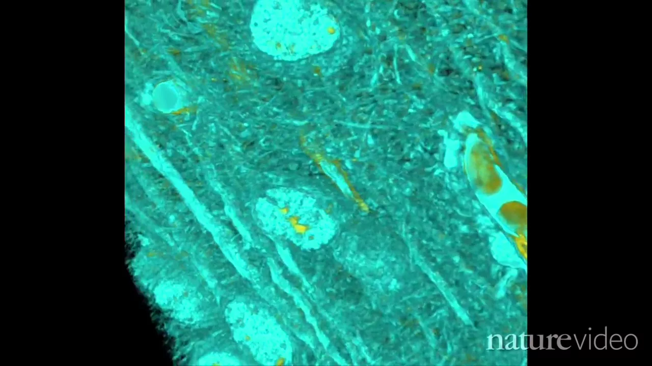
okay thank you everybody for joining our webinar with the today's episode Landscapes of the human mind this is already the 16th webinar that we are offering from the human brain project organized in this lecture project of the European commission but it's really our idea to give you everybody a broad overview of what is happening in research and today we are focusing on the Landscapes of the human mind and as you can see we have a exciting program put together hopefully for you exciting as well and I will introduce the speakers in a second but let me start with a notion that the brain is of course not only one of the most fascinating systems that we can approach in science but it's also one of the most complex systems and this complexity can be seen for example in the way how the brain is organized on different spatial scales but also on different temporal skills that means that when we are looking to brain organization then we can do it from the level of single molecules at the scale of certain angstroms or nanometers and molecules are for example involved in neurotransmission and receptors are important molecules that by neurotransmitters that transmit information from one cell to the other at synopsis at the level of a very few micrometers synopsis connect neurons and sometimes these neurons have very very long branches axons several centimeters long and they connect cells in Far distant regions in the brain cells from networks with very complex architecture this is an example of the hippocampus and the architecture of course has to do something with a function of a certain brain structure like here is a hippocampus and the hippocampus is only one part of many structures that we have in the human brain finally we should be aware that of course the brain is not acting by itself the brain is part of a body we as human beings have a body which is extremely important for the way how we are perceiving information and processing information and how we are communicating with the outside world possesses what we would like to understand in human brain research and the question is how to link the many different aspects of brain organization to each other and one of the answers is well let's do it in an atlas and we know atlases for for several centuries but probably we need some kind of new Atlas and sometimes we compare our human brain Atlas that we are developing in the human brain project with Google Maps because there's a browser there's a web-based access there are different options to zoom in and zoom out but I feel that the atlas approach for the human brain goes beyond that and poses question to us that are very specific and that are also very complex so one of the aspects why I feel that it's so complex this is inter-subject variability and brain organization brains differ in their size and shape as human beings differ with respect to hate or color of their eyes or color of hairs and so on and so forth and this is an example of intersubject variability of two areas in the visual cortex area 17 and 18 and 10 human brains and the red and green areas are always the same and you can see that these brain areas vary between the brains and shape size or the position but also the side side that is a folding pattern of this region varies between the brains and the relationship between borders and side varies as well and I can add that in the subject variability can also found at the level of the microstructure and when we started more than 25 years ago to map the human brain we introduced an algorithm that helps us to identify borders between cytoseotactonic areas in the brain using image analysis and statistical tools here you see such an example that we map cell bodies stay in sections here again in the visual cortex and using these quantitative tools we are able to identify borders between brain areas in a reproducible way and in order to capture and the subject variability we are mapping not one brain but 10 brains and bring this in a so-called reference space to make this site or archetectonic areas available as an anatomical reference for data coming from functional Imaging studies for example the methodology we have published uh two and a half years ago and describes the details and the work flows that lead to this Atlas how can it be used well this is one example of many so you see here on the right top three areas in the pre-motor cortex and in the middle you find again these three areas which are called 6r16v1 and V2 and the little spheres they indicate clusters of activation of functional Imaging studies coming from quite different functional Imaging tasks for example fulfilling a certain hand movement or having a grasping movement or Orient yourself in the space or observing a movement and we see that there's a certain dissociation between the areas in the three d space and this is a possibility to link data and findings from the living human subject that we can measure using neural Imaging with the high resolution postmortem maps that we have acquired in the past years and this Atlas now has grown and does not only provide site or Azure tectonic Maps as we can see here but also a lot of tool to get access to other data for that can be found for each of the areas and Timo dictionary later will show some applications so there are different ways to look to the brain to zoom in but also to get access to different types of data to get description of these data to learn more where it has been published and to better understand for example how a certain brain area is related and connected to another brain area and we are going also one step towards higher spatial resolution not only to large systems involved in a certain function like um like for example grasping or reaching but we also wanted to go into more microscopical detail and what you see here are images obtained with polarized light Imaging this is a technique that allows to image axons that is Somalian sheets surrounding axons using an optical technique and in the same images we have stained and labeled different types of cells here binding and Cal retinine cells and the cell specific parameters makes it also possible to link this type of Atlas data to Atlas data coming for example from the U.S brain initiative or coming from um from the work in which Katie is involved so the idea today is to show you what are the tools to approach Atlas data in the human brain this is done by Timo diction and then Tim zaidet will join us he is a physicist and coming from a different perspective analyzing microscopical details of tissue structure that are below even the one micrometer range and that could benefit from the integration into a larger spatial framework we will have today here Katie Bronner from the US who speaks about the perspective of the human body and I'm really excited today also to to have her here because as neuroscientists we sometimes forget that's not only the brain but the body is equally important and at the end Christian will close our journey to the Landscapes of the human mind he's an expert and functional Imaging and were speak more about from brain structure to cognition and then going back so having said this I am happy now to introduce and hand over to Timo dixia it was a professor of informatics at Dusseldorf University and working as a research center ulage and he is the head behind the Edna's technology Timo please thanks a lot Catherine I was muted for introducing me happy to present the infrastructure more or less in a nutshell um so um what we are doing in eprints in terms of the Applause is to provide infrastructure and tools and services to access our classes of different species you see here um some screenshots of a human brain Atlas the red brain Atlas Mouse brain Atlas and um a monkey Atlas which is coming up and of course in this webinar I will be focusing about the concepts on the concept of the Human Atlas and what we want to do here has already been outlined by Katrin I will try to show this a bit more uh from an infrastructure and and Technical perspective so um we are trying to build here a system uh powered by by software and infrastructure that connects um different facets of brain organization and of course we start with uh maps of the brain in terms of their regions um but the system is built to address the variability of the individual regions in the same system Katrin has shown how this varies for example in the visual system but from there to be able to switch over to different spatial scale down to the microscopic level to see in more detail how an individual brain region uh what it looks like at the cellular level and what the what the variance inside each region so we're trying to build a system where you can go all the way from the whole frame down to Cellular detail crossing the scales and then there's another dimension to this this is the dimension of different modalities or different principles of brain organization not only cells but fibers receptors Catherine has shown that and the system is is designed to build links also across those modalities Um this can be in the form of connectivity data that has been computed for a given brain passulation and shows the connection strength between different regions of such a population but this could also be measurements inside different brain regions characterizing how for example the molecular architecture in a specific brain region is organized you see here some example images of receptor densities and such information can be linked to a brain structure conceptually to the name of a brain structure it can be linked to a space to a coordinate of that space or range of coordinates there are different ways to build such links and we try to build a system that is very flexible in terms of these uh of such links so this needs to become accessible and usable uh by many people and since we are here including large data sets especially when we go down to the cellular detail what you see here on the right we need to put that on an infrastructure and that's what we have been doing um in in the last years in in the frame of building the infrastructure e-brains here we have a used um a data sharing Services a database in e-prince is called a knowledge graph that describes the metadata of all those different uh contents of the atlas the different Maps the different reference spaces and the data sets that are linked there are fair data sets shared in e-brains they have a DUI they can be openly accessed we are also using um an infrastructure that is called Phoenix that provides virtual machines storage and of course also a cluster Computing Services which we use to host the data to compute the brain Maps things like this we're also building links to other repositories that that provide structured interfaces such as the Allen brain map or on neuromorpho which is a um quite well-known online repository and and others to to make links that point even outside of our own framework here um all of this needs to become accessible and we are trying to Target here a range of different use cases which means uh that we have to provide both interactive access uh like you know a low threshold way of just exploring what we see here in a visual interactive way that's what you see here on the left we have for example implemented a 3D viewer that allows access to this framework that's called zebra Explorer I will show that in a in a second um but uh but also um serving use cases that provide programmatic access and that allow users to build data analyzes workflows based on atlases or to connect our classes uh to their own software or simulation workflows for this we have a python Library that's called cpropyton that allows full computational access scripting access to uh to the framework um I'm going away from my slides for a second to show you this interactive way the first way that's the zebra Explorer this is just an online viewer that is accessible for everybody on the web and here you see this this how to architectonic Atlas that cartoon was mentioning and very briefly in in the interest of time you can here click a brain region you see the probabilistic map and you see here a list of linked Regional features I'm here showing for example a receptor density in the selected brain region we won yeah all of these things that we are seeing here are themselves data sets um that you that you can find on the on the eprints knowledge graph um they're they they are linked from here so you can each element that you find here you can also find it as a well-described well-structured data set on e-prains and download the data independent of the atlas ecosystem of course um and as I said we want to cross the scale so in the same Atlas viewer you can also select the big print model which is based on on microscopy um and here you can now zoom in into much more detail so this is a this is a 20 Micron model to which we are constantly linking data at even higher resolutions of one micrometer and here you can see that I can I can zoom in here in the browser to the level of individual cells while still having the the 3D context of this whole plane available and having links to the three regardless in the system um I was here very brief in in the interest of time um you can do the same thing with this python Library I'm not going into detail here this is this is openly accessible you find a link here but the idea is really that you can with a few lines of code uh do the same things and access passillations reference spaces sample from the big brain at high resolution and also find such region Regional data sets here you see also neurotransmitter receptors cell body densities and so on which you would then programmatically access and have them in in the form of common python data types such as pandas data frames Nifty images common data structures now all of this is not uh um is is not worth so much if it is not a living system to which we can integrate more data and we are implementing several strategies to link new data continuously to this ecosystem and only I'm only mentioning a few of them now there's for example a two that's called quickly developed in Oslo that allows um to Anchor to the histological sections to um a brain Atlas space and we are developing a tool called uluba which allows to Anchor high resolution volumetric data to high resolution templates such as the big brain and um I only want to briefly mention and show this so This uluba tool can be used if you have a um a tissue block from a histological experiment such as as this piece of tissue here it can be used to upload it um in into a into such a cloud service and then interactively manipulate the position the scale also add landmarks to to place it at an appropriate location in a reference space to give it a more context yeah to clarify the 3D orientation design is the closeness and spatial relationships to other structures in the atlas ecosystem from here you can then once you are there you you basically have access to the whole Atlas functionalities that I have shown you can open this same data set then in the atlas viewer see information about close by Regency in that case sts2 for example has been discovered as being closed by region and you can access all this information in the context of this data sets and this these are strategies that we are using uh to um to basically enrich the system continuously with more data and more measurements this has also been used already by by some researchers also some who are here with us in the webinar like Tim cell that has beautiful data sets like like this one which we have started to integrate with the big brain model Tim will certainly mention this as well and with this I will close my part of the uh um of the presentation to pass over to the next speaker thank you yes uh hello um uh I'm Tim saldet here from uh Gottingen and um thank you for for having me and for the opportunity to share some of our results by face contrast x-ray tomography uh on post-mortem mapping of of the human brain and um if you if we talk about the Landscapes of the human mind uh um irregardless of of the level an atomic level cyto architecture or maybe in future increasingly on the connectomic level what you want since Landscapes are three-dimensional you want three-dimensional Imaging and this is of course a challenge and the classical Paradigm is is really based on sectioning and whether for a basic Neuroscience or for pathology neuropathology um you can reconstruct three-dimensionality by uh by adjacent sectioning but you cannot do this in high throughput and not at isotropic resolution and this is a very limiting factor if you wanted to really zoom in and out and and approach uh really a multi-scale three-dimensional reconstruction What If instead of that you had the opportunity to go through sections just virtually from a whole data set this is a a movie that plays through a face contrast reconstruction of one millimeter cross-section tissue post-mortem paraffin embedded tissue of human hippocampus and here we see a higher resolution region of interest you see as we go through the dented gyrus on one side we see a very strongly contrasted plaques so as you can guess it's from an Alzheimer's patient here we have segmented the vasculature and we can see how in this case mineralized plugs are positioned with respect to vasculature or the dental gyrus or other regions and features of interest and the nice thing about this modality is that it's non-destructive you can do this on a volume and afterwards so you can still use the full capability of immune listochemistry and conventional histology that you can do on sections to identify what contrast you have in your X-ray data set and with with the information of specific proteins on the left I show you an example of a recent work that we were able to do in with in-house micro City so that's the topic today and it really has two uh two aspects the the the the atlas uh the and the the enhanced capability of having digital Twins and representatives but also um this usefulness for for for clinical histopathology um where you know that essentially we're still working with the sectioning and Optical microscopy that that is has been around for quite some time as opposed to that we now take a volume and we use x-ray Imaging but not by this contrast mechanism of attenuation but simply by the property of of matter that that x-ray wave gets propagates at different speeds and hence there's a phase contrast phase delay of the X-ray wave at the exit of the object and then this distorted waveforms interfere and give you a measurable signal that still needs to be reconstructed so this is a lot of the work that we have to do as physicists to really find the best way to calculate a sharp image and if you wanted to do it at high resolution nanoscale let's say voxel sizes between 50 and 200 Nano a meter you have to use a magnification scheme and this is where x-ray Optics and Brilliant x-ray radiation comes in we need large synchrotron sources and for that purpose as a university group we've been able to to work together with Daisy and and put up an x-ray microscope for for this type of of Imaging when we started to work on this that was all Mouse is the ideal way to to approach uh uh brain um tissue Imaging neural Imaging we work with slices different embeddings we saw that you could can get different contrasts depending on whether you have it in PBS or in alcohol or in paraffin and since the clinical material is to a large degree all stored in archived in if former formerly fixed paraffin embedded tissue blocks um this is what we then selected so tissue is unstained and we teamed up with Christina stadelman he had neuropathology to with this Imaging capability also tackle uh questions on a small cohort and ask how can we for specific specific brain region like human hippocampus see and monitor pathological changes in the cyto architecture in in the context of neurodegeneration for example for the case of Alzheimer's disease and as opposed to just Imaging plugs or investigating a particular hypothesis how about just measuring and then ask the data where where you find changes and where changes come about and this is what we set out to do this is with the projects spearheaded by Marina Eckerman in her PhD and of course it all starts with the 2D map conventional histology then we can select the region of Interest we can stitch tomogram by tomogram for an overview let's say of eight millimeter cross-section pixel size is 650 nanometer in that modality and we can go layer by layer and then locate things that we want to see and the structures that we want to represent and and quantify so um here's a small movie that shows how we select the region of interest and the gyrus and how we can reconstruct features in this case you see uh the segmentation of nuclear granules uh the nuclei of granules uh the the holes in that that structure is where the vasculine comes in and we have the ability to to zoom in further using Divergent x-ray beams and then really look into certain features here plugs again or a a particular nucleus and and find about about the 3D distribution of densities in in in these structures then you can do this on many samples let's say 20 different uh samples 10 from Alzheimer's um disease from subjects that suffered from Alzheimer's disease and H match controls and you're using machine learning you can segment for instance in this case the nuclei of the granules and then if we analyze that without a prior class attribution we can see that um the major there was a major change from uh in this this cohort where you can see that these nuclei become more compact with more heterogeneous and more dense as we as we move along the main axis of change and then if we switched on the neuropathological staging afterwards you can see that there's really a pathway from let's say physiological to pathological um structures and if you want to interpret this it looks as if this this is a manifestation in fact of cellular senescence the nuclei shutting up to a more compact state so that's just an example how we can use this High throughput large volume high resolution scans and then ask questions and and and eval you had that data now towards higher resolution still I have a first result here by my current student Jacob eishman where we were interested in Louis body dementia and looked at a sample of substantia in the same In The Same Spirit we take a first a larger overview one millimeter cross-section we then zoom in into particular if it's not easy to find these uh Louis bodies and then we go at a resolution of 90 nanometer it this using extremely radiation at the European Central facility beamline ID 16 at esrf and we get really um what we think is support data so these are just examples there's more on the way we want to scale these these methods to both to higher resolution and to larger brain regions that are meaningful and that could be inspected and and uh thank you for for uh for for listening today I want to credit um my former PhD students Marina eckermann now a postdoctoral scientist uh scientist in Grenoble Marika tepevin who has started neuroimaging in the group and my current um PhD student Jacob heismann and of course none of this would be possible without this collaboration with my colleague Christina stardoman and her team in neuropathology we also work together with colleagues from mathematics in in trying to find morphometric parameters and to evaluate this this very well thank you for your attention thank you thank you Tim and everybody this is really a fascinating data I'm happy to share my screen so thank you for the invite to present um this is collaborative work across many different consortia and also including hubmap the ambition to Maps human body at single cell resolution it's my great pleasure to present um here in this forum on the human reference Atlas some of you might be part of this effort um some of you might be new to it but we would like to accomplish is to create a 3D reference space that can be used to map major anatomical structures and cell types ultimately also identified by characterizing biomarkers all of this should be linked to existing ontologies and we are in the process of extending existing ontologies so that they properly capture healthy human structures across organ systems um second we would like to have a mapping that helps us map new data to this reference adolescent team already showed examples for that and we would like to have an atlas that's authoritative that's computable so you can run API queries against it not just a book an atlas books that you hold in your hands it should be published as linked open data so that it connects with other data sets for instance on diseases on food items it should be open so that anyone can use it for research and teaching and clinical practice and it will be continuously evolving as almost every other Atlas is and so there is a paper which is linked from here where a number of consortia are now together in this because it will take a village to build such an atlas and the different um teams really focus also on different organs and you have here the list of all the organs that were in Focus as of last year I think there are a few new ones in the mix now and you see that the brain is one of the early studied organs and also one that enjoys quite a bit of funding so it's wonderful to see what can be done for the brain but keep in mind that every organ is actually quite different and has different opportunities for creating an atlas and a mapping process to map new data to it um you might know our work from mapping science Technology Innovation job market Landscapes educational Landscapes so we have created a number of atlases and have an extra land uh exhibits that goes around the globe again some of you might have encountered it at libraries at science museums at National academies and we can learn much from the history of making maps of cartographic spaces but also of abstract spaces such as collaboration networks or science itself in general it typically takes quite a bit of time here on the right hand side you see a map of India and it took Generations literally generations to create this network of repeating sight line triangles that was ultimately used to measure these space and extent of India and give it a true a shape we hope it doesn't take decades to get to an atlas because we all believe certain Atlas would be very very useful for understanding how the human body works but also helping us Pro search across different efforts that are now all mapping the human body as single cell resolution just as first maps of our world here map from less than 300 years ago we are not perfect the initial maps of our human body will not be perfect or complete they will focus on major organs and I think brain is one major organ that is the focus typically of this talk series um in order to map something systematically across organs across Labs it is important to have a system in place that helps unify data in the human reference Atlas effort we look at anatomical structures that are nested or branching we look at cell types that are commonly located in these anatomical structures and we also record characterizing biomarker sets and we connect all of this to a 3D references at the organ level as you saw earlier in terms of our brain but also it's a functional tissue unit level and it's a single cell level this so-called anatomical structure cell types biomarker tables can be mapped and visualized so that you can interactively explore them and here you see a network for the spleen and you see the corresponding 3D spleen organ and white pulp of spleen one of the functional tissue units but then also a biomarkers genes proteins lipids metabolites that are characterizing certain cell types inside of certain anatomical structures and many of you might also use cell type annotation tools such as asimos or cell typist or V to annotate um genetic data sets that are now available at the Single Cell level using these annotation standards helps unifies unify itself type annotation across different efforts and of course there's also one for the human brain and also for Mouse brain the motor Aquatics especially this in hubmap we have been developing so-called omaps validated antibody mapping panels and here you see one for the skin aligned with the ASAT plus b for the skin and then you give you see the affinity reagents for different types of cells and there's a lot more information about all maps behind this link and I think all these slides will be shared Jeremy Miller last week presented in the ASAT plus b working group a new extension of caic plus b table for the human brain you might like to check that one out there's a lot of major improvements from the previous digital objects that we have for the human brain and we also now have first registrations in to the 3D human brain that actually is adopted from the Allen 3D brain for the male and female human body and here data from a very specific study from the Allen Institute were registered in 3D Timo also just showed how you can use the eulish brain Atlas to map new data and we are very interested to create projections from the usage Atlas to the Allen brain for Atlas Construction it takes a very detailed um careful data collection processing ideally using entire organs which is not always feasible but here you see our military process for using a reference organ to construct is really cutting device to ultimately cut entire organs in 100 or 200 pieces and to then register them inside of the 3D reference organs and these are the instruments you would use um this is the butterflying of the kidney this is how the tissue blocks are preserved and Frozen and this is how they would be registered typically one by one by hand but using the melaton they all would be registered semi-automatically happy to present more information we now have uh Collision events with more than 1500 anatomical structures which then automatically serve as anatomical structured tax for the tissue block so that you can retrieve them based on these tags you can then explore all these um anatomical structures and cell types and registered tissue blocks via the exploration user interface and I think there are links now in the chat that you can play with these tools yourself and you can then zoom into certain areas you can explore different types of data that are associated with these tissue blocks and you can also run spatial search you can click on a data block and you can see all similar especially similar tissue blocks but you can also take this golden sphere to identify a volume that is of particular interest to you and then retrieve all data and download it for offload computation you can also use CV test um tissue browser to see details on different types of assays we have more than 30 assay types Within hubmap and you can also of course explore umap or tsne plots of a cell type populations generated via the gene by sound matrices or protein by seller cell matrices um beyond the process of implementing interactive user interfaces for functional tissue units so that you can explore what cell type populations exist for which populations but also what Gene protein and lipid biomarkers are common and what Expressions expression levels are common for different sex ethnicity and age groups and yeah this is the set of the initial 19 functional tissue units we are focusing on non for the brain maybe in the future this is more information on how we actually get the data which is specific to a functional tissue units also using Target competitions and engaging thousands of teams from around the globe and um even though the initial hop map um and ultimate human reference Atlas will not focus on all organs it will be multi-scale really helping you to zoom into an organ to a functional tissue unit two cells and their interactions down to the here protein biomarker level and in some very exciting collaborations we have been able to show how that might work for our skin here for instance where you register a specific tissue blocks reconstruct tissue in 3D and then also start to look at using the vasculature as a latitude longitude system for the human body and I would be happy to answer questions and also there's a lot of training and Outreach that is available for uh hubmap and the human reference Atlas it's part of the atlas portal uh it's part of the working group which uh you might like to uh join it's every first Wednesday of the months um the next one is on April 5 and there are many standard operating procedures now on how to build this Atlas and how to use it properly and there is a visible human mooc that is free for anyone to enjoy we also have 24-hour events which you might like to check out and I think we will host another one not this year but in December 2024 thank you okay fascinating um so let me share my screen to sort of get going okay so basically sort of we're changing gear a little bit so um as Catelyn already pointed out in her presentation we are now going to research that basically uses these atlases and uses these ideas to sort of put functions of the human brain into a space so what I would like to do is to take you on a journey through the human pain system and this is a very let me get my pointer this is a very old indication of a certain idea of a system that sort of uh from the skin efforts go through a tube into the brain from Rainy Day card many many years ago but actually not that bad if you look at this little more contemporary atlases we have a brain system where you have uh peripheral efference entering the spinal cord and the dorsal horn crossover branching off at various stages in the brain stem or midbrain then uh basically synapse in the thalamus and then project onto cortical targets this is the classical paint system what is less known is that we have a so-called descending pain modulatory system and I want to talk a little bit about why we have that and how it works coming from cortical midline areas the singular cortex but also from the insular having a major Hub in the midbrain the periquiductal gray then projecting over the roster of intermedia medulla onto the dorsal Horn of the spinal cordial exert it's modulatory influence on the pain that's coming in here so why is this important and how does this work so we have investigated together with urica Binga whom you see here uh who was a postdoc in the lab many years ago the idea that expectation can change how you feel pain sometimes this is also known as Placebo analgesia and in the first functional Imaging studies we could identify that we get an activation increase in the roster anterior Cingular cortex and an increased coupling to a PhD this is actually a very old slide and you've seen much more beautiful Atlas slides and I probably should change this so but then we asked the question how is this mediated how does the brain actually sort of down regulate so we basically wanted to know the sort of how is this implemented and the idea was that it's probably endogenous opioids endorphins that are released by expectation and thus get you a decrease in pain perception so what we did is we treated one group with uh basically saline and another group was actually treated with naloxone and as you can see here this difference in the saline group is the placebo energy the effect and it's much smaller if you block endogenous opioids basically showing you that the effect is mediated by the release of endorphins and this is basically showing you that the activation in the restaurant here singular cortex is also diminished and importantly this also involves opioidergic structures like the periquiductor grade furthermore the connectivity between the Russell interior singular cortex and the pag that we saw to be sort of elevated in Placebo energies is sort of a network effect is actually blunted in the group where we have used this very selective mu opioid antagonist naloxone so telling you that it's the effects of endogenous opioids that mediate the analgesia by expectations now we've taken this further because as I've shown you in the very first slide that pain is Media or basically sort of uh also happening at the spinal cord so we have an evolved fmri to also do imaging of the spinal cord so this is a human spinal cord and many studies that we've done in our lab actually show very precisely a location of the dorsal horn activation in the spinal cord and we've also taken back the idea that maybe in Placebo analgesia by expectation this already happens at the first synapse of the central nervous system namely here the spinal cord and like iPad who did this study could actually show that the activation to a placebo-treated painful stimulus is very reduced as compared to a stimulus that has the same intensity and basically showing that a modulation of this already happens at the first synapses of the central nervous system in the spinal cord but we also wanted to look at the interaction between the brain and the spinal cord so we're very lucky that our physicists could develop a sequence where you can image the brain and the spinal cord at the same time did the first study with this sequence so here you see main activations of pain that we've seen before and Main activations of the dorsal horn spinal cord but more importantly he could show that the connectivity the coupling between the pag and the spinal cord is nicely correlated with perceived intensity of the same temperature so the temperature was 48 degrees and the rating of how much you perceive in pain was correlated with the activation or the coupling between the pag and the spinal cord so in the final part that I want to share with you is that instead of making things nicer by expectation Placebo we also unfortunately have the opposite that things get go worse by expectation and that's called the nocebo effect and it's often basically accompanied by drugs where you think you have a side effect although that side effects not coming from the drug so Alexandra Timmerman who was a PhD in the lab created two creams they were invented they don't exist and they're basically she told people that they were against itch with neurodermatitis and the two creams one was called to be more expensive than another one which was confirmed by behavioral study and we actually wanted to see whether these creams can buy expectation increase pain that's what we told them it's good against itch but it unfortunately gives you a little bit more pain and this works so you see this more expensive cream gives you more side effects than the cheap cream this leads to activation of the periacroductor gray it also leads to deactivation of the restaurant here singular cortex that actually scales with the individual effect of how many side effects you get that means how much pain you get by this cream and this is sort of basically also shown in the spinal cord we've done another study to look at that very similar location and to finish off my talk I want to show you why we are so keen on looking at spinal Imaging and brain Imaging at the same time because we could show that the coupling between the spinal cord and the pag and the pag and the cortex basically can nicely dissociate between the two effects of side effects namely the cheap and the extensive cream or a sort of bigger side effect more pain as compared to the other one and I'm very sorry for the technical glitch I hope you can still or you still have heard most of my present rotation and also as everybody else I'm happy to take thank you very much Christian this was really an exciting uh journey through through the pain system and I think you are one really of a few people uh that that I know that is taking care about activations in the spinal cord and look how precisely you can you can image and uh and visualize it so we have received already some questions uh but we could have more so go to the F and a and place your questions to the event a section and I would simply start uh to read from from the very beginning and the first question were are these functional connections based on task-based fmri or resting States this was related to my presentation and indeed uh these data come from the Thousand brain studies and we have many data in it but this was a resting state uh fmri there are diffusion diffusion data as well and also task related data I have everything is so to say in the I plus and then did them ask is a large area delineating the doors a lateral prefrontal in the segmented 3D brain shown actually a single monolithical area outside or is it just different from other areas and it's going to be segmented further into sub area areas so what I was showing these were maps of three pre-motor areas so some researchers would not include it into his dorsal lateral prefrontal cortex in a strict sense so this was pre-motor cortex only the ventral part and we see three areas there are three more areas in the dorsal part of the pre-mortar cortex and there are also um three areas I would say in the Musical part um when you think about the whole prefrontal cortex or lateral prefrontal cortex we have an estimate of a few dozens of different cytosite tectonic areas then Timo we have a question to you um is this framework going to be extended in the future in which respect and by whom yeah oh that's a that's a good question so yes it is it is designed from the ground up to be extended and they are maybe just different levels how people would contribute so of course we are working with labs and inviting labs to contribute data the accuration teams that help to uh to link more data in into this system share your data with this but um people can also extend with in terms of functionality for example the the atlas viewer provides a plug-in architecture where people could could write functionalities that are integrated into this interactive system and there are structured ways of of also adding functionality on on the python side to these systems so yes it is it is fully thought to be extended in terms of functionality and data thank you Tim we have a question for you to reconstruct your own circuits tremendous efforts are undertaking by electron microscopy do you think x-ray tomography can reach the required resolution this is an excellent question and I think it's too early to to uh answer that what I showed you today was really unstained tissue and that also means that there's a tremendous Dimension now to to Really exploit um uh radio contrast agents optimized for phase contrast tomography rather than what happens now occasionally that we take protocols from em and say okay that's probably gonna give us the uh a boost in contrast what was not necessarily you know it can also be overly contrasted once that we understand how to how to correctly label a larger 3D volumes I think there's there's uh just uh based on on the Optics and the photons and and the information that is in in the data potentially I think there's there's good reason believe to believe that we can scale the method and hopefully go to the synaptic uh a level and then since it we would would probably have a a completely different wood product capability still I think larger longer projections could be traced yeah probably the image quality will always lag behind Yem but if it's good enough to to trace larger projections and see synapses that would be great um not shown yet it's it's speculation but it's definitely something that we uh and others must and will try thank you Jim now two questions to uh to you Katie so you have an orphan how can I contribute to the human reference Atlas perhaps you can share a link and perhaps also the second question um Haiti India mapping very true GTS took 55 years through plan for five years brain mapping what sort of time frame is expected so if you can comment yeah so I can put the link to the working group again so if you have your own 3D ideally Atlas also if it's just a Bondi Atlas so for instance for the um gut we actually have a one-dimensional atlas which is very valuable for understanding how deep in you are into the colon so um the imaginality is different for different organs but if you have this really reference Atlas for instance for the brain please do uh come join us for the working group um the link is in the chat again and find out how you can project from your Atlas to our human reference Atlas because then you automatically can search across um 26 plus organs and you can identify what cell types are commonly found in which organ and where and ultimately there are also many experts interested to ask her for a certain biomarker expression values um for again genes proteins lipids metabolites across organs I think we are all in this together the organs are in this together they cannot exist on their own they depend on each other and I think we want to have a moralistic understanding of the human body so um that's maybe for that part so please come in and also look at the works that has already been done for the brain we are always watching out for our viewers and I think there might be quite a few experts on the brain in this session and they would be very welcome to serve as raviors for the existing human reference Atlas for the brain um the next question was uh time scales yeah it took a long time to map the human genome and that was a one-dimensional structure that we all pretty much share there's very um there's not as much human diversity for the human genome as there is for our human bodies and the human body is very much a 3D system each cell exists in a 3D environment and it matters what it experience to get there right from the initial one cell 2z37 trillion cells so we have to understand that better and I don't have a good um guesstimate but I believe if we all work and collaborate together it will be way faster than if we try to do this in parallel and that's really what the human reference Atlas is trying to do to bring good experts together to help each other understand how to do this best across organs across scales yeah I can I I think we cannot more agree because having now built an infrastructure or an informatics system here where where the things can be included and this is the same for for teamers work uh now it can go much faster than when we started and nothing nothing was there to uh to build Christian for you now the effect of Chief Rosa's expensive medication could should also be related to the basal orbital frontal cortex as well have you investigated their relationship in terms of similar regressions with pain perception and that's a good question um so actually we looked at this area across different tasks Placebo and nocebo and we found there's basically a one-dimensional mapping so we would even argue that this area would map the sort of value of treatment where in Placebo you see an increase it's it's sort of a high activation whereas no sibo you get basically a negative Activation so the better your treatment or the value of treatment the more activation you get and that nicely aligns with all other decision making tasks that look at this area okay thank you Christian so I would say this time for last question we are approaching the end of the seminar uh already a little bit over but if there are burning questions we would of course address it nothing burning I would say um so thank you very much all the speakers uh for making this seminar possible and thank you also very much to the team behind from the uh from the human brain project and for you who we are joining our webinar have questions so it's great that you are so curious if you have any question that come into your head after this meeting no problem just contact us and having said that I thank you again for participation and wish you a very nice evening and hope to see you perhaps at the summit of the human brain project which takes place in the last week of March come and join us for at the next webinar that we will have here very soon see you and and have a nice week
Up Next

78:16
How Placebo Effects Alter Our Biology & Psychology
@hubermanlab
186.9K views•2024-03-04

21:55
Deciphering Neural Circuits Controlling Anorexia in Mice | Richard Palmiter
@AllenInstitute
2.3K views•2011-10-10

4:10
Vagus Nerve (CN X): Anatomy, Nuclei & Functions Explained
@Alilamedicalmedia
305.2K views•2022-10-31

13:03
How Exercise Benefits Your Brain: Science Explained
@TED
11.4M views•2018-03-21
Related Study Plans & Knowledge Roadmaps
Structured learning paths in Neuroscience
neuroscience
Appetite Regulation: Brain Circuits & Hormones
01Foundations of Endocrinology and Brain Anatomy
02Introduction to Energy Homeostasis
03Peripheral Signals: Leptin, Ghrelin, and Insulin
+ 2 more learning modules
14h5 modules
Explore Roadmap
neuroscience
Neuroeconomics: Value, Risk & Brain Mechanisms
01Foundations of Neuroscience and Brain Anatomy
02Introduction to Neuroeconomics and Subjective Value
03Dopaminergic Prediction Errors & Reinforcement Learning
+ 3 more learning modules
18h6 modules
Explore Roadmap
neuroscience
Spatial Navigation: Cells, Grids & Brain Maps
01Foundations of Brain Anatomy and Memory
02Place Cells and Head-Direction Cells
03Grid Cells: The Brain's Coordinate System
+ 2 more learning modules
12h5 modules
Explore Roadmap
neuroscience
BCIs: Neural Decoding & Prosthetic Control
01Neuroscience Foundations: How the Brain Signals Intent
02Signal Acquisition: Recording the Brain's Activity
03Neural Decoding: Translating Brain Waves into Data
+ 2 more learning modules
25h5 modules
Explore Roadmap