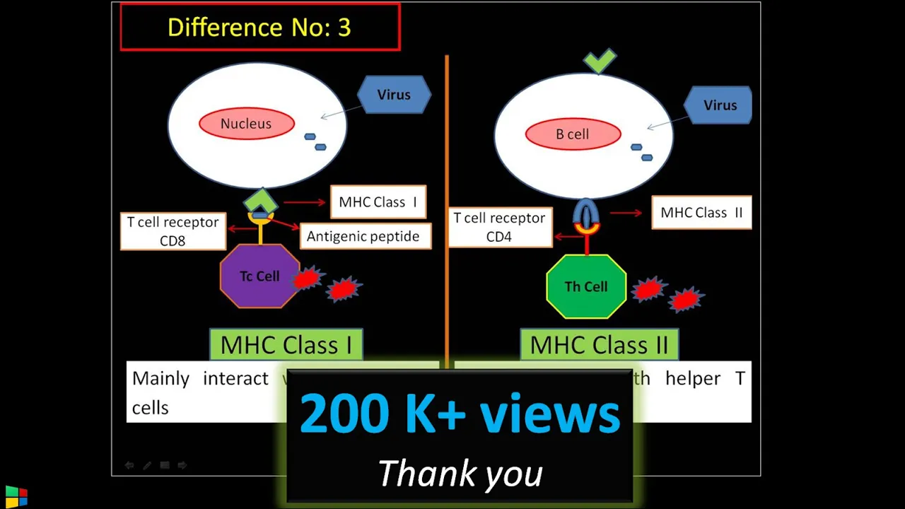












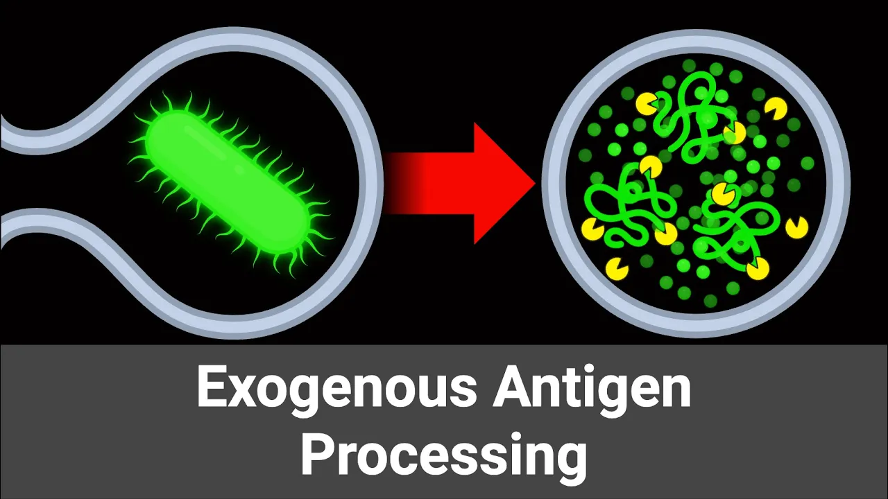




In the exogenous pathway of antigen presentation, extracellular pathogens are internalized through phagocytosis, forming phagosomes that fuse with lysosomes to create phagolysosomes where acidic conditions activate hydrolytic enzymes that degrade pathogens into peptides; simultaneously, MHC II molecules are synthesized in the ER, associate with invariant chain (Ii/CD74) to prevent premature peptide binding, are transported to the Golgi, and then to endosomes where cathepsins degrade the invariant chain to leave CLIP; HLA-DM then removes CLIP to expose the MHC II binding site, allowing antigen peptides to load onto MHC II before the complex is transported to the cell surface for recognition by CD4+ T helper cells.
Exogenous Antigen Presentation Pathway via MHC II Processing
Added:Basic structure and function of the Major Histocompatibility Complex (MHC), specifically distinguishing between Class I and Class II molecules.
The role and identity of professional Antigen-Presenting Cells (APCs) such as dendritic cells, macrophages, and B cells.
Fundamental cellular processes of endocytosis, phagocytosis, and vesicle fusion (phagolysosome formation).
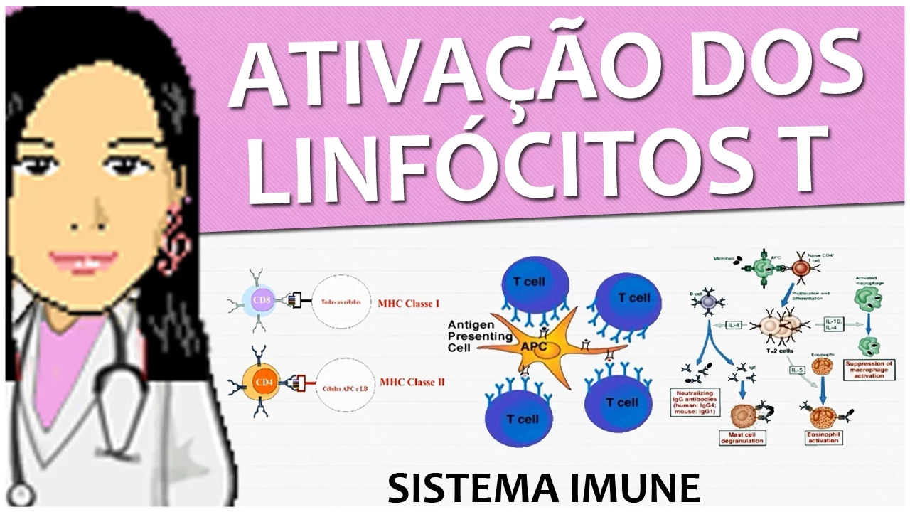
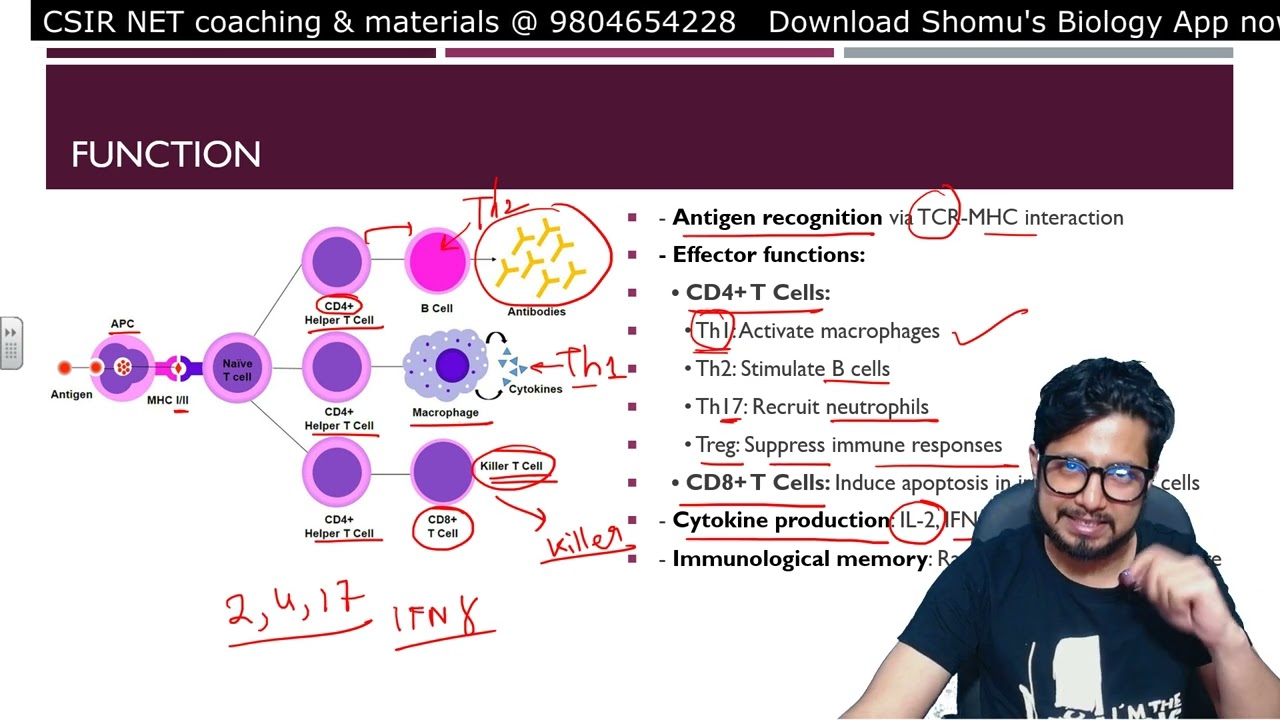
An introduction to T-lymphocyte biology, specifically the distinction between CD4+ helper T cells and CD8+ cytotoxic T cells.
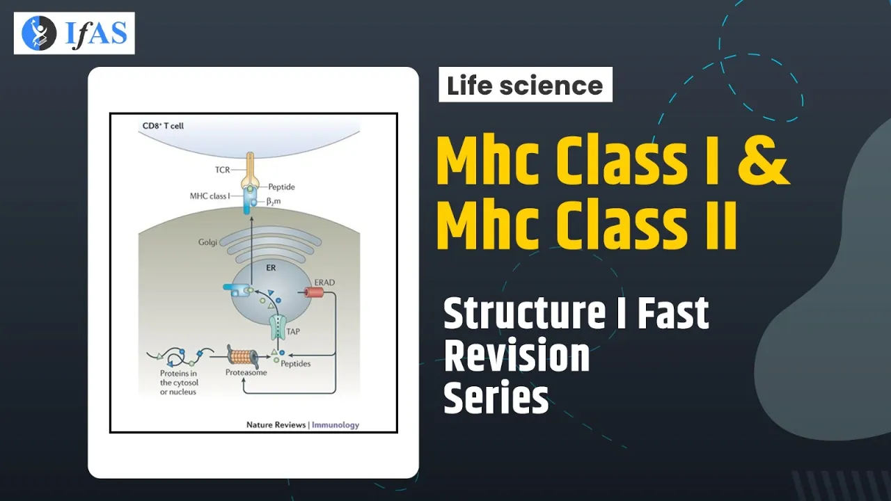
The endogenous antigen presentation pathway via MHC Class I to CD8+ T cells for comparative analysis.
The downstream activation mechanism of CD4+ T cells, including TCR binding, co-stimulation (e.g., CD28/B7), and subsequent cytokine secretion.
The phenomenon of 'cross-presentation', where exogenous antigens are diverted to the MHC Class I pathway.
Medical and clinical applications, including vaccine design strategies that target MHC II pathways and pathogen evasion mechanisms (such as blocking phagolysosome fusion).
94.6K views2.6Klikes4:57Original Release: 2019-07-23
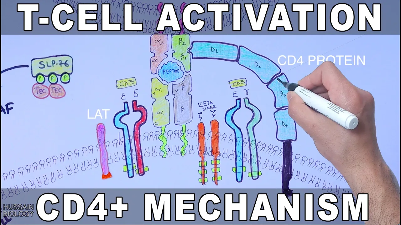
Oh in the previous video we discussed about the MSU and processing and endogenous pathway up and teaching presentation if you want to watch that video first - the link is in the description now in this video we'll be discussing about the MSC - processing and the exogenous pathway up and teaching presentation in exogenous pathway of antigen presentation the exogenous antigen or pathogen is processed within the cell and then it's present - towards the T cell by the help of MHC 2 molecule here in this part a we see we have a endoplasmic reticulum surrounded by plasma membrane as shown in the diagram and we also have Golgi apparatus in exogenous pathway the antigen is taken inside the cell when it makes contact with the plasma membrane in the diagram we can see the pathogen comes in and the cell drives the pay-go psychosis of the pathogen the cell membrane produced inside and pons the peg Azul so now we had this pathogen inside the phagosome as shown in the diagram then in the next step the lysosome within the cell uses with phagosome and we get the peg or lysosome and we know in pega lysosome the protons are pumped inside it by the help of the ATP's pumps and by this the pH is lowered with in the pagolis resume the lower pH means acidic medium in the Pago lysosome and this acidic environment brings about activation of acid hydrolases of pango lysosome remember the acid hydrolysis aka lysosomal hydrolysis works best in acidic medium that's why we need to create acidic medium first now with the activity of acid hydrolysis the bacteria is broken down into small peptides as shown in the diagram so this is how the exogenous pathogen is processed within the cell now let's pause it here and get to the endoplasmic reticulum here in the endoplasmic reticulum we see the ribosome produces both the light and heavy chains of MHC 2 molecule these chains are then stabilized into final structure of MHC 2 by chaplains then the ribosome also produces another protein molecule called the III protein or we can say in Marin chain also known by the name of cd70 for molecule it is a trimer protein this I approach in or invariant chain assemblers with the MHC 2 molecule and occupies the peptide binding groove of MHC 2 molecule as shown in the diagram now with this the MHC 2 cannot bind any antigen till high protein is bound now we see the invariant chain has the endocytic sorting signals in its cytoplasmic tail for the MSC 2 molecule along with the I a protein is exported out of endoplasmic reticulum and is sent to the Golgi apparatus the MSC 2 molecule with invariant chain exits the goal here breads in the form of endo zoom as shown in the diagram now from here the things change in the lid and the room here the cat absence more specifically there cadets in s degrade the inherent chain but only a tail and off ihn or invariant chain is degraded and it leaves a small portion bound to the MSC 2 group called the clip this clip still holds back the MSC 2 grew to bind the antigen now after that the end ozone fuses with pega lysosome the paralyzer zoom has bacterial antigen in it now after fusion we need to load this antigen or bacterial antigens on MHC 2 grew but we still have this clip bound to the MHC 2 grew so for this the pega lysosome has its own several molecules by the name of HLA DM the agile ADM is a chapel molecule within the lysosomes and it removes the clip molecule from MSC 2 grew and exposes the binding site of MHC 2 molecule as shown in the diagram now the antigen is loaded onto the MSC 2 grew as shown in the animation and this MHC 2 mole tool with antigen has to be presented to the t-cell on the side we have a cd4 T cell it has a cd4 receptor which rep Alma's that we get the MHC 2 molecule nor the MSC 1 molecule so after recognition that TCL also has a TCR which checks the antigen and transform the signal inside the cell while from the signal transduction kicks in and the cd4 name T cell gets transformed into T helper cell so this is how the MHC 2 processing is driven along side with the exogenous pathway app and teaching presentation I hope you liked the video if you liked it give it a thumbs up do consider supporting my work on patreon and also make sure to subscribe this channel Thanks
Up Next
4:35
How Vaccines Work: The Science Behind Immunity Explained
@TEDEd
3.4M views•2015-01-12

50:12
Bacterial Communication: Quorum Sensing and Biofilm Formation Explained
@JHUAAP
12.6K views•2011-06-28
3:29
Autophagy and Mitophagy: Pathway and Mechanisms Explained
@hussainbiology
102.1K views•2020-03-11

7:08
Bacteriophages: Earth's Deadliest Killers and Future Antibiotics
@kurzgesagt
34.6M views•2018-05-13
Related Study Plans & Knowledge Roadmaps
Structured learning paths in Biology
biology
RNAi: Biogenesis, RISC & Gene Silencing
01Molecular Biology Foundations: DNA to Protein
02Introduction to RNA Interference (RNAi)
03Molecular Biogenesis of miRNA and siRNA
+ 2 more learning modules
10h5 modules
Explore Roadmap
biology
Mycorrhizae: Signaling, Biophysics & Ecology
01Foundations of Mycorrhizal Symbiosis
02Molecular Signaling and Root Colonization
03Biophysics and Bioenergetics of Nutrient Exchange
+ 1 more learning modules
11h4 modules
Explore Roadmap
biology
Circadian Clocks: TTFL, SCN & Periphery
01Foundations of Molecular Biology & Gene Expression
02Introduction to Chronobiology and Actograms
03The Core TTFL: Molecular Architecture of the Clock
+ 3 more learning modules
14h6 modules
Explore Roadmap
biology
Population Genetics: Math, Drift & Selection
01Genetic & Probability Foundations
02The Hardy-Weinberg Principle and Baseline Models
03Mathematical Modeling of Natural Selection
+ 3 more learning modules
18h6 modules
Explore Roadmap