

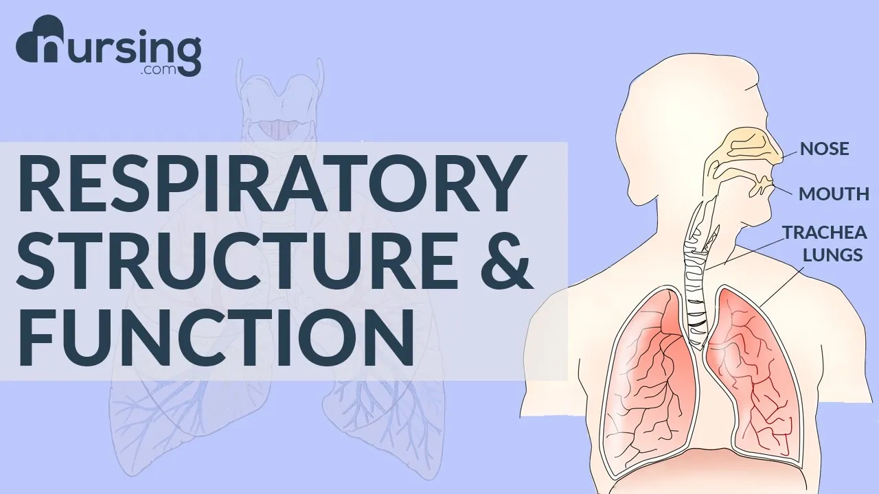







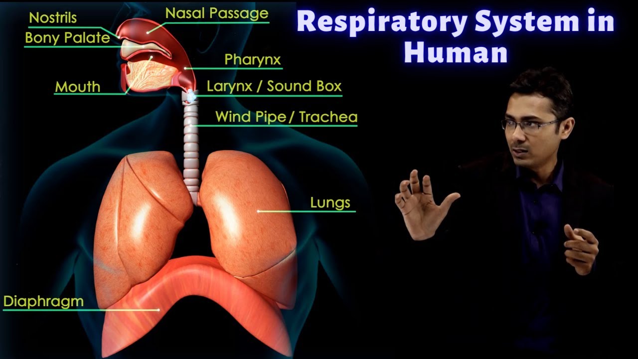

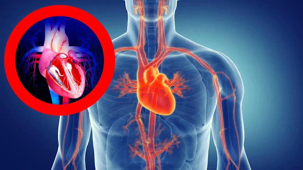


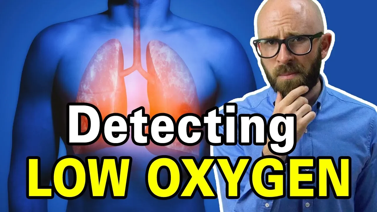




The respiratory system consists of the upper respiratory tract (nasal cavity, pharynx, larynx) and lower respiratory tract (trachea, bronchi, bronchioles, alveoli), where the nasal cavity filters, warms, and humidifies air through ciliated pseudostratified epithelium and turbinates; gas exchange occurs in alveoli with their thin walls and large surface area; breathing mechanics follow Boyle's law where diaphragm and intercostal muscles change thoracic volume to create pressure gradients that drive air movement; neural control involves the medullary respiratory centers (dorsal group for inspiration, pneumotaxic center for rate control) and peripheral chemoreceptors that respond primarily to CO2 levels.
Respiratory System Anatomy, Physiology & Gas Laws | Dr Mike
Added:Basic cellular respiration, including how cells utilize oxygen and produce carbon dioxide as a waste product.
Fundamental physics concepts of pressure, volume, and temperature relationships.
The principles of simple diffusion and how substances naturally move down concentration gradients.
An introductory understanding of the circulatory system, specifically how blood transports gases to and from tissues.
The neural and chemical regulation of breathing, including the roles of the brainstem and peripheral chemoreceptors.
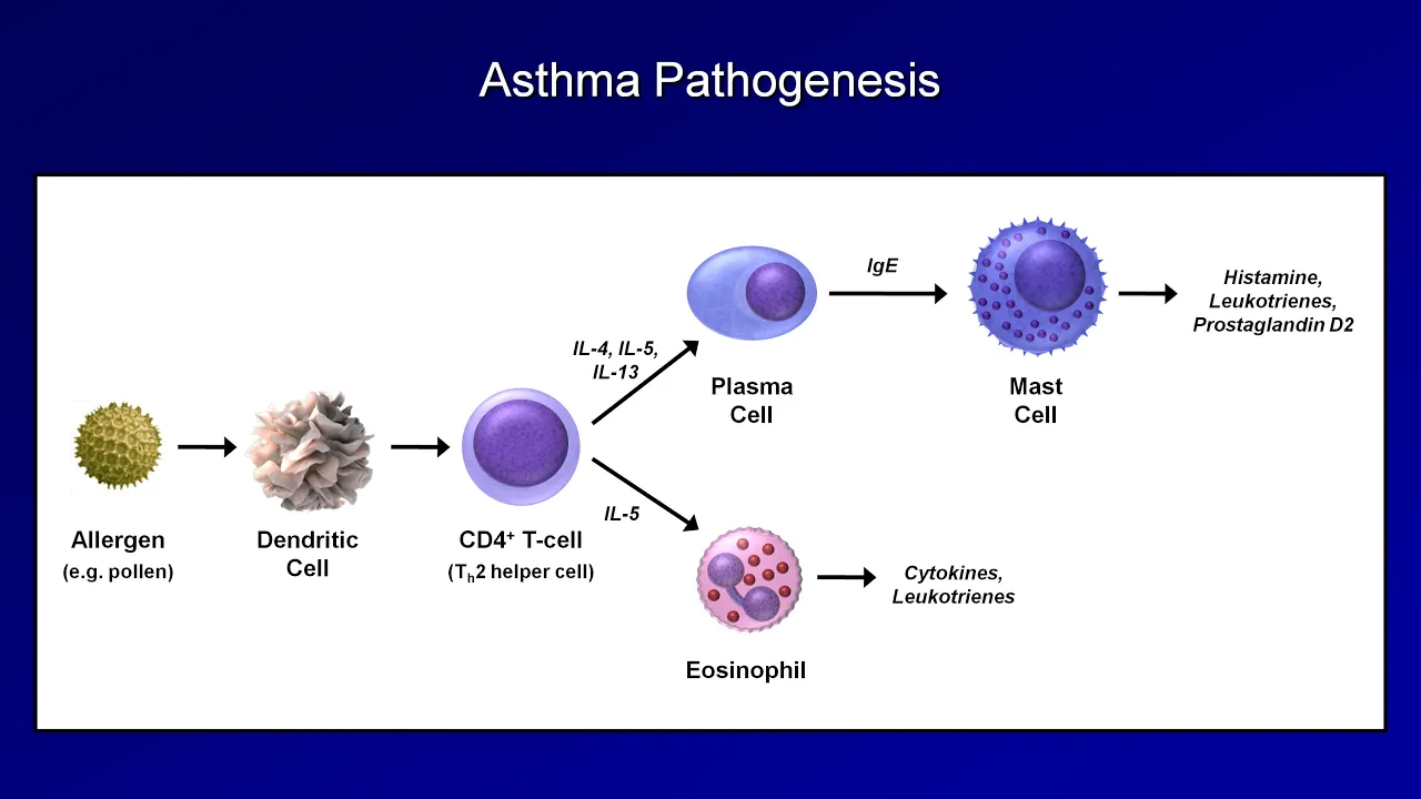
Pathophysiology of common respiratory disorders such as asthma, COPD, and respiratory acidosis/alkalosis.
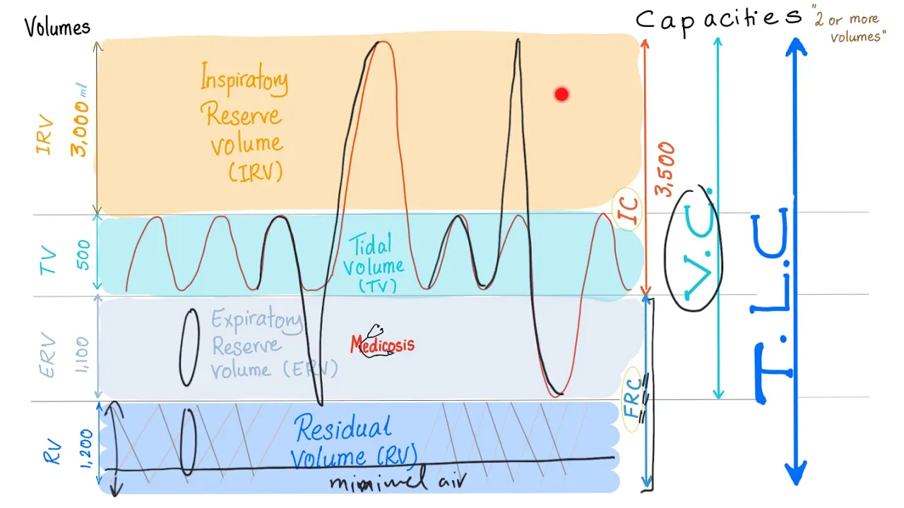
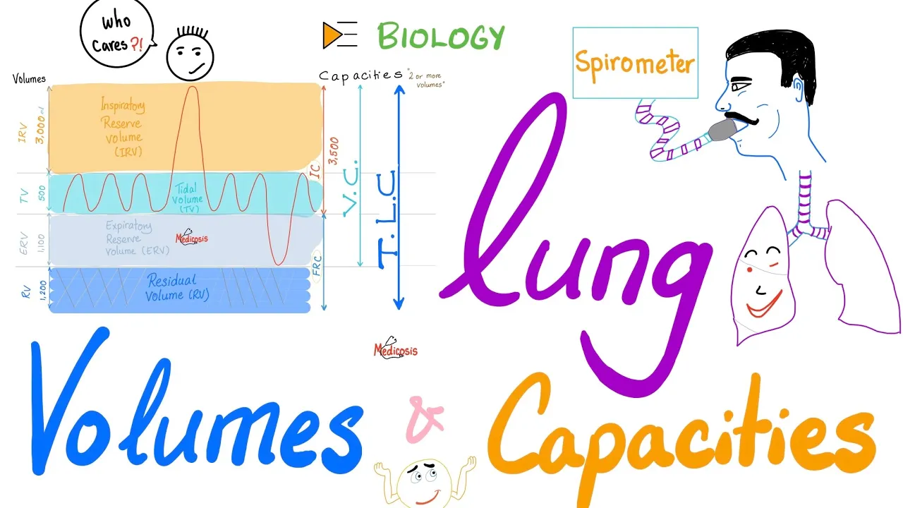
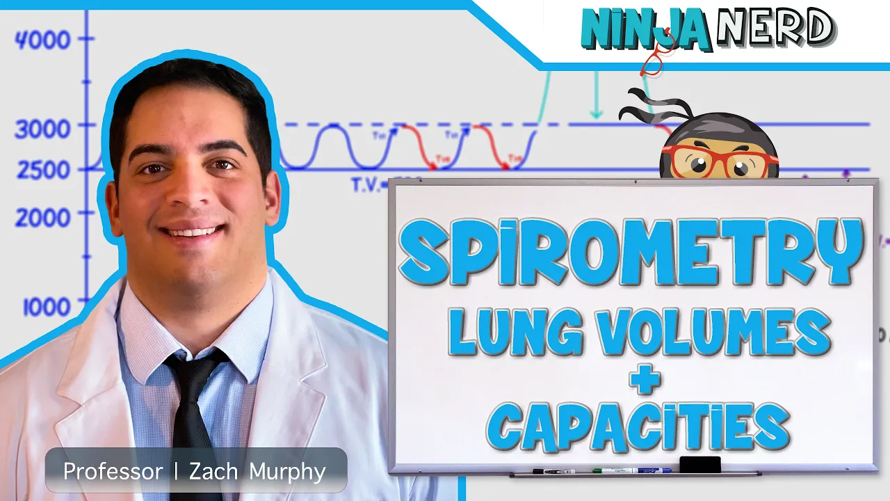
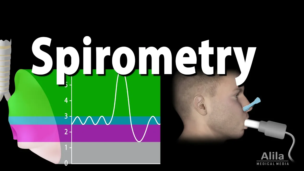
Clinical pulmonary function testing (PFTs), including spirometry and the analysis of lung volumes and capacities.
Physiological adaptations to extreme environments, such as high-altitude hypoxia and deep-sea diving decompression sickness.
46.6K views733likes1:07:10Original Release: 2020-06-23
now what we need to talk about is what happens once we bring that air in what happens once it gets inside of our nasal cavity and then goes to our oral cavity and our pharynx which is basically the back of our throat our larynx which comprises of the voice box down through our trachea bifurcates to our bronchi continues to split off into Branka further smaller bronchi and bronchioles and then ultimately to our alveoli for gas exchange so we're going to take a quick look in this video at the process starting at the nose or Nerys and moving down to the alveoli so we've taken that first breath in we've inspired that first bit of air what do we have well we take that air into our Nerys which is our nostrils around nose and we inspire that air now we know that we have what's called vibrissae which are basically nose hairs and this vibrissae is that first line of defense to protect the inside of our respiratory cavities from the external atmosphere so vibrissae are those nose hairs now as we get in we end up hitting the nasal cavity now the nasal cavity has some very important anatomical structures or features associated with it which impart very important physiological properties so first thing is that if you to have a look and feel that nasal cavity this is further than bringing your finger inside your nose this is still in your nostrils as you do this you need to go back inside your nasal cavity you'll find a couple of things one it's quite moist that's the first thing and two there are these very fine hairs called cilia that line your nasal cavity now the tissue which is going to be epithelial tissue because we know epithelial tissue lines cavities okay the epithelia that lines our nasal cavities what's called pseudo stratified ciliated epithelium so pseudo stratified when we look at epithelium we break it down according to its structure and that structure tells you something about its function so for example if you have one layer of epithelial cells okay that's a simple layer that's what we call it if you have many layers of epithelial cells packed on top of each other it's called stratified if you have a single layer of epithelial cells but actually looks like many layers that's called pseudo stratified so we have pseudo stratified epithelia inside our nasal cavity I also said that it was ciliated now these are even finer hairs than that vibrissae within our nose and this cilia again captures particles the other important point about them this olfactory epithelium or the nasal cavity epithelia I should say olfactory epithelium a bit further up this nasal cavity epithelia is that it is highly vascularized so there's a lot of blood vessels and just underneath this epithelia we have what's called goblet cells now these are cells that secrete mucus so we've got ciliated mucosa that's releasing all this mucus and it's highly vascularized so you need to remember that because as you bring that air in the cilia and the mucus capture any particulate or particles or pollutants that should not be in the nasal cavity okay furthermore should not get all the way down into the alveoli because that is quite dangerous that's the first thing second thing is you can see I've drawn these weird structures here in the nasal cavity these structures are important they're the con key and me toises very strange names konkey is the bump-out that you'll see and then meatus azar the groove in so that actually look bump out groove in bump out groove in bump out prevent it like I said bump out of the konkey and the meatus azar the groove in and what this creates is something called turbulence so they're also known as the turbulence of the nasal cavity and when you inspire air as the air comes in through your nostrils it will hit these scroll like structures and the air will start to spin hence the termination so the air starts to spin around and becomes turbulent within the nasal cavity why do we want this to happen well as that air comes in the more air that spins around the increased likelihood that that air will touch that olfactory mucosa that means the more likely filtration will occur so particles or particulate SAR going to stick to that mucosa and the cilia able to grab it and throw it now where do the cilia throw these particles welcome throw it out through the nostril or can throw it to the back of the throat for us to swallow and we actually swallow quite a fair bit of this particulate Laden mucus every single day okay so that's one thing is that these turbinates increase filtration what else well I told you that our nasal cavity is highly vascularized so there's a lot of blood vessels there and this is important because it warms up all that air that's coming in which means it doesn't matter if it's minus two degrees outside as you bring that cold air in it touching or this vascularized tissue will heat it up because as that air gets down to the lungs ultimately we want it to be 37 degrees so fill filters the air this is what this mucosa does filters the air heats up the air and it does one more thing it humidifies the air so that means it starts to attach all these water molecules starts to make that air quite moist and again that's important for when that air makes it down to the lungs for appropriate gas exchange to occur so these are some important properties of the nasal cavity now there's some other things that you can see associated with the nasal cavity such as these little holes here now these are called sinuses now what we have up here is actually the cranial cavity that's where the brain sits and you can see the spinal cord would sit within here and you've got the vertebrae either side so you've got the bodies of the vertebrae and you've got the spinous processes of the vertebrae here so this isn't parlor respiratory system obviously it's just for perspective so these two areas here are called sinuses and they're hollowed out areas of bone within our skull now these sinuses are also covered with this epithelium okay with this mucosa so it's vascularized but not as vascularized as the nasal cavity but again has mucous cells and cilia and so fourth there's little areas of conduction so that the air can get in and that air can get out now why do we have these sinuses well it's a contentious point we don't necessarily know exactly why but we do know that these sinuses helpful resonation okay so when it comes to vocalizing or vocalization they help for resonating sound that's one point the second point is that if you've got numerous holes in this very thick skull it will lighten the skull so it makes our head a lot lighter for us but obviously problems arise because this tissue similar to the epithelium within the nasal cavity is prone to infection and when something is prone to infection inflammation occurs and inflammation always leads to excess secretion in this case it'll be mucus secretion and it blocks up and it forms air pockets like vacuums and this is what leads to those headaches those sinus headaches that you get okay so this airs come in it's been terminated by those conky meatus is its touch the old factor so the nasal mucosa and it's filtered it's warmed up and it's humidified a couple of other things is that what you'll find at the very top here of the nasal cavity has some holes through this bone okay now this is called the cribriform plate part of the ethmoid bone and neurons project through called olfactory neurons now these olfactory neurons that project through give us that sense of smell so that when all these particles come through the more air that turbinates the more likely some of those particles or chemicals that we've been held touch these olfactory neurons this chemical signal gets transduced into an electrical signal goes up to the olfactory bulb and the brain and we make sense of some smell that has come in this is important clinically because some people may come in and they've lost their sense of smell and this may be because they've had some sort of knock to the head now the first question wouldn't be have you had a knock to the head it may be do you have some sort of nasal infection or a cold because we know when this happens it reduces or we even can sometimes lose for a short period of time our sense of smell and not to the head can actually shift this cribriform plate in this F my bone and it can actually shear off those neurons that are projecting through and hence losing that sense of smell okay the air that's come in that would just inspired has warmed up filtered humidified so forth it's going to come back through to the pharynx okay now the pharynx is basically the back of the throat and there's three parts of the pharynx you've got the nazo pharynx you got the RO pharynx and you've got the Lehren Joe pharynx so nasopharynx oropharynx and Lorenzo pharynx so nasopharynx is basically the back of the throat with at the nose although folks is the back of the throat at the mouth and lo and Joffe Eric's at the back of where the larynx is basically our voice box and we'll do a whole lecture talking about the voice box and creating sound information and so forth so these are all different parts of the pharynx which is just basically this muscular conduction system moving all the way down now as we get to the back let's just say we've gone past the nasopharynx are going down to the oropharynx and we know we can inspire air through the mouth that's just the tongue there we can inspire air through the back of the mouth go into the oropharynx and we start to move our way down now we've got this important structure here called the epiglottis now the epiglottis is this Cadillac genus structure which when we go to swallow food the tongue moves up and back it's part of the swallowing process called the buccal phase of swallowing the tongue moves up and back pushes on the uvula which is the punching bag at the back of the throat which blocks the nasopharynx aspect the tongue moves back pushes down an epiglottis and blocks the Loranger pharynx aspect and blocks the trachea so that when we swallow food there's only one place for it to go and that's right to the back of the throat down this tube here could the esophagus and we know the esophagus leads to the stomach but we're talking about inhaling and we don't necessarily want much air inhaled into our esophagus even though inevitably that happens the majority of our burps are actually just inspired air coming back out so we've inspired this air it's coming in and we want it not to go down the esophagus because the esophagus is predominantly closed due to these esophageal sphincter x' that's smooth muscle and it's predominantly closed and sometimes it will open but we want this air to go down this tube that sits anterior or in front of the esophagus called the trachea okay so here we're at the start of the trickier well let's just say most superficial or not superficial most superior I should say to the trickier is the larynx now the larynx is where our voice box sits and there's numerous cartilage tendons and so forth associated with the larynx and also vocal cords as well which are sort of drawn up the opening here which are associated with creating sound now again this is going to be the focus of another lecture so we're just going to pass the larynx now as we hit the larynx we're going to start to move down into our trickier so what I've drawn up is the larynx here with the laryngeal prominence now this is also known as our Adam's apple sorry up here the laryngeal prominence that's the Adam's apple which both male and females do have it's just more prominent in males because it's known as a secondary characteristics and hormones can start to produce that prominence more so than others low and joy prominent Adam's apple and we go down you can see the bulk of the trachea this is where the air starts to move down there air can be inspired or expired through this tube one important thing that you're going to find is that this tube is surrounded or has numerous C shaped rings associated with it that are made of cartilage so the cartilaginous C shaped rings all the way down how many of the mother of the trachea Wren about 16 to 20 cattle a genus C shaped rings the opening of the C shaped ring is at the back what's at the back as well the esophagus so the opening of the C shaped ring actually tickle eights with the esophagus and this is important because as you swallow food the esophagus begins to distend now some of us don't chew as well as we should and swallow like ducks big particles of food bringing it down and that distends the esophagus so much that it can potentially impinge on these c-shaped rings imagine if they were fully closed the food will get caught and unfortunately we wouldn't be able to propagate your genetic material to the next generation so we've evolved have these C shaped rings so that they can descend with the esophagus if need be 16 to about 20 of them as we move down now that's the cartilage of the esophagus if you look inside we've still got that pseudostratified starting to become more columnar cuboidal epithelium still has cilia associated with it and still has goblet cells secreting mucus now this is important again because once we inspire more air we will start to capture particles in this mucus and the cilia which has movement properties start to push this these particles captured in the mucus upwards this is known as the mucosal iary escalator and it's important because as we inspire all this air we start to inspire crappy material that we just don't want down in our lungs luckily it's captured by the mucus and thrown up what happens once it gets thrown up it gets pushed right back to our esophagus and we swallow it why would we want to swallow it because then our stomach we have very acidic chemicals we got hydrochloric acid there and other lasers arms and so forth which can break all this stuff down okay so we've got all this now another important point clinically is that smokers people who smoke cigarettes and so forth inhale that smoke it starts to destroy the cilia so the cilia begin to die off what does that mean it means the particles get caught in the mucus and because gravity sucks pulls things downwards where does it go into the lungs hence the reason why smokers early in the morning tend to cough and cough and cough trying to bring up all that crap the cilia that they no longer have couldn't bring up so it's down inside their lungs okay as we go down that your care you'll see that the trachea bifurcates this is also known as the Carina and it bifurcates at around about t45 so thoracic vertebra for thoracic vertebra five is there it bifurcates or splits into two this is now what we call the bronchi so I've got the trickier here and now it's split off into the right main stem bronchi also known as the right primary bronchi and the left main stem bronchi okay so as it splits up you can see it still has cartilage but this cut this cartilage isn't necessarily C shaped anymore because there's no esophagus going behind it so you can see it has this non uniform pattern to it so it splits off now another clinically important point the right mainstem bronchi compared to the left mainstem prong bronchi is a little bit different one it's wider in diameter and two it's more vertical why is this important well it's important because sometimes children or even adults can inhale objects which they potentially shouldn't look maybe like a peanut and it may get caught down in their respiratory system somewhere what he thinks the most likely place for this peanut to be lodged it's going to be the right mainstem bronchi because it's wider and more vertical now splits off into two and you can see that this splitting off into the left and right mainstem bronchi it will split off again now this splitting off into secondary bronchi okay splits off on the right hand side three times one two three and on the left hand side splits off two times okay into the superior and inferior and this is superior mid and inferior and these will go to the various lobes of the lungs which means that on the left-hand side the left lung has two lobes and on the right-hand side the right lung has three lobes why is this the case is because you're gonna find that the left lung actually has an area cut out of it okay now this is called the cardiac notch cardiac notch tells you about the heart this is to make room for the heart because we know the Hutt's it's predominately to the left-hand side within our chest okay so it's that left lung is a little bit smaller or reduced because of that cardiac notch so there's only two lobes the right lung has three lobes now this branching continues continues continues about 23 to 25 more times like a tree branch branching branching branching and as it branches it gets smaller in diameter these tubes they go from bronchi and once they hit about half a mil they turn into what's called bronchioles so trachea left and main stem bronchi secondary bronchi and then branch about 23 more times till they're about half a millimeter in diameter and now their bronchioles now what's the difference structurally or anatomically going from trachea - Branka all the way down to bronchioles a couple of things one the cartilage reduces once he hit bronchioles there's no cartilage okay but what does increase as you move down is smooth muscle so here in the trick here and bronchi we have very little to no smooth muscle as we start to go to the smaller bronchioles we have huge amounts of smooth muscle now this is important because of asthmatics okay smooth muscle as muscle can contract relaxed contract relax it actually can alter the diameter of those Airways when somebody has an asthma attack it can lead to constriction or contraction of that muscle that ultimate leads to constriction of those Airways which means in an asthma attack it doesn't happen at the trachea doesn't happen at the bronchi happens all the way down at the smaller bronchioles and that's where the airway start to close up okay so a lot of smooth muscle the bronchioles are not much up at the turkey and bronchi not much movement smooth muscle a lot of cartilage okay as we continue through we also get reduced amounts of goblet cells and reduced amounts of cilia once we at the bronchioles no goblet cells no cilia you may be thinking then how do we capture all those particulates well luckily in our alveolar sacs this is where gas exchange occurs we actually have important cells called macrophages these are cells that eat up big eaters actually macro meaning big or large far your phage meaning to eat so they're big eaters and they help pick up anything that's left over hopefully by then there's not much that's getting past all those systems so as we move down we'll ultimately at the bronchioles half a millimeter in diameter and it gets smaller there's two major types of bronchioles there are as we move down let's just say we'll draw up some branching here okay so what we've got here is how the bronchial bronchi turned into bronchioles and we're gonna have let's say these are bronchi now these bronchi they may be secondary or tertiary or maybe the tenth branching or twentieth branching maybe and then we get down to half a millimeter in diameter we're gonna have terminal bronchioles and then at the very end we have respiratory bronchioles okay what's the difference well terminal bronchioles are the very end of what we call the conducting pathways meaning basically the tubes these are just tubes carrying air all the way to the terminal bronchioles okay the respiratory bronchials tells us something important because the terminal respiration clinically refers to gas exchange across a membrane okay it's actually ventilation which means bringing air in and out not respiration respiration is gas exchange at a membrane so here where we have the respiratory bronchials this is also where we have our various alveoli and alveolar sacs okay now these LVO lie and like bunches of grapes and their bunches of grapes like this because they increase the surface area why do we want to increase the surface area if you to unfold all of these alveoli be the size of a tennis court okay all inside of these pleural cavities that in case our lungs these alveoli are there for gas exchange that means oxygen will jump out into the bloodstream that surrounds for the pulmonary arterial or the pulmonary circulation and the carbon dioxide will jump back in so for us to breathe out so gas can jump across them very easily these alveolar sacs are 15 times thinner than a piece of paper okay so that membrane is very very thin so I think that will do us for going through the respiratory tract broken up into the nasal cavity oral cavity pharynx which is oropharynx nasopharynx low angio pharynx then the pharynx which has the voice box and then we've got the trachea we've got the left and right main stem bronchi which branch 23 times ultimately into these structures called terminal bronchioles the respiratory bronchioles and then we hit the alveoli which is the side of gas exchange that's a very quick run-through of the respiratory tract so what is dalton's law and what is the clinical significance of Dalton's law when it comes to respiration so Dawn's law states that in a mixture of gases the total pressure is equivalent to the sum total of the individual gases now what does that actually mean well we can use a very simple easily relatable example such as that of us living in our atmosphere so if you go down to the beach and you were to take your container and capture some of the air around you you would find that the pressure of that air probably surprisingly is a pressure of around about 760 millimeters of mercury so like I said this is called atmospheric pressure at sea level now you've probably heard of millimeters of mercury as a measurement of pressure before when we look at blood pressure the systolic pressure at the strongest contraction of the heart is 120 millimeters of mercury within the arterial system and if you look at the diastolic pressure when the heart relaxes the pressure is around about 18 millimeters of mercury so how can the atmosphere around us be greater than that 760 well because we'll born into this pressure so we don't feel this atmospheric pressure around us and it has to do with the column of air immediately above us obviously all this gas on top of us is pushing their way down due to gravity and placing some sort of pressure upon us that we don't necessarily feel and that pressure is 760 millimeters of mercury now the atmosphere even though it is 760 millimeters of mercury is made up of a number of individual gases these individual gases include nitrogen include oxygen include carbon dioxide and some trace gases one of these trace gases or these trace gases could be argon could be water vapor could be a number of different gases now if we were to take a look on the 760 millimeters of mercury water pressure around us around about 78 percent of it is actually composed of nitrogen around about 21% of it is oxygen approximately 0.05 percent is carbon dioxide and the rest is made up of the trace gases so if we were to do the calculations to find out what the individual pressures are for each of these well you do 78% of 760 millimeters of mercury is around about 597 millimeters of mercury and this is for nitrogen if you look at oxygen well that's 21% of 760 and that equals around about 159 millimeters of mercury like I said that's for oxygen carbon dioxide 0.05 percent of 760 is approximately zero point three millimeters of mercury and like I said that's for carbon dioxide so Donald's law states that a mixture of gases the pressure of which is the sum total of all the individual gases which means if we were to add these gases up the 597 of nitrogen 159 of oxygen and the 0.3 of carbon dioxide it will approximately be equivalent to 760 millimeters of mercury now why is this relevant clinically well it's relevant clinically when it comes to looking at something called Henry's law of course there's a number of different laws that you need to remember clinically Henry's law states that when you look at an individual gas or even looking at a mixture of gases that the partial pressure so that's what these are called these pressures here are the partial pressure of these gases within a total gas so while the atmospheric pressure is 760 millimeters of mercury the partial pressure of nitrogen is 597 the partial pressure of oxygen is 105 jinan the partial pressure of carbon dioxide is 0.3 millimeters of mercury these are the partial pressures and when it comes to Henry's law it states that when it comes to these pressures of these gases dissolving into a liquid it has to do with their individual partial pressures and they will only dissolve into or down a concentration gradient so what that means is is if for example oxygen being 159 millimeters of mercury in the atmosphere if you were to have a container so this is now moving into Henry's lorry a little bit but if you were to have a container with water and I 159 millimeters of mercury outside of the container but only 40 millimeters of mercury in the container this oxygen would diffuse or dissolve into this liquid down its concentration or down it's pressure gradient even if for example even if in addition to this the pressure outside for carbon dioxide is 0.3 and the pressure inside the liquid for carbon dioxide was 20 does that mean that because it's lower of carbon dioxide outside and higher inside its mixture together would balance it out no of course no it means that the carbon dioxide is going to come out on that liquid okay so it's talking about dissolve ability has to do with the very specific partial pressure of that gas it goes down its own pressure graining doesn't care what the pressure gradient is of any other gas around it even when it's within a mixture like this so what is dalton's law dalton's law states that when there's a mixture of inert gases that it's pressure is going to be an equivalent to the sum total of all the individual pressures and if you know the percentage of those gases you can easily calculate what's termed the partial pressure of those individual gases what is Boyle's law and what is its clinical relevance when it comes to respiration well first we need to define what Boyle's law is Boyle's law states that as the volume of a container increases the pressure within that container decreases and vice versa as the volume of that container decreases the pressure of gas within that container increases so what that means is there's an inverse relationship between the volume of the container and the pressure of gas inside of that container so if we were to write that down shorthand would state that as we increase the volume of a container it results in a decrease of gas pressure within that container and vice versa decrease the volume equals increase in pressure so let's first graphically describe what that really means and then we'll refer to what it means clinically so if we were to draw up a container and let's just say we were to put a piston in this container and inside this container we have gas particles let's just say we have oxygen inside of this container let's just say we have 1 2 3 4 5 6 7 8 9 10 11 12 13 14 15 16 17 18 9 20 gas particles of oxygen 20 oxygen molecules within this container and we'll to measure the pressure inside of this container let's just say that the pressure is 10 kilo Pascal's now don't really worry about the unit's here but remember that it's 10 okay 10 kilo Pascal's all right now let's just say so there's a particular volume of this container all right now let's just say that we would have pushed down on this syringe now it's a closed container remember this is very important it's a closed container what if we're now to push down on this syringe what would happen so we've pushed down on the plunger and what ends up happening is how many molecules of oxygen did we begin with 20 we now have 1 2 3 4 5 6 7 8 9 10 11 12 13 14 15 7 18 19 20 oxygen molecules still within this container but they now have less space to move around so let's think about this for a second let's think about what pressure actually is when we talk about blood pressure for example we talk about the force that the blood puts on the walls of those vessels when we talk about pressure in this case the pressure of gases again it's the force that these molecules put on the walls of its container so here where we have a larger container these 20 oxygen molecules as they bounce around they're bouncing off the walls bouncing off each other they are putting some sort of force on the wall of that container and that's measured as pressure in this case it was 10 kilo Pascal's of pressure okay as it's bouncing around now think about as we decrease the volume of that container or decrease the size of that container we still have 20 molecules bouncing around but they have less room to move around so does that mean statistically I think is it going to be an increased likelihood of these molecules hitting walls and hitting each other or decrease likelihood it's going to be an increased line so you have more of these reactions in which it bounces off the walls bounces off each other and what this does is it increases the pressure inside of that container because again pressure is going to be a measurement of the force that's generated by these molecules inside the container on the walls of that container okay so this is that inverse relationship decrease the volume of a closed container increase the pressure of the gas within that closed container now what does this have to do with respiration and breathing well let's think about if this container was open at one end all right let's look at the first syringe we have gas 20 oxygen molecules and let's say that out in the atmosphere we're also going to have some oxygen molecules now with this syringe or plunger not moving the amount of gas inside of that syringe compared to the amount of gas outside would end up balancing each other out okay because we know that as these oxygen molecules are bouncing off the walls there's going to be some probabilistic likelihood that it's going to bounce out side of the syringe and the same notion some are going to bounce in to the syringe and over time this exchange will be equivalent and the pressure inside that syringe will balance out with the pressure outside of that syringe if you were to just leave it and not even touch that plunger okay so it balances itself out now think about what happens when you push on that plunger as you push down think about as you push down you're decreasing the volume and increase in the pressure see increasing the amount of times that these molecules bounce off the walls and increase how many of these oxygen molecules are going to bounce out of the syringe you're going to get an increased likelihood of oxygen molecules bouncing outside of this syringe and what does that mean it means that as you push down you force air or oxygen out of the syringe into the surrounding atmosphere now this makes sense if you get a syringe and you've put your thumb on the end of it and push on the barrel of that syringe what's gonna happen is you feel that pressure pushing onto your thumb you remove your thumb the gas flows out now what that's telling you is that pressure always moves down a concentration gradient always moves from high pressure to low pressure always from higher pressure to low pressure okay a high pressure system is moving in they may say when when you're watching the weather it's always going to go from a high pressure system to a low pressure system now what does this mean when it comes to respiration well think about what we have if we were to compare this syringe to our respiratory system well let's take a quick look if I were to draw a very simplistic version well we've got our trachea coming into our lungs and obviously we have a left and right lung but this is what we've got here and we're going to have our pleural cavity and we're going to have our ribs either side obviously we have more than six ribs and we're gonna have our diaphragm down the bottom as well let's color that diaphragm in red because we know it's skeletal muscle and we also know that our external intercostals are going to be lining the outside of our ribs here and these are the two muscle groups I want to focus on first of all now when you so when you take a breath in what's actually happening is that diaphragm contracts and moves down because it's this dome shape as that diaphragm contracts and moves down what happens is it opens up it increases the volume of this thoracic cavity now remember Boyle's law as you increase the volume you're going to decrease the pressure and that means the pressure inside of the lungs is going to be less than the pressure outside of the lungs and I also told you that gas likes to go from high to low so if the pressure in the lungs is lower than the pressure outside what's going to happen gas will inevitably be dragged in to the lungs that's how we inspire that's how we bring this air in now we've also got our rib cage with our external intercostals attached and they can also contract too now if they're contracting they're going to be contracting up and out if they can track up and out they're going to further increase the volume of this thoracic cavity making the pressure inside of the thoracic cavity even lower which means there's now a greater differential of the pressure from outside to inside so it's going to be higher outside compared to inside and gas always goes from high to low so more gas will rush in so what I'm basically telling you is the more muscles we recruit to increase the thoracic volume the more air we will inspire into our lungs now obviously when we relax these muscles the volume will decrease and that air will be pushed straight back out this is the clinical relevance of Boyle's law so in this video we're going to take a quick look at the muscles involved in respiration so these are the muscles that we use to allow air to rush into our lungs and also to push that air back out of our lungs so the muscles use for inspiration and expiration now before we begin you need a basic understanding of the physiology so just very quickly you need to understand something called Boyle's law now Boyle's law simply states that the volume of a container is inversely proportional to the pressure within that container so what I mean is this that sounds quite confusing but it's simple if you have a container of a fixed volume inside of that container there's going to be a number of gas particles and those gas particles are going to be bouncing around at all times as gas particles do and they're going to be bouncing off the walls of that container if you would have measured the pressure inside of that container you're simply measuring the force generated by those gas particles bouncing off the walls of the container now let's say we take that container and we were to squish it down we keep the same amount of particles of gas inside but we now have its size well what now happens is that the same amount of gas particles have less space to move around and they're more likely to bounce off the walls which means when you take a measurement of the pressure inside the pressure is going to be greater because they're going to be more likely hitting off the walls at a greater force okay so we've just decreased the volume and increase the pressure hence the inverse relationship again if you were to take that same container with the same amount of gas inside and you were to double its size by keeping the same amount of gas particles inside you've now got these gas particles floating around a larger area and there's a there's less likelihood that those gas particles are going to bounce off the walls of that container therefore the pressure will be reduced so by increasing the volume you're decreasing the pressure now this is important and it's also important if you take this piece of information in consideration - this piece of information that gas will always move from an area of high gas to an era of low gas pressure okay you watch this on the news or the weather I should say all the time the weather man or woman will say that there's a high-pressure system moving in this is simply saying that high-pressure gases are coming towards an area where there's low pressure gases now take these two pieces of information together and what that means is this our lungs sit within our thoracic cavity so here's our thoracic cavity okay now our lungs are adherent to that thoracic wall so if we were to increase the volume of our thoracic cavity we are effectively decreasing the pressure inside of our lungs that means that the lungs the pressure inside the lungs is decreased or reduced compared to the external environment and therefore gas wants to rush towards that negative pressure so all that air will come in to our lungs through our nasal cavity through our oral cavity down to the lungs if we were to decrease the volume of our thoracic cavity we're increasing the pressure in our lungs and air wants to rush out so that's how we breathe in and out and the way we do this is by contracting muscles so let's first look at the muscles used in inspiration breathing in the major muscle you need to be aware of involved in inspiration is the diaphragm now the diaphragm is the muscle that separates the thoracic cavity from the abdominal cavity it is basically the anatomical barrier between those two cavities it's an unpaired skeletal muscle that dome shaped okay now a couple of pieces of information in order for you to understand the diaphragm a little bit better I've drawn up this picture now you may look at this picture and think this is making things worse that's a terrible picture but let me try and orientate you to what we're looking at so what we've just done is we've done a transverse section threw me right at the thoracic and abdominal Junction or barrier which is right where the diaphragm is we cut straight through and now we're looking up through using your eyes you're looking up through to my thoracic cavity which means what you're looking up at is my diaphragm okay now you can see that there's going to be the front and the back anterior-posterior that's the zip void or or part of the sternum and here's the ribs coming around towards the back and then you can see some of the lumbar vertebrae telling you exactly where we're looking at the very top of the lumbar vertebra well one two three and four now couple of pointers should be aware of for the diaphragm is that you can see that the diaphragm is attached by these ligaments to the lumber portion of our spine this is called the cross of the diaphragm okay and it's attached there so there's Anchorage points the diaphragm is also attached to the thoracic wall which you can see here and the sternum as well now there's three major holes in the diaphragm okay now two true holes one which isn't really a hole let's talk about that one first at the very back of the diaphragm which you can see here pretty much against our vertebrae you've got a blood vessel that's coming down from the heart through the diaphragm down to the rest of the body this blood vessels called the aorta and it's the major artery coming away from the heart that feeds the body with oxygenated blood now this a order doesn't actually go through a hole in the diaphragm it actually goes through a gap behind the diaphragm posterior to the diaphragm this is important because if the aorta went through a hole in the diaphragm at me it means that any time the diaphragm contracts it may grossly impede blood flow and we obviously don't want that happening coming from a major arterial blood vessel the other two holes is one here and one here this one here is what's called our esophageal hiatus basically the hole that allows the esophagus to come through this one up here is a cavity or a hiatus that allows for the vena cava to move through so I said the aorta is the major artery coming down from the heart the vena cava is the major vein going back to the heart so it's actually coming back up through the diaphragm okay now the diaphragm is innervated by a nerve called the phrenic nerve and the phrenic nerve comes survival regions three four and five so phrenic nerve phrenic nerve comes from survival 3 to survival 5 and you can remember that see 3 4 5 keeps you alive so this is now you might be thinking the diaphragm is down there I know that see 3 4 & 5 are up here so why is it that there's a nerve or a nerve plexus that's coming from the neck innovating the diaphragm and that's simply because embryologically if you've looked at the embryology lectures the diaphragm originated up in the neck okay and so it's basically pulled those nerves down so the diaphragm when it contracts it's pulling on these tendons it's pulling on all its connections to the thoracic cavity and what it does is it pulls on this one thing here probably thinking what is this white thing that I've drawn it's not another hole in actual fact this white thing is filled with collagen and it's actually a tendon okay so I've drawn of these red lines here to indicate that that's muscle that's skeletal muscle of the diaphragm this big white part right in the middle is a tendon called the central tendon so when the diaphragm contracts all of this skeletal muscle pulls and it pulls on that central tendon and what that means is I told you it's a dome shape as it pulls it flattens that central tendon down which means it increases the thoracic volume that decreases the pressure and air rushes in so the diaphragm when it contracts is the major or main muscle for inspiration okay now there are other muscles used in inspiration what we call accessory muscles and why they call accessory muscles because we basically just need the diaphragm to to bring air in and out when we do what's called quiet breathing so the breathing you're doing right now when you're sitting down when you're relaxing it's called quiet breathing and you're breathing in about half a liter of air out half liter of air in half liter out half liter simply by the diaphragm contracting relaxing contracting relaxing but there's other muscles called accessory muscles that allow us to do what's called forced inspiration breathing more than that 500 mils of air so let's talk about those muscles now I've drawn a picture of the body thoracic area the external anatomy basically and you can see some bones and some of the musculature so let's orientate you so this is these are the clavicles up here this is the sternum coming down here here we've got the first five ribs attach to the sternum okay that's the other side too but hidden by this muscle which I'll talk about a sec and then six seven eight nine and ten coming out here and now having got the floating ribs or anything like that and then we've got the red stuff of all muscles that are attached to it so let's talk about it now when you take it breath in and it's forced if we need to bring more air in than that quiet breath in that five hundred mils we need to increase the thoracic volume even more the more you increase the thoracic volume the less the pressure gets inside so the more negative it gets inside and the more air that rushes in which means the more muscles you recruit to increase the thoracic volume the more air will come in okay so let's talk about some of these muscles that we're going to recruit for inspiration all right so probably the first muscle I should state because it also is involved in quiet inspiration but is definitely involved in forced inspiration is what we call the external intercostal muscles now costal means cartilage inter means between intercostal means between cartilage what we referring to is the muscles that sit between the ribs okay now external intercostals tells you that it's it's more externally to another set of intercostal muscles called the internal now the internal used for forced expiration so don't get those two too confused so firstly for forced inspiration external intercostals and you'll see that they are oriented tailored like this okay these are the external intercostals and when they contract they pull up the rib that's below them okay which means they pull up and out the rib cage what does that mean if you're pulling up and out the rib cage again you're increasing the thoracic volume so these are the external intercostals external intercostals okay now next muscle is all Grouper muscles are the pectoralis muscles so the pecs now you've got the PEC major PEC minor so let's have a look what I've drawn here is obviously clavicles here's the deltoid so the shoulder and the start of the bicep tricep here now that's part of the lat so I just want to show you basically coming down here this is the PEC this is actually PEC major the largest PEC muscle and you can see coming in attaching to the clavicle attaching to the sternum and so forth so when the pectoralis major muscle contracts it pulls on the sternum and ribs and again lifts it up so think when you take a big deep breath in you can track the pectoralis major muscles and it lifts it up and out again increasing thoracic volume decreasing lung pressure air rushes in inspiration okay now this is innervated by the pectoralis nerve and this is coming from from around about c3 to c8 okay survival three survival eight and a bit of t1 thoracic one and like I said comes from the pectoralis nerves these nerves also innervate pectoralis minor now pectoralis minor is this muscle right here and what you can see is pectoralis minor is coming in and is attaching to rib three four and five which means when you contract pectoralis minor it's going to lift rib three four and five again increasing thoracic volume so we can then state that we've also got PEC minor involved and PEC major what else is involved in forced inspiration well we've also got the serratus muscles the serratus muscles are serratus anterior these muscles here that you can see look like they're attached to the ends of the rib and again when you can track them they're going to shift the ribs as well increase in thoracic volume so these muscles here are the serratus anterior you've got a muscle called the sternocleidomastoid muscle now the sternocleidomastoid muscle sterno sternum Claddagh or clear do' means clavicle mastoid referring to the jaw so this is a big strap-like muscle which you can see attaches to the clavicle here and to the sternum as well now let's write it down first because it's such a big word I can't talk and write it at the same time sternocleidomastoid and again when this muscle contracts it's going to lift everything up and out okay again increasing thoracic volume now this muscles innervated by the cranial nerve 11 I wanted to bring that up because maybe you've gone through cranial nerves already maybe not cranial nerve 11000 to touch and feel very good velvet ah heaven it starts with a it's the spinal accessory nerve sometimes also known as the spinal nerve so this nerve helps too it's not the only nerve that innervates it but helps to innervate the sternocleidomastoid muscle now another muscle that's involved is this muscle which sort of sits next to on behind the sternocleidomastoid which is called the scalings and you've got the anterior middle and posterior scalenes and you can see that these scalenes are attached to the first and second ribs again contracting lifting them up increasing thoracic volume so these are the major muscles that are involved in accessory muscles I should say involved in forced inspiration so quiet inspiration diaphragm and external intercostals forced inspiration definitely external intercostals the PEC muscles the sternocleidomastoid serratus anterior and scalenes these are the muscles involved in inspiration now in this video we're going to take a quick look at the muscles involved in expiration in the previous video we looked at the muscles for inspiration and very quickly stated that they were the diaphragm external intercostals the scalenes the serratus anterior and the sternocleidomastoid and the pectoralis muscles major and minor just to name a couple now in this video let's have a look at those muscles that are involved in getting that air out of the lungs so remember Boyle's law states that the pressure of a container is inversely proportional to the volume of the container which means if we need to get air out of the lungs we need to compress that container to increase the pressure so that air goes out from a high pressure to a low pressure so what can we do well we know that when we breathe out we compress our abdomen and that's basically what we need to do we need to try and reduce the volume of the thorax and compress the abdomen and you can really have a good understanding of what muscles are involved in this when you actually do the process breathe out forcefully first thing you know is your abdominals are contracting so let's have a look at what abdominal muscles are involved first of which you have the rectus abdominal muscles so here are the rectus abdominals now the rectus means straight and you can see that these abdominal muscles are straight up and down so these are the rectus I've done in addition to the rectus you've got the external obliques now the external obliques one externals telling you its most superficial oblique means it's at an angle okay now you can see the external obliques here attached to the fifth sixth seventh ribbon in actual fact the external obliques are attached to the lower eight ribs so that's five six seven eight nine ten eleven twelve okay so the external obliques are attached to the lowest eight ribs and when you contract it it pushes the ribs up and in and what does that do it compresses the abdomen allows you to breathe out by increasing that pressure so these are the external intercostals sorry the external obliques talking about the external intercostals I said that they were involved that that's these ones here I said that when they contract they lift the rib below them increasing your thoracic cavity the internal intercostals which are actually these ones here you can see they internal intercostal so they are deeper to the external and you can see that they go in the opposite direction the fiber runs in the opposite direction when they contract what they do is they push the rib down and in and again reducing thoracic volume so these are the internal intercostals now going back to the oblique we've got the external obliques which I said lower eight eight ribs that it's attached to that's its origin site if you go a bit deeper to the external obliques you have the internal obliques and you can see again the internal obliques the fiber moves in the opposite direction and they're actually attached to the lower three ribs and you can see here the internal obliques also involved in reducing thoracic size by compressing the abdomen even deeper to the external obliques is the transversus abdominus muscles and again when they contract compress the abdomen transversus abdominus and the last thing I want to talk about is this muscle coming down here which is the latissimus dorsi the lats when that contracts you can see what it does is it brings forward your thoracic cavity and again compresses that thoracic cavity forcing you to exhale so you can see that there are a number of other muscles involved in expiration that's bringing air out and their goal is when they contract they reduce the thoracic volume and they can press the abdomen to bring it up compressing that thoracic volume making the pressure inside the thoracic cavity and lungs higher and that is forced to run out so you can see we've got the internal intercostals external internal obliques transversus abdominus and the rectus abdominis and the latissimus dorsi these are the muscles for expiration hi Ron dr. Mike here in this video we're going to talk about neural regulation of breathing this is how does the brain control our respiratory rate so first thing you need to be aware of is the fact that we've got our lungs here and we know that we need to bring air into our lungs and back out of our lungs and the way that we do this is because of pressure changes now simply put anytime you increase the volume of a container you decrease the pressure inside and so for example if I were to increase the volume of our lungs by contracting the diaphragm and pulling it down contracting the external intercostal muscles of our ribcage and pulling it up and out the lungs get bigger now because you're increasing the volume the pressure inside gets lower that's called Boyle's law and when pressure is low gas will always move from a high to a low pressure so air rushes into the lungs now when we want that air or gas to go back out we simply relax the diaphragm it snaps back up we relax the external intercostals the ribcage moves back down and the elastic recoil of the lungs snap back into place therefore decreasing the volume increasing the pressure inside and gas moves back out again so that's the basics that's breathing mechanics but how do we tell the muscles of the external intercostals urn the muscle that's the diaphragm how do we tell it to contract well this has to come from our brain and specifically it comes from our brain stem then we know our brain stems made up of the midbrain the pons and the medulla but specifically we're referring to the pons and the medulla when it comes to breathing now another thing you need to be aware of is we have what we call this basal breathing rate it's normal inspiration expiration okay there's quiet breathing and this actually originates at the back of the medulla that most dorsal region of the medulla takes up most of the medulla as well and this is the dorsal inspiratory region and what happens is these neurons spontaneously fire off they don't need any signals coming from anywhere else in the body they will spontaneously fire off they're timed to do so in actual fact you can cut everything below the medulla cut everything above the medulla and they'll still fire off now they won't be far enough to anything but they'll still fire off when they fire off and you've got a nice intact spinal cord what they'll fire - is they'll send signals down to c3 c4 c5 so these are the cervical nerves 3 4 5 it right at the level of the neck and I send these signals out and they will send a signal through c3 c4 c5 and these signals go down and they innervate the diaphragm so c3 c4 c5 keeps you alive that's what we say because they innervate the diaphragm via a nerve called the phrenic nerve now in actual fact for quiet breathing just breathing in breathing out quietly 500 milliliters in 500 milliliters out you only need the diaphragm to contract which means the dorsal aspect of your medulla is firing these neurons off through c3 5 and is doing it in a patent way fire stop fire stop contract relax contract relax and that's normal quiet breathing but sometimes you need a little bit more air coming in and so this dorsal region of the medulla can also send signals down to the thoracic area that's here through a couple of nerves and these nerves are going to come out and they will innervate the external intercostal muscles right and if you innervate the external intercostal muscles you'll tell them to contract and if they contract they bring the ribcage up and out for increasing the volume further decrease in the pressure inside further pulling more air in right so it's all the way if you want more air in you're just going to increase that thoracic volume alright so I've spoken about these inspiratory neurons here at the medulla that's spontaneously fire off but we can also depending on what's happening in our body we can alter that ok there's actually an area above it in the pons again the dorsal region at the back of the pons and this is called the new Mattacks ik area and what this does is it's like a switch okay like a light switch because these neurons in the medulla are going to constantly fire off sometimes you need to tell them to stop and if you tell them to stop it shortens the breath in so instead of you can fire the pneuma toxic area and you go so you can increase your respiratory rate again through this pneumo toxic area and like I said it sends these inhibitory signals to these inspiratory neurons that are spontaneously firing off now other things can happen in our body for example if we have increased levels of co2 decreased levels of ohto will increase concentration of hydrogen ions in the blood these can all trigger this inspiratory center in the medulla and one of the ways that it can do it in the peripheral portion of our body okay so not centrally not in the brain or brain stem but it can do it peripherally at the aorta so here's the aortic arch so you can have the heart here the otic arch coming out here's the three branches and two of them turn into carotid x' and you're going to have chemo receptive neurons okay so these are chemo receptors they're based at the aortic arch they're also based in the carotid and they will pick up concentrations of increased carbon dioxide decreased oxygen and increased hydrogen ion concentration and they will send signals to this inspiratory center now if they're doing it via the carotid that's going to be through the glossopharyngeal nerve and if it sends it via the aortic arch its via the vagus nerve and like I said it stimulates these neurons and tells you let's breathe a little bit more so what we've got here is the neural control of breathing its stimulated predominately by increased carbon dioxide and predominately from decrease hydrogen ions and basically the last scenario is decreased oxygen that's right increased carbon dioxide mainly because we know the equation co2 plus h2o gives you h2 co3 carbonic acid which hates itself splits itself apart turns into hydrogen ions and bicarbonate ions but the most important thing here is co2 turns into acid in the blood carbon turns to acid we don't like acid we die if we have too much of it so one way of getting rid of it is breathing out breathe out co2 breathe out acid therefore the body's most receptive to co2 and acid as stimulators to breath and that is the neural control of breathing
Up Next
5:11
Viral Replication: Lytic vs Lysogenic Cycles | MCAT Biology
@khanacademymedicine
676.3K views•2015-01-21

50:12
Bacterial Communication: Quorum Sensing and Biofilm Formation Explained
@JHUAAP
12.6K views•2011-06-28

5:35
Enteric Nervous System Explained: The Gut's Brain | Neurobiology Lecture
@alumniu6029
438 views•2018-09-12

7:08
Bacteriophages: Earth's Deadliest Killers and Future Antibiotics
@kurzgesagt
34.6M views•2018-05-13
Related Study Plans & Knowledge Roadmaps
Structured learning paths in Biology
biology
RNAi: Biogenesis, RISC & Gene Silencing
01Molecular Biology Foundations: DNA to Protein
02Introduction to RNA Interference (RNAi)
03Molecular Biogenesis of miRNA and siRNA
+ 2 more learning modules
10h5 modules
Explore Roadmap
biology
Mycorrhizae: Signaling, Biophysics & Ecology
01Foundations of Mycorrhizal Symbiosis
02Molecular Signaling and Root Colonization
03Biophysics and Bioenergetics of Nutrient Exchange
+ 1 more learning modules
11h4 modules
Explore Roadmap
biology
Circadian Clocks: TTFL, SCN & Periphery
01Foundations of Molecular Biology & Gene Expression
02Introduction to Chronobiology and Actograms
03The Core TTFL: Molecular Architecture of the Clock
+ 3 more learning modules
14h6 modules
Explore Roadmap
biology
Population Genetics: Math, Drift & Selection
01Genetic & Probability Foundations
02The Hardy-Weinberg Principle and Baseline Models
03Mathematical Modeling of Natural Selection
+ 3 more learning modules
18h6 modules
Explore Roadmap