Magnetic bead-based extraction is a nucleic acid purification technique that uses small particles with paramagnetic cores to bind to nucleic acids; the process involves adding magnetic beads to a cell lysate, applying an external magnetic field to capture the beads while removing other cellular components, washing the beads with buffer to remove contaminants, releasing the nucleic acids with elution buffer, and finally collecting the purified nucleic acids by reapplying the magnetic field.
Magnetic Bead-Based Nucleic Acid Extraction Explained
Added:Deep Dive
The basic chemical structure of nucleic acids (DNA and RNA), particularly their negatively charged phosphate backbone.
HARPER'S BIOCHEMISTRY | Chapter 34 | Nucleic Acid Structure & Function
149
DNA nucleotides consist of deoxyribose sugar, phosphate group, and nitrogenous base (A, G, C, T). The 'deoxy' means missing an oxygen at the 2' carbon—this single atomic deletion makes DNA chemically inert and stable for millennia, unlike RNA which hydrolyzes easily. Phosphodiester bonds connect sugars in a directional chain (5' to 3'), creating polarity essential for all cellular machinery. The negatively charged phosphate backbone creates electrostatic repulsion that must be overcome for stability.
Click to watch →

Molecular Biology A Review of the Basics Part 1
7.4K
Nucleic acids (DNA and RNA) are polynucleotides composed of linear polymers of nucleotides. Each nucleotide contains three components: nitrogenous bases (purines: adenine, guanine; pyrimidines: cytosine, thymine, uracil), five-carbon sugars, and phosphate groups. Nucleotides link through phosphodiester bonds forming the backbone. DNA uses deoxyribose sugar while RNA uses ribose, differing at the 2' carbon (hydroxyl vs hydrogen). The phosphate group creates the strong negative charge of nucleic acids. Both DNA and RNA share adenine, guanine, and cytosine, but DNA contains thymine while RNA contains uracil.
Click to watch →

L'ARN aux Origines de la Vie sur Terre ? — Le Monde à ARN
321.1K
Both DNA and RNA are polymers made by linking monomers (building blocks) together. DNA contains four nucleotide bases (A, G, T, C) that can be arranged in any order, allowing it to store genetic information like an alphabet. Each nucleotide consists of a base attached to a sugar (deoxyribose in DNA, ribose in RNA) and a phosphate group. The key differences between DNA and RNA are: (1) RNA has ribose instead of deoxyribose (one less oxygen atom), and (2) RNA uses uracil (U) instead of thymine (T). These structural similarities suggest RNA could have preceded DNA in evolution.
Click to watch →

Tudo sobre Estrutura dos Nucleotídeos
884
Nucleotides can have one, two, or three phosphate groups attached to the 5' carbon, forming monophosphates, diphosphates, or triphosphates respectively. The phosphate groups are designated as alpha (closest to sugar), beta, and gamma. The phosphate group is responsible for the acidic nature of nucleic acids because it donates a proton (H+) at physiological pH, becoming negatively charged. Both DNA and RNA carry strong negative charges due to these deprotonated phosphate groups, which is essential for molecular biology techniques like gel electrophoresis.
Click to watch →

Structure of DNA | DNA की संरचना | मात्र 15 मिनट में | DNA का वाटसन और क्रिक मॉडल | Rahul Sir
2.1K
The basic unit of DNA is the nucleotide, consisting of phosphate, sugar, and nitrogenous base. DNA has acidic nature due to phosphate groups, giving it a negative charge. The sugar is Deoxyribose (5-carbon pentose sugar). Nitrogenous bases are classified as Purines (double-ring: Adenine, Guanine) and Pyrimidines (single-ring: Thymine, Cytosine). The genetic code has 64 codons, with 61 coding for amino acids. DNA and RNA share three bases but differ in the fourth: DNA has Thymine while RNA has Uracil.
Click to watch →
Deep Dive
The fundamental concept of cell lysis and how chemical buffers are used to break open cell membranes and denature proteins.

Como fazer uma miniprep?
1.8K
Solution 2 (lysis buffer) contains SDS and NaOH. SDS is an amphiphilic surfactant with a polar sulfonate head and non-polar hydrocarbon tail, similar to phospholipids in cell membranes. NaOH raises pH to 10-12.5, destabilizing membrane proteins and phospholipids. Together, they rupture the cell membrane and denature proteins by disrupting hydrogen bonds, ionic interactions, and hydrophobic interactions that maintain protein structure. The alkaline environment also denatures DNA by breaking hydrogen bonds between nitrogenous bases, separating the double helix. This alkaline lysis process is the key step for plasmid isolation.
Click to watch →
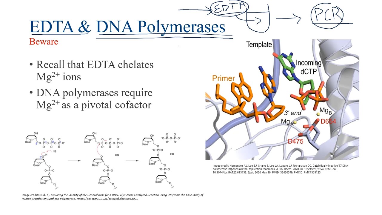
Intro To Cell Lysis Buffers
328
Cell lysis buffers are essential solutions used to break open cells and release their contents for further analysis. They are categorized into two main types: detergent-based buffers that use amphipathic molecules to disrupt cell membranes, and non-detergent buffers that rely on methods like hypotonic solutions. Detergents can be further classified as non-denaturing (which preserve protein structure) or denaturing (like SDS, which unfolds proteins and adds negative charges for gel electrophoresis). Key components of effective lysis buffers include salts to maintain physiological ionic strength (~200mM), glycerol to stabilize proteins by preventing aggregation, chelating agents like EDTA and EGTA to inhibit proteases and DNases by binding metal ions, protease inhibitors to prevent protein degradation, and buffers like Tris-HCl to maintain pH stability. The specific composition varies based on the target protein (cytoplasmic vs nuclear) and whether denatured or native protein is desired.
Click to watch →

Minipreps (alkaline lysis)! A practical guide to what goes on in each of the steps
731
Buffers maintain stable pH using conjugate acid-base pairs that donate or accept protons. Tris buffer maintains pH around 8 for initial resuspension. Glycerol prevents premature cell lysis by balancing osmotic pressure—without it, water would flow into cells and burst them. Bromothymol blue serves as a pH indicator, appearing blue at alkaline pH and colorless at neutral pH. Understanding these chemical principles explains why specific buffers are used at each step and how they protect DNA until it is ready to be released.
Click to watch →
Técnicas de Purificación de proteínas
7.7K
Cell lysis is the process of destroying cells to release internal components including proteins. Common methods include: (1) freeze-thaw cycles using liquid nitrogen, (2) sonication using high-frequency sound pulses, (3) high-pressure homogenization using French presses, and (4) chemical methods using organic solvents. Each method carries risks of protein denaturation or structural alteration. Sonication uses cavitation bubbles but generates heat and free radicals. French press homogenization uses high pressure without denaturation risks. Lysozyme can be used as an adjunct to degrade bacterial cell walls. After lysis, centrifugation separates cellular components based on size and density. The buffer typically contains buffering agents, salts (approximately 150 mM NaCl), and additives like glycerol, detergents, or charged amino acids that enhance protein stability and solubility.
Click to watch →

Minipreps (alkaline lysis plasmid purification) behind the scenes
7.5K
Bacterial cell membranes contain phospholipids and lipopolysaccharides with hydrophobic tails and negatively charged outer regions. Magnesium and calcium ions normally neutralize these negative charges. EDTA chelates these metal ions, causing negative charges to repel each other. SDS detergent then penetrates the membrane, denaturing embedded proteins and breaking down the lipid-protein structure, effectively lysing the cells and releasing cellular contents.
Click to watch →
Deep Dive
Basic principles of physics regarding magnetism, specifically the distinction between permanent ferromagnetism and induced paramagnetism.

Ferromagnetism & curie temperature | Magnetism & matter | Physics | Khan Academy
148.8K
Materials with unpaired electrons exhibit magnetic behavior, but most show only slight attraction (paramagnetism). Four elements—iron, cobalt, nickel, and gadolinium—show incredibly strong attraction (ferromagnetism). The key distinction lies in atomic arrangement: paramagnetic atoms are randomly oriented, canceling each other's magnetic effects. Ferromagnetic materials contain groups of billions of atoms that spontaneously align in the same direction, forming magnetic domains. This spontaneous alignment is a quantum mechanical phenomenon that occurs without external influence, giving ferromagnets their exceptional magnetic strength.
Click to watch →

How do Magnets & Magnetic Fields Work?
750.1K
Paramagnetic materials can be weakly attracted to magnets when placed in an external magnetic field, but they lose this magnetism when the field is removed. Unlike ferromagnetic materials, paramagnetic materials cannot maintain permanent magnetization because their electronic structure prevents stable alignment of electron spins. The weak attraction occurs because the external field causes partial alignment of electron magnetic moments, but thermal motion quickly randomizes these alignments once the field is removed. Examples include aluminum, tungsten, and lithium. This phenomenon demonstrates that magnetism is fundamentally a quantum mechanical effect dependent on the specific electronic configurations of materials.
Click to watch →
Física - Magnetismo: ferromagnetismo, diamagnetismo e paramagnetismo
82K
Magnetism originates from the magnetic moments of atoms, where electron movement generates magnetic fields; materials are classified into three categories based on their magnetic behavior: ferromagnetic materials (like iron) have domains that strongly align with external magnetic fields, producing strong attraction; paramagnetic materials (like aluminum) show weak alignment in the same direction as the external field, producing minimal attraction; and diamagnetic materials (like gold or silver) exhibit weak alignment in the opposite direction of the external field, producing weak repulsion. The key distinction is that ferromagnetic materials can retain permanent magnetization because their domains remain aligned even when the external field is removed, while paramagnetic and diamagnetic materials only show temporary magnetic effects.
Click to watch →

8.02x - Lect 21 - Magnetic Materials, Dia- Para- & Ferromagnetism
344.1K
This lecture explains three fundamental types of magnetic materials: diamagnetic materials oppose all external magnetic fields through induced dipoles (quantum mechanical effect), paramagnetic materials have permanent atomic dipoles that align with external fields but return to chaos when the field is removed, and ferromagnetic materials contain domains of aligned atomic dipoles that can retain permanent magnetization; the magnetic field inside a material is proportional to the vacuum field through relative permeability, with ferromagnetic materials achieving internal fields thousands of times stronger than vacuum fields, and ferromagnetic materials lose their permanent magnetic properties above the Curie temperature (e.g., 770°C for iron).
Click to watch →
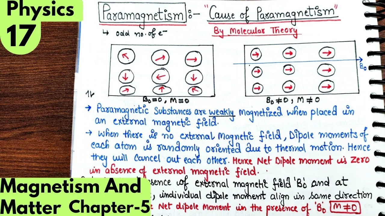
17) Cause of Paramagnetism and Ferromagnetism| Magnetism and matter Class12 Physics #neet #jee
6K
Paramagnetism arises from unpaired electrons in atoms, which create individual magnetic moments that align with an external magnetic field, producing weak magnetization; ferromagnetism involves spontaneous alignment of magnetic moments within domains, leading to strong permanent magnetization even without an external field.
Click to watch →
Deep Dive
The general purpose of nucleic acid isolation and purification in molecular biology workflows.
DNA Extraction | Biotechnology | Vivek Srinivas | #Molecularbiology #Biotechnology
19.8K
Nucleic acid isolation is a fundamental molecular biology technique used for molecular-based diagnosis. The process involves five key steps: cell lysis using SDS and proteinase K to break membranes and release cellular contents; extraction using phenol-chloroform mixture (pH 7-8) to separate nucleic acids from proteins and lipids; precipitation with cold ethanol/isopropanol to concentrate DNA; washing with 70% ethanol to ensure purity; and resuspension in Tris-EDTA buffer for storage. The phenol-chloroform method relies on phase separation where nucleic acids remain in the aqueous phase while proteins and lipids partition into the organic phase. This technique enables routine DNA/RNA isolation for downstream applications in molecular biology research and diagnostics.
Click to watch →

Extração de DNA e RNA - aula prática
43.8K
Nucleic acid extraction and purification follows three fundamental phases: (1) Extraction breaks open cells using physical, chemical, or enzymatic methods to release DNA/RNA; (2) Isolation separates nucleic acids from cellular debris using techniques like phenol-chloroform extraction or column-based methods; (3) Elution recovers purified nucleic acids in solution. Column-based purification uses a solid-phase matrix that selectively binds nucleic acids while allowing contaminants to pass through. The process involves loading the lysed sample onto the column, washing away impurities with alcohol-based solutions, and finally eluting the purified nucleic acids with buffer or water. This methodology enables high-purity nucleic acid preparations suitable for downstream molecular biology applications.
Click to watch →

isolation and purification of Nucleic Acid part 1
1.9K
Nucleic acid isolation forms the cornerstone of molecular biology research. Genomics encompasses the comprehensive study of entire genomes, including gene sets, nucleic acid sequences, organizational structures, and inter-species interactions. To manipulate or study nucleic acids, DNA and RNA must first be isolated through systematic processes. The fundamental workflow involves three essential steps: rupturing cell membranes and walls to release cellular contents, separating nucleic acids from other cellular components, and purifying the extracted nucleic acids. Cell lysis employs detergents in lysis buffers that break lipid membranes, while enzymatic treatments using proteases and ribonucleases inactivate unwanted macromolecules. Nucleic acids are subsequently precipitated with alcohol and quantified using spectrophotometric or fluorescent methods. RNA presents unique challenges due to its inherent instability and susceptibility to ubiquitous RNases. Plant cells complicate isolation due to rigid cellulose cell walls. Microbial culture preparation requires careful growth monitoring through visual turbidity assessment, followed by centrifugation and resuspension at optimal concentrations to ensure consistent starting material quality for downstream extraction procedures.
Click to watch →
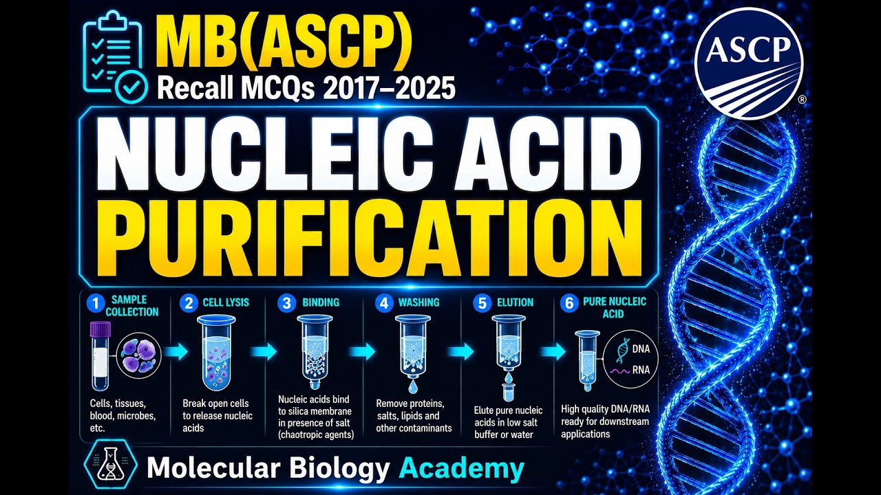
24. DNA Purification SECRETS for MB(ASCP) Success! 🧬🚀
813
Nucleic acid purification is a cornerstone technique in molecular biology for isolating DNA or RNA from biological samples including cells, tissue, blood, and environmental specimens. The process involves three essential steps: lysis (breaking open cells to release nucleic acids), separation (removing contaminants like proteins, lipids, and carbohydrates), and recovery (concentrating purified nucleic acids). Three main methods exist: chemical extraction using phenol-chloroform for protein removal, solid-phase extraction using silica columns or magnetic beads under specific salt and pH conditions, and enzymatic treatment using RNase or proteases. Common contaminants including protein, lipids, and carbohydrates must be removed as they inhibit downstream applications. Understanding these fundamentals is essential for successful molecular biology experiments.
Click to watch →

Purification techniques of DNA and RNA
160
Nucleic acid isolation and purification is essential for genetic experiments, requiring pure DNA or RNA. The process involves three steps: cell breakage to expose nucleic acids, separation from cellular components, and recovery in pure form. Cell lysis methods vary by cell type: bacterial cells use enzymatic (lysozyme) and chemical (EDTA, SDS) approaches; animal cells use SDS detergent plus EDTA; plant cells require harsher methods like mortar and pestle due to strong cell walls. This foundational step determines the success of downstream purification.
Click to watch →
Prerequisite Knowledge
- Concept 01The basic chemical structure of nucleic acids (DNA and RNA), particularly their negatively charged phosphate backbone.
- Concept 02The fundamental concept of cell lysis and how chemical buffers are used to break open cell membranes and denature proteins.
- Concept 03Basic principles of physics regarding magnetism, specifically the distinction between permanent ferromagnetism and induced paramagnetism.
- Concept 04The general purpose of nucleic acid isolation and purification in molecular biology workflows.
Subsequent Learning
- Step 01Downstream applications of purified nucleic acids, such as Polymerase Chain Reaction (PCR), quantitative PCR (qPCR), and Next-Generation Sequencing (NGS).
- Step 02High-throughput laboratory automation and the use of robotic liquid handlers for automated magnetic bead-based extraction.
- Step 03A comparative analysis of alternative extraction methods, such as silica-gel membrane spin columns and phenol-chloroform liquid-liquid extraction.
- Step 04Clinical and diagnostic applications of magnetic bead extraction, including viral load testing, non-invasive prenatal testing (NIPT), and forensic DNA profiling.
Method intro
Binding step
Magnetic capture
Wash process
Elution step
Final collection
Method intro
0:11Playing Section
- 1
Explains magnetic bead-based extraction basics
- 2
Highlights paramagnetic core binding to nucleic acids
- 3
Mentions common use in automated systems
Silica-Membrane Columns and Organic Phase Separation as Superior Alternatives
While magnetic bead-based extraction is highly praised for its automation compatibility, alternative methods like silica-membrane spin columns and traditional liquid-liquid extraction (e.g., phenol-chloroform) offer distinct advantages. First, magnetic beads are significantly more expensive and require specialized magnetic separators. For budget-constrained laboratories, spin columns remain the cost-effective industry standard. Second, magnetic bead protocols are susceptible to 'bead carryover'—where residual beads escape elution and inhibit downstream PCR assays—whereas spin columns physically retain the matrix. Lastly, for complex sample types like plant tissues or soil, traditional organic extraction remains the gold standard for high-molecular-weight (HMW) DNA recovery, as humic acids and polyphenols can clog magnetic beads or cause non-specific binding. Consequently, magnetic beads are often viewed as an operational convenience for high-throughput workflows rather than the universally superior method for purity and yield.
Deep Dive
Downstream applications of purified nucleic acids, such as Polymerase Chain Reaction (PCR), quantitative PCR (qPCR), and Next-Generation Sequencing (NGS).
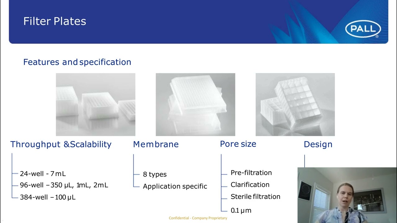
Tips and Tricks - When Using Filter Plates or Spin Devices with Nucleic Acid Protein Purification...
525
Nucleic acid binding membranes enable high-quality plasmid DNA, genomic DNA, and RNA extraction suitable for downstream applications including PCR/qPCR, NGS sequencing, Northern blotting, and Sanger sequencing. Using filter plates and spin devices with commercially available reagents or in-house developed protocols produces nucleic acids comparable to or exceeding commercially available kits in quality and concentration. The quality is sufficient for demanding applications like restriction digestion and cloning.
Click to watch →

NCERT Reading | Biotechnology | L6 | Unacademy NEET | Seep Pahuja
40.5K
PCR (Polymerase Chain Reaction) amplifies specific DNA sequences exponentially through three temperature cycles: (1) Denaturation at 94°C - separates double-stranded DNA into single strands; (2) Annealing at 55-65°C - primers bind to complementary sequences; (3) Extension at 72°C - Taq polymerase synthesizes new DNA strands. Each cycle doubles the DNA, so n cycles produce 2^n copies. Downstream processing involves: (1) Separation - isolating the desired product from the host cell; (2) Purification - removing impurities; (3) Preservation - adding preservatives; (4) Clinical trials - testing safety and efficacy for drugs/vaccines; (5) Marketing - commercial distribution after regulatory approval.
Click to watch →
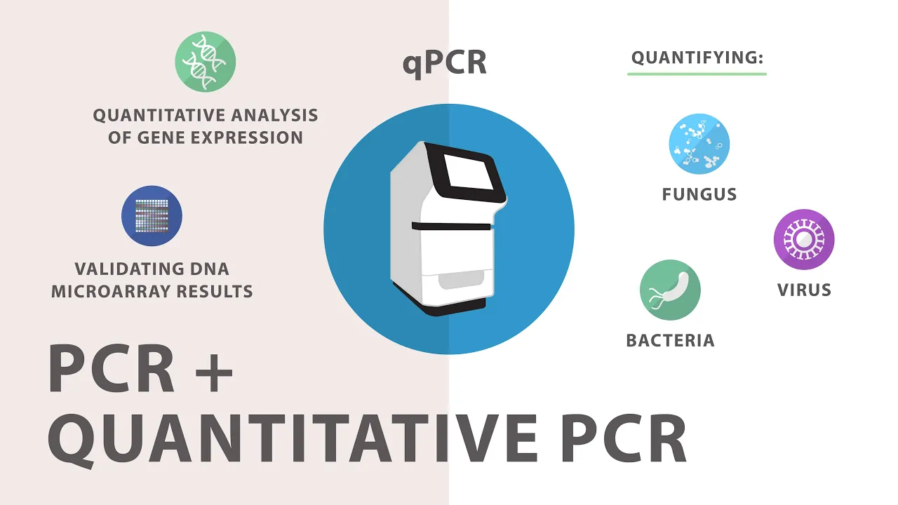
3) Polymerase Chain Reaction (PCR) - Quantitative PCR (qPCR)
230.6K
Quantitative PCR (qPCR) is a technique that simultaneously amplifies and detects changes in amplicon concentration by collecting fluorescence signals during amplification; it uses two main methods: SYBR Green-based detection, which binds to all double-stranded DNA and requires melting curve analysis for specificity, and TaqMan probe-based detection, which uses hydrolysis probes with fluorescent reporter and quencher dyes that only bind to target sequences, enabling multiplex reactions and better specificity.
Click to watch →
Extração de DNA e RNA - aula prática
43.8K
Elution is the final step in nucleic acid purification where the purified DNA or RNA is recovered from the binding matrix. A small volume of elution buffer or water is added to the top of the column, and after centrifugation, the nucleic acids are released from the matrix and collected in the collection tube below. The choice of elution volume affects the concentration of the final product—smaller volumes yield higher concentrations but may reduce recovery efficiency. The eluted nucleic acids are now free of alcohol and ready for downstream applications such as PCR, sequencing, or library preparation.
Click to watch →

Plus Two Model Exam One Shot Biology | Biotechnology - Principles And Processes
16.6K
PCR (Polymerase Chain Reaction) is used for amplification of genes in DNA technology, producing millions of copies from small samples. Taq polymerase, derived from Thermus aquaticus (a bacterium from hot springs), is used because it can withstand high temperatures required for DNA denaturation. Downstream processing prepares bioreactor products for marketing through: (1) Separation - isolating desired products from cell culture, (2) Purification - removing impurities, (3) Formulation - adding preservatives and stabilizers, (4) Quality Control Testing - ensuring safety and efficacy standards, (5) Clinical Trials - testing products in humans before approval. This process ensures products meet regulatory standards before reaching patients.
Click to watch →
Deep Dive
High-throughput laboratory automation and the use of robotic liquid handlers for automated magnetic bead-based extraction.

Vazyme Automatic Nucleic Acids Extraction Instruments | Smart automation for every lab
548.7K
Automated nucleic acid extraction instruments use magnetic bead-based technology with UV sterilization and pre-filled reagents to efficiently isolate DNA/RNA from various sample types (blood, plants, cells, tissue, soil, bacteria) in high-throughput formats (32 or 96 samples), reducing hands-on time to 13-60 minutes while ensuring contamination-free results through splash-free liquid handling and transparent chamber monitoring.
Click to watch →
Automating Nucleic Acid Extraction (Part 1): Basic Chemistry and Robotics
2.2K
Automated nucleic acid extraction using magnetic beads follows four core chemical steps—cell lysis, binding to magnetic particles, washing to remove impurities, and elution with water—and relies on robotic systems that perform liquid handling, magnetization, mixing, and temperature control to automate this process efficiently.
Click to watch →

Implementation of a High Throughput Wastewater SARS CoV 2 Detection Method Providing Actionable...
270
Traditional wastewater virus concentration methods (filtration, centrifugation, ultrafiltration, PEG precipitation) require 6-8 hours per sample and limit processing to 10-20 samples daily. The innovative solution uses magnetic nanotrap particles that specifically bind viral particles, combined with Kingfisher liquid handling robot's magnetic head. This automation reduces concentration time from 6-8 hours to 45 minutes while processing 24 samples simultaneously, achieving approximately 100-fold time reduction. The magnetic separation cleanly isolates viral particles from solids and contaminants.
Click to watch →

Matthew Nesbitt - Automation of High Throughput Agarose Gel Electrophoresis with the NIMBUS Select F
1.4K
The NIMBUS Select workstation automates high-throughput agarose gel electrophoresis by combining a 96-channel liquid handler with Ranger technology, enabling simultaneous size selection and analytical electropherogram generation with high recovery yields (80-90% vs. 10-20% for magnetic bead methods), resolution under 25 bp at 1,000 bp, and processing up to 96 samples in approximately 2 hours at $2/sample.
Click to watch →
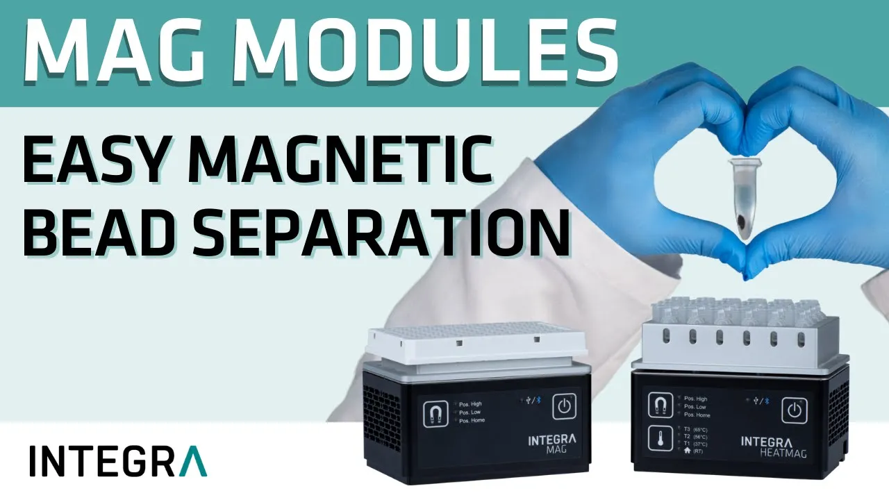
MAG and HEATMAG – Modules for magnetic bead separation
1.9K
Magnetic bead separation modules automate the purification of biomolecules such as DNA, RNA, and proteins by using magnetic forces to collect beads from solution; these modules feature adjustable magnet heights for different volumes, powerful magnets to minimize bead loss, and can be combined with heating functions (up to 65°C) for applications like nucleic acid extraction, while also integrating with various pipetting platforms for high-throughput automated workflows.
Click to watch →
Deep Dive
A comparative analysis of alternative extraction methods, such as silica-gel membrane spin columns and phenol-chloroform liquid-liquid extraction.

Ekstraksi DNA Metode PCIA (Phenol Chloroform Isoamyl Alcohol)
407
DNA exists in two forms: chromosomal DNA (in nucleus of eukaryotes or cytoplasm of prokaryotes) and extrachromosomal DNA (mitochondrial DNA in humans, plasmid DNA in bacteria). Chromosomal DNA is packaged with histone proteins to form nucleosomes, which condense into chromatin fibers and chromosomes. DNA extraction methods are classified into liquid-based (phenol-chloroform, salting-out, Chelex) and solid-phase (silica columns, magnetic beads) approaches. The phenol-chloroform extraction method is based on differential solubility: polar molecules dissolve in polar solvents, while nonpolar molecules dissolve in nonpolar solvents. DNA, being polar and negatively charged, remains in the aqueous phase while proteins and lipids partition into the organic phase.
Click to watch →
Turning plastic gloves into hot sauce
24M
Silica gel chromatography separates compounds based on polarity differences. Load the crude mixture onto silica gel packed in a column, then elute with increasing polarity solvents. Polar impurities elute first, followed by less polar compounds. This technique effectively removes colored impurities and improves product purity beyond simple liquid-liquid extraction.
Click to watch →
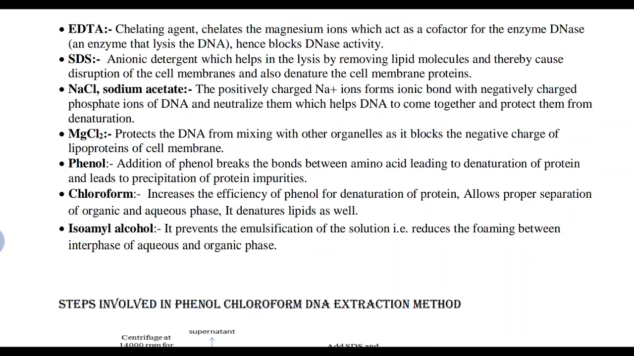
Phenol chloroform DNA extraction method
29.8K
The phenol-chloroform DNA extraction method is an organic DNA isolation technique based on liquid-liquid extraction principles, where different biomolecules separate based on their solubility in immiscible polar (aqueous) and nonpolar (organic) solvents; the method uses Tris buffer to maintain pH and lyse cells, EDTA to chelate magnesium ions and inhibit DNAse enzymes, SDS to disrupt cell membranes, phenol and chloroform to denature proteins and separate phases, and isopropanol to precipitate DNA, followed by ethanol washing and TE buffer resuspension to obtain purified DNA suitable for downstream applications like PCR.
Click to watch →

DNA extraction
853
DNA extraction is a laboratory technique for isolating DNA from cells, involving cell lysis (breaking cell membranes using detergents or sonication), protein precipitation (using phenol-chloroform), centrifugation to separate phases, and purification using silica membrane columns; the extracted DNA can be either intrachromosomal (from inside human cells) or extrachromosomal (from bacterial plasmids), and its purity is assessed using spectrophotometry (260/280 ratio of 1.8 indicates pure DNA) and gel electrophoresis.
Click to watch →
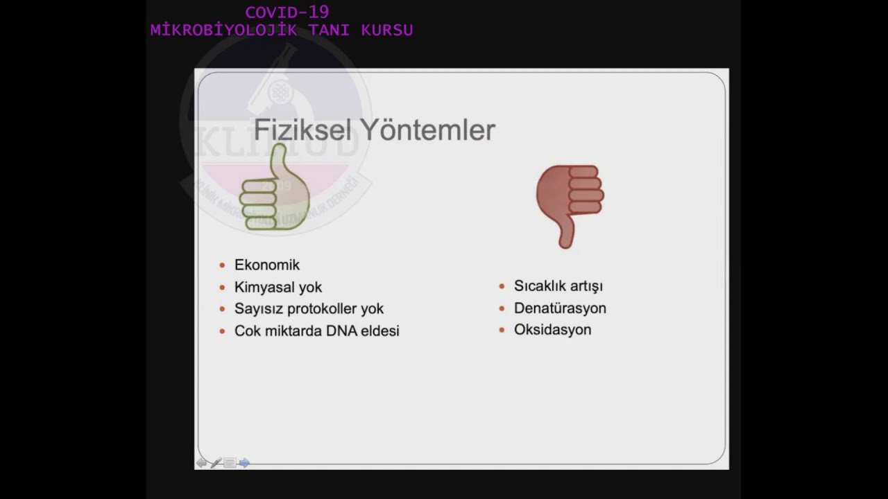
Nükleik Asit İzolasyon Yöntemleri- Dr. Canan Külah
1.8K
Solid phase extraction uses matrices including silica, glass, diatomaceous earth, and magnetic beads. Spin column extraction uses silica-based membranes where nucleic acids bind under high pH and ionic strength conditions while contaminants pass through. After washing, nucleic acids are eluted using low pH, low ionic strength buffer. This method provides fast, high-purity DNA extraction with effective inhibitor removal. Magnetic bead extraction uses DNA-coated beads captured by magnets, allowing washing without liquid handling. These methods are compatible with automated systems and provide high-purity DNA.
Click to watch →
Deep Dive
Clinical and diagnostic applications of magnetic bead extraction, including viral load testing, non-invasive prenatal testing (NIPT), and forensic DNA profiling.

ProtonDx Breathe and Learn
252
The SmartLid system from ProtonDx enables rapid viral DNA and RNA extraction from cell-free fluids using magnetic bead technology, where a disposable lid with a removable magnet collects magnetic beads that bind nucleic acids, allowing complete extraction protocols to be completed in under 10 minutes for 1-12 samples, with comparable or better results than market-leading competitors.
Click to watch →
TANBead - Company Profile (English)
1.2K
TANBead is an integrated service provider specializing in nucleic acid extraction technology, using patented magnetic beads with gear-driven stirring to achieve high extraction efficiency while minimizing cross-contamination risk, offering a range of products from hand-carried M8 to high-flow M9600 models for various molecular diagnostics applications.
Click to watch →
NIPT – Teste pré natal não invasivo: uma avaliação para risco genético - Dr. Giuliano Bedoschi
12.3K
Non-invasive prenatal testing (NIPT) analyzes cell-free fetal DNA circulating in maternal blood, primarily derived from the placenta. By 9-10 weeks of gestation, sufficient fetal DNA is present for testing. NIPT detects Trisomy 21 (Down syndrome), Trisomy 18 (Edwards syndrome), Trisomy 13 (Patau syndrome), and sex chromosome abnormalities. It serves as a secondary screening tool, not primary, due to cost (approximately R$ 2,000, not covered by Brazilian health insurance) and technical limitations. Indications include maternal age over 35, family history of genetic diseases, or abnormal ultrasound/blood screening results.
Click to watch →
DNA and RNA extraction with magnetic beads – How it works
18.5K
Magnetic bead-based nucleic acid extraction is a solid-phase purification method that utilizes superparamagnetic iron oxide beads coated with ligands capable of reversibly binding nucleic acids; the process involves four sequential steps: (1) cell lysis to release DNA/RNA from intact cells using chemical or physical methods, (2) binding where magnetic beads and binding buffer are added to the lysate and incubated to allow specific nucleic acid-bead interactions, (3) washing where ethanol-containing buffers remove contaminants while magnetic separation retains the bead-bound nucleic acids, and (4) elution where nucleic acids are released from the beads using elution buffer or nuclease-free water for downstream applications; this technique can be adapted for either nucleic acid extraction from whole-cell samples or purification of pre-amplified PCR products without requiring cell lysis.
Click to watch →

Extração de DNA e RNA por beads magnéticas
6.3K
Magnetic bead-based extraction is a rapid and efficient method for isolating DNA and RNA from biological samples, utilizing positively charged magnetic beads that bind nucleic acids during lysis, followed by magnetic separation, washing steps to remove contaminants, and final elution to recover pure nucleic acids; this technique offers advantages of speed, reduced contamination risk, and automation compatibility, though it requires specialized equipment and is more expensive than alternative methods.
Click to watch →
Method intro
Binding step
Magnetic capture
Wash process
Elution step
Final collection
Method intro
0:11- 1
Explains magnetic bead-based extraction basics
- 2
Highlights paramagnetic core binding to nucleic acids
- 3
Mentions common use in automated systems
Silica-Membrane Columns and Organic Phase Separation as Superior Alternatives
While magnetic bead-based extraction is highly praised for its automation compatibility, alternative methods like silica-membrane spin columns and traditional liquid-liquid extraction (e.g., phenol-chloroform) offer distinct advantages. First, magnetic beads are significantly more expensive and require specialized magnetic separators. For budget-constrained laboratories, spin columns remain the cost-effective industry standard. Second, magnetic bead protocols are susceptible to 'bead carryover'—where residual beads escape elution and inhibit downstream PCR assays—whereas spin columns physically retain the matrix. Lastly, for complex sample types like plant tissues or soil, traditional organic extraction remains the gold standard for high-molecular-weight (HMW) DNA recovery, as humic acids and polyphenols can clog magnetic beads or cause non-specific binding. Consequently, magnetic beads are often viewed as an operational convenience for high-throughput workflows rather than the universally superior method for purity and yield.
The basic chemical structure of nucleic acids (DNA and RNA), particularly their negatively charged phosphate backbone.
The fundamental concept of cell lysis and how chemical buffers are used to break open cell membranes and denature proteins.
Basic principles of physics regarding magnetism, specifically the distinction between permanent ferromagnetism and induced paramagnetism.
The general purpose of nucleic acid isolation and purification in molecular biology workflows.
Downstream applications of purified nucleic acids, such as Polymerase Chain Reaction (PCR), quantitative PCR (qPCR), and Next-Generation Sequencing (NGS).
High-throughput laboratory automation and the use of robotic liquid handlers for automated magnetic bead-based extraction.
A comparative analysis of alternative extraction methods, such as silica-gel membrane spin columns and phenol-chloroform liquid-liquid extraction.
Clinical and diagnostic applications of magnetic bead extraction, including viral load testing, non-invasive prenatal testing (NIPT), and forensic DNA profiling.
21.8K views177likes1:42@CDCOriginal Release: 2022-03-09
[Music] magnetic bead based extraction is a method that utilizes small particles with a paramagnetic core that binds to nucleic acid magnetic bead-based extraction is commonly used in automated nucleic acid extraction systems add the magnetic beads to a cell lysate to capture nucleic acid in a solution apply an external magnetic field to capture the beads while the solution containing the other proteins and cellular components are removed add a wash buffer to the magnetic beads to wash off additional contaminants remove the magnetic field and the aleutian buffer to release the nucleic acid from the magnetic beads apply an external magnetic field to capture the beads in order to collect diluted nucleic acid in the solution [Music] you
Up Next

18:32
STR Analysis and DNA Fingerprinting Explained | Genetics
@DrWD40
34.8K views•2020-04-14

50:12
Bacterial Communication: Quorum Sensing and Biofilm Formation Explained
@JHUAAP
12.6K views•2011-06-28

5:35
Enteric Nervous System Explained: The Gut's Brain | Neurobiology Lecture
@alumniu6029
438 views•2018-09-12

7:08
Bacteriophages: Earth's Deadliest Killers and Future Antibiotics
@kurzgesagt
34.6M views•2018-05-13
Related Study Plans & Knowledge Roadmaps
Structured learning paths in Biology
biology
RNAi: Biogenesis, RISC & Gene Silencing
01Molecular Biology Foundations: DNA to Protein
02Introduction to RNA Interference (RNAi)
03Molecular Biogenesis of miRNA and siRNA
+ 2 more learning modules
10h5 modules
Explore Roadmap
biology
Mycorrhizae: Signaling, Biophysics & Ecology
01Foundations of Mycorrhizal Symbiosis
02Molecular Signaling and Root Colonization
03Biophysics and Bioenergetics of Nutrient Exchange
+ 1 more learning modules
11h4 modules
Explore Roadmap
biology
Circadian Clocks: TTFL, SCN & Periphery
01Foundations of Molecular Biology & Gene Expression
02Introduction to Chronobiology and Actograms
03The Core TTFL: Molecular Architecture of the Clock
+ 3 more learning modules
14h6 modules
Explore Roadmap
biology
Population Genetics: Math, Drift & Selection
01Genetic & Probability Foundations
02The Hardy-Weinberg Principle and Baseline Models
03Mathematical Modeling of Natural Selection
+ 3 more learning modules
18h6 modules
Explore Roadmap