Bacteriophages are virus-like particles that exclusively infect bacteria; they attach to bacterial cells using leg-like fibers, which trigger conformational changes that drive a needle-like shaft into the bacterial envelope, allowing the phage to inject its genetic material contained within an icosahedral protein capsid head.
Bacteriophage T4 Attacking E. coli: Nanoscale Infection Mechanism
Added:Deep Dive
Basic anatomy of bacteria and viruses, specifically the structural differences between Gram-negative bacteria (like E. coli) and bacteriophages.
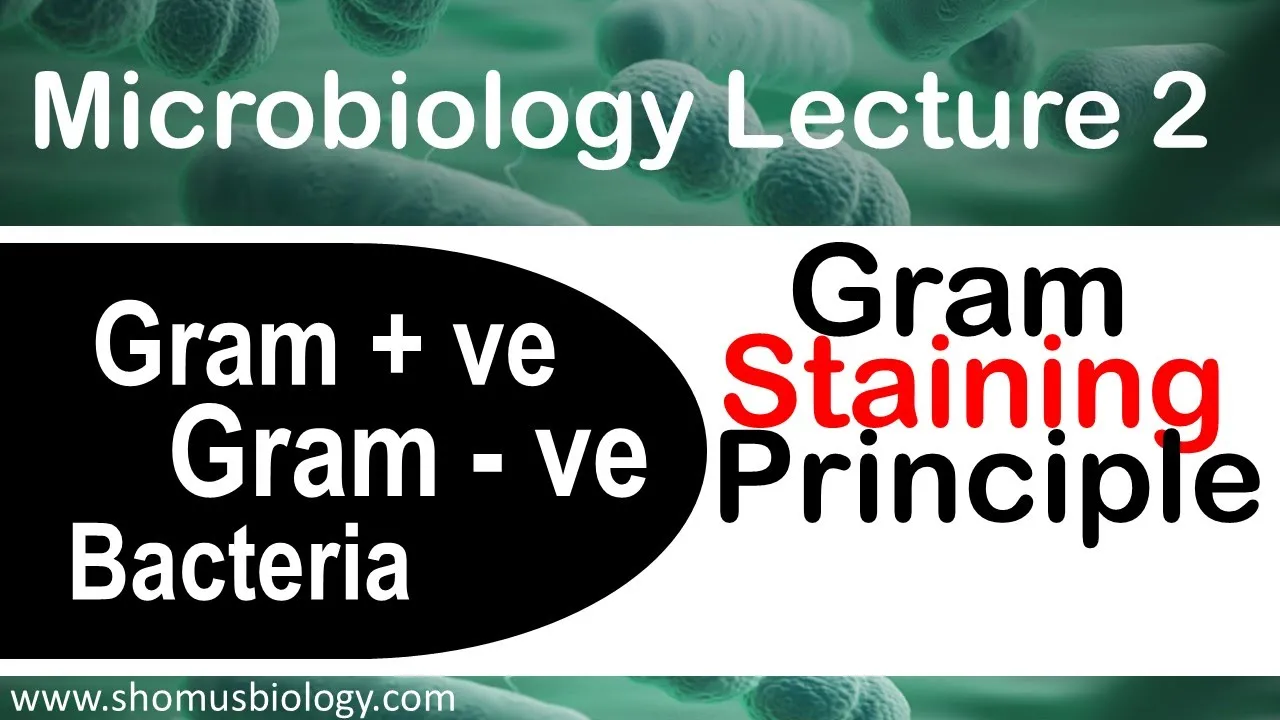
Microbiology lecture 2 | Gram positive and Gram negative bacteria Gram staining principle
354.5K
Gram staining differentiates bacteria into gram-positive (purple) and gram-negative (pink/red) based on their cell wall structure: gram-positive bacteria have a thick peptidoglycan layer (20-80 nm) with teichoic acids, while gram-negative bacteria have a thin peptidoglycan layer (8-11 nm) surrounded by an outer membrane containing lipopolysaccharides (LPS). During gram staining, crystal violet binds to peptidoglycan; the alcohol decolorizer removes the stain from gram-negative bacteria by dissolving their outer membrane but leaves it intact in gram-positive bacteria due to the thick peptidoglycan layer. Safranin counterstain then colors only the decolorized gram-negative cells.
Click to watch →
التكاثر بدون تعقيد! | مراجعة الفصل الثالث – المحاضرة الأولى
38.9K
Bacteriophages are viruses that specifically infect bacteria, particularly bacteria of the genus E. coli. They have a distinctive structure with a protein coat (capsid) containing genetic material (DNA), a tail structure, and tail fibers. The tail fibers attach to specific receptors on the bacterial cell surface, while the capsid protects the viral genetic material. Bacteriophage reproduction occurs through two overlapping cycles: the lytic cycle and the lysogenic cycle.
Click to watch →
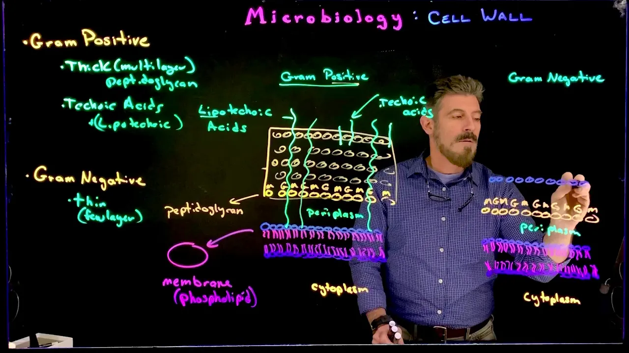
Gram Positive vs Gram Negative
8.2K
Gram-positive bacteria have a thick peptidoglycan layer with lipoteichoic acids extending from the cell membrane through the peptidoglycan, while gram-negative bacteria have a thin peptidoglycan layer surrounded by an outer membrane containing lipopolysaccharides (LPS) and a larger periplasmic space; these structural differences determine how each type of bacteria interacts with the environment, transports molecules, and responds to antibiotics and immune defenses.
Click to watch →

Lab 3-7: Gram Stain
28.2K
Gram-positive and gram-negative bacteria have fundamentally different cell wall structures. Gram-positive bacteria have a very thick peptidoglycan layer (~8x thicker than gram-negative), teichoic acids that cross-link the peptidoglycan, and lipoteichoic acid embedded in the cell membrane. They lack an outer membrane. Gram-negative bacteria have a thin peptidoglycan layer located in the periplasmic space between two phospholipid bilayers (inner cell membrane and outer membrane). The outer membrane contains porins that regulate substance passage and lipopolysaccharide (LPS) containing lipid A, an endotoxin.
Click to watch →
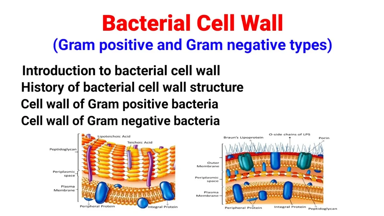
Bacterial Cell Wall | Gram positive & Gram negative bacteria cell wall| Structure|Salient features|
63.7K
Gram-positive bacteria feature a thick peptidoglycan layer (20-80 nm) directly outside the plasma membrane, while gram-negative bacteria have a thin peptidoglycan layer (2-7 nm) sandwiched between an inner plasma membrane and an outer membrane. The periplasmic space exists between the plasma membrane and peptidoglycan in gram-positive bacteria, and between the outer membrane and plasma membrane in gram-negative bacteria. The cell envelope encompasses all structures outward from the plasma membrane, including the plasma membrane, cell wall, and capsule (not present in all bacteria).
Click to watch →
Deep Dive
The fundamental stages of the viral lytic cycle, including attachment, penetration, biosynthesis, maturation, and lysis.

المراجعة المركزة - الفصل الثالث - الجزء الاول
131.7K
The lytic cycle of viruses consists of five sequential stages: (1) Attachment - the virus attaches to the host cell surface, (2) Penetration - the viral genetic material enters the host cell, (3) Biosynthesis - the host cell machinery is hijacked to produce viral components, (4) Maturation - new viral particles are assembled, and (5) Release - new viruses are released from the host cell.
Click to watch →

(الحلقة الثالثة ) من الفصل الثالث مادة الاحياء السادس العلمي الجديد
95.7K
The lytic cycle of viral reproduction consists of five distinct stages: (1) Attachment - the virus attaches to the host cell, (2) Penetration - the viral genetic material enters the host cell, (3) Biosynthesis - the host cell machinery is used to produce viral components, (4) Maturation - new viral particles are assembled, and (5) Release - new viruses are released from the host cell, often causing cell lysis.
Click to watch →

مراجعة نصف السنة || الفصل الثالث التكاثر || احياء السادس العلمي
25.7K
The lytic cycle consists of five stages: attachment, penetration, biosynthesis, maturation, and release. During attachment, viral fibers bind to specific receptors on the host cell. During penetration, viral DNA enters the host cell. During biosynthesis, the host cell's machinery is redirected to synthesize viral components. During maturation, new viruses are assembled. During release, the host cell bursts, releasing new viruses to infect other cells.
Click to watch →

أحياء السادس العلمي | منهج 2026 | الفصل الثالث | 3| التكاثر في الفايروسات
74.5K
The lytic cycle of virus reproduction consists of five distinct stages: (1) Attachment - the virus attaches to the host cell surface using specialized tail fibers that bind to specific receptor sites on the bacterial cell wall, (2) Penetration - the virus injects its genetic material into the host cell by secreting enzymes that weaken the cell wall and create a hole, (3) Biosynthesis - the host cell's machinery is hijacked to produce viral components, (4) Maturation - new virus particles are assembled from the synthesized components, and (5) Release - new viruses are released from the host cell, often causing the cell to burst.
Click to watch →

الفصل الثالث | المحاضرة 2|تكاثر الفايروسات والتكاثر اللاجنسي للبكتريا
551
The lytic cycle of viral reproduction consists of five distinct stages: 1) Attachment (adsorption), 2) Penetration (entry), 3) Biosynthesis (replication), 4) Maturation (assembly), and 5) Release (lysis). These stages occur in sequence to produce new viral particles.
Click to watch →
Deep Dive
Key molecular biology processes, particularly how host cell machinery is hijacked for viral protein synthesis and genome replication.

BBYCT-131 full lecture (part-3) | Unit-1 Virus:-Genral account and economic importance|#ignou #virus
2.8K
Viruses hijack host cell machinery for replication and protein synthesis. The viral genome is replicated using either host or viral polymerases depending on the virus type. Viral proteins are synthesized on host ribosomes using viral mRNA. The viral replication cycle includes attachment to host cell receptors, entry into the cell, uncoating, replication, assembly, and release. This hijacking process can be destructive to the host cell, leading to cell death and release of new viral particles.
Click to watch →

chapter 6 video 6
276
Viruses lack their own cellular machinery and must infect host cells to replicate. The viral replication cycle involves: (1) Attachment to specific host cell receptors, (2) Entry of viral genetic material into the host cell, (3) Hijacking of host cellular machinery for genome replication and protein synthesis, (4) Assembly of new viral particles from replicated genetic material and synthesized proteins, (5) Release of new viruses from the host cell. This process allows viruses to produce multiple copies of themselves within the host cell.
Click to watch →

Lo 8 g10 bio STEM virology and HIV study case
624
Viruses hijack host cell machinery through a process called hijacking, using the host's ribosomes, enzymes, and other cellular components to synthesize viral proteins and replicate viral genetic material. Host cell proteins assist viral replication through multiple mechanisms: receptor proteins help viruses attach and enter cells, nuclear proteins transport viral genetic material into the nucleus, and host ribosomes translate viral mRNA into viral proteins. These host proteins are exploited by viruses without the host's intention, essentially turning the host cell into a factory for producing new viruses.
Click to watch →

Biological Classification l lecture 11 l Biology l NEET
63.1K
Viruses lack their own metabolic machinery and must hijack the host cell's ribosomes to replicate. The viral genome enters the host cell and takes control of the cellular machinery. The host cell's nucleus (the 'boss') is essentially destroyed, and the viral genome takes over, directing the cell to produce viral proteins and replicate the viral genome.
Click to watch →

Virology Lec6 part 1
182
Viruses hijack host cell machinery to replicate their genetic material. The viral genome enters the host cell and directs the cell's ribosomes and other machinery to produce viral proteins. Positive-sense RNA genomes can be directly translated by host ribosomes, while negative-sense RNA genomes must first be transcribed into positive-sense RNA by viral polymerases. The host cell's resources are redirected to produce viral components, effectively turning the cell into a viral factory. This process allows viruses to replicate efficiently while evading host immune responses.
Click to watch →
Deep Dive
An understanding of antibiotic resistance and how 'superbugs' evolve to survive traditional antimicrobial treatments.

Open Mics Episode 121 - Superbugs - Senator Roger Marshal
182
Superbugs are antibiotic-resistant bacteria that have evolved to survive common antibiotics. Antibiotics kill or stop bacterial growth by targeting reproduction or other cellular processes. Resistance develops when bacteria survive antibiotic exposure and pass resistant genes to offspring. Common examples include resistant pneumonia, urinary tract infections, and skin infections. The problem worsens as bacteria adapt to frequent antibiotic use, creating a cycle where resistant strains proliferate while effective treatments diminish.
Click to watch →

6 Superbugs to Watch Out For
690
Superbugs are drug-resistant bacterial infections that have evolved to survive antibiotic treatments. Antibiotic resistance develops over time as bacteria constantly evolve and adapt to new environments. Some microbes swap genes with each other, making them drug-resistant. The bacteria that survive antibiotic treatments eventually outnumber susceptible bacteria. Six major superbugs are highlighted: Klebsiella pneumoniae (resistant to carbapenems via beta-lactamase enzyme), MRSA (resistant to methicillin and spreads through contact), Clostridium difficile (causes severe diarrhea, linked to 14,000 deaths annually), XDR-TB (resistant to multiple antibiotics), drug-resistant gonorrhea (resistant to penicillin, tetracycline, and ciprofloxacin), and STEC (resistant to multiple antibiotic classes). Prevention includes strict hygiene procedures like hand washing and wearing hospital gowns.
Click to watch →

MEDICAMENTOS en TÚ DE CIENCIAS Y YO DE LETRAS con @Linguriosa y @Lahiperactina COMPLETO 1x08
292
Antibiotic resistance occurs when bacteria evolve to survive antibiotic treatment. When antibiotics are used, they kill sensitive bacteria, leaving resistant bacteria to multiply. This is a process of natural selection. The overuse of antibiotics accelerates the development of resistant bacteria, creating 'superbugs' that are difficult or impossible to treat.
Click to watch →
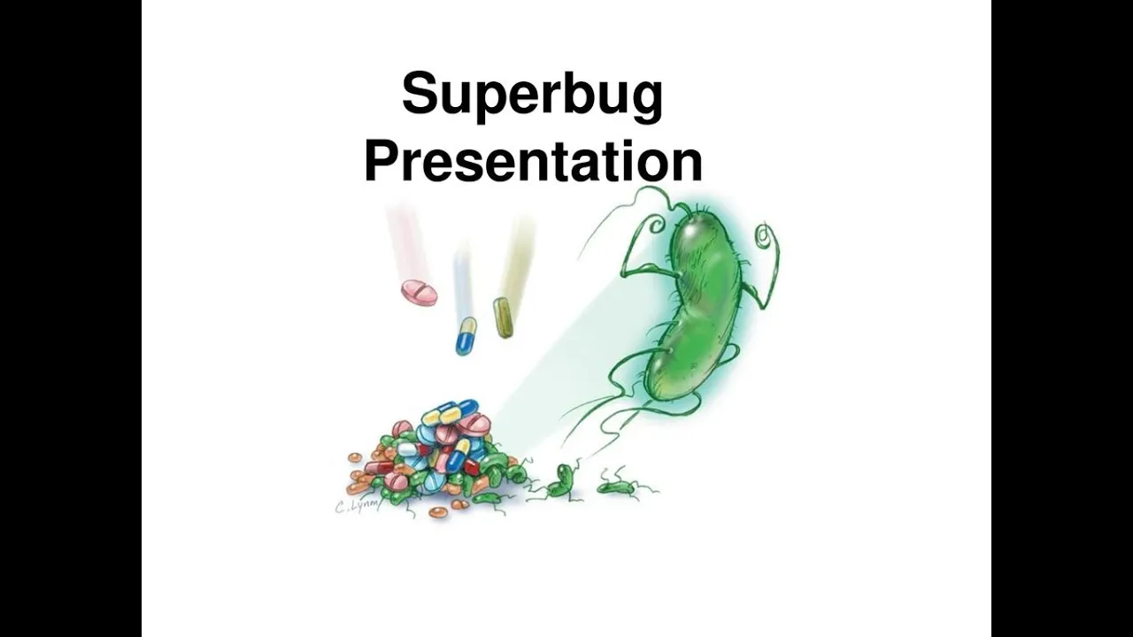
My Student's Paper Presentation on Super Bugs | #superbugs | #oldvideo | Ravi Prasath S
450
Superbugs are antibiotic-resistant bacterial strains that survive antibiotic treatments because they lack the enzymes that normal bacteria produce to neutralize antibiotics; this resistance develops through random mutations that reduce bacterial death rates, and can spread through gene transfer between bacteria, making infections like urinary tract infections and tuberculosis increasingly difficult to treat.
Click to watch →

AULÃO ENEM 2016 - Biologia - Prof. Paulo Jubilut
680.6K
Antibiotic resistance occurs when bacteria evolve to survive antibiotic treatment. This happens when bacteria with resistance genes survive antibiotic exposure and reproduce. Overuse and incomplete antibiotic courses accelerate resistance development. Resistant bacteria (superbugs) can become untreatable, representing a major global health threat.
Click to watch →
Prerequisite Knowledge
- Concept 01Basic anatomy of bacteria and viruses, specifically the structural differences between Gram-negative bacteria (like E. coli) and bacteriophages.
- Concept 02The fundamental stages of the viral lytic cycle, including attachment, penetration, biosynthesis, maturation, and lysis.
- Concept 03Key molecular biology processes, particularly how host cell machinery is hijacked for viral protein synthesis and genome replication.
- Concept 04An understanding of antibiotic resistance and how 'superbugs' evolve to survive traditional antimicrobial treatments.
Subsequent Learning
- Step 01The clinical applications, history, and current regulatory challenges of Phage Therapy as an alternative to antibiotics.
- Step 02Bacterial defense mechanisms against phage attack, such as the CRISPR-Cas system and restriction-modification systems.
- Step 03Genetic engineering of bacteriophages (synthetic biology) to enhance their host range, stability, and efficacy in medical treatments.
- Step 04The concept of Phage-Antibiotic Synergy (PAS) and how combining phages with sub-lethal antibiotic doses can prevent resistance.
Phage Discovery
Killer Looks
Prey Selectivity
Attachment Phase
Injection Action
DNA Delivery
Phage Discovery
0:00Playing Section
- 1
Félix d'Hérrelle identified bacteriophages over a century ago.
- 2
Phages are viruses that specifically target and destroy bacteria.
- 3
They are named for their bacteria-eating capability.
Limitations of Phage Therapy: Resistance, Specificity, and Delivery Barriers
While the nanoscale mechanism of bacteriophage T4 highlights the therapeutic potential of phages, significant hurdles challenge their viability as a universal solution to superbugs. A primary limitation is the rapid evolution of bacterial resistance. Bacteria can quickly mutate surface receptors—such as OmpC or lipopolysaccharides targeted by T4—rendering the phages ineffective. Additionally, phages exhibit extreme host specificity; a treatment tailored for one strain of E. coli may have no effect on another closely related strain, necessitating complex 'phage cocktails' and rapid, precise diagnostics. From a clinical perspective, the human immune system often recognizes phages as foreign proteins, generating neutralizing antibodies that clear them before they can reach the target infection. Finally, the pharmacokinetics of administering live biological viruses are highly unpredictable compared to traditional small-molecule antibiotics. These factors suggest that phage therapy faces major evolutionary, biological, and translational barriers that may limit its widespread clinical efficacy.
Deep Dive
The clinical applications, history, and current regulatory challenges of Phage Therapy as an alternative to antibiotics.
TWiV 1279: Let hepatitis b
3.6K
Phage therapy was discovered in 1917 by Felix d'Herelle but fell out of favor after the 1934 AMA report concluded insufficient evidence for clinical use. Post-WWII antibiotic development further marginalized phage therapy until renewed interest emerged from antibiotic resistance concerns. Modern regulatory frameworks vary significantly: the EU classifies phages as biological products requiring GMP compliance; Canada uses single-patient clinical trial authorizations; the UK employs a one-health approach with pharmacist exemptions; the US requires IND applications with comprehensive CMC requirements. A fundamental challenge exists: phages are highly specific to bacterial hosts, preventing large-scale testing needed for regulatory approval, creating a chicken-and-egg problem where regulations block implementation without data, and data cannot be collected without regulatory flexibility. With over 4.95 million deaths worldwide associated with antimicrobial resistance in 2019, coordinated international efforts are essential for advancing phage therapy as a complementary strategy to extend antibiotic usable life rather than replace antibiotics entirely.
Click to watch →
Why Infections Are Getting More Dangerous | Virus VS Bacteria | Spark
143.2K
Translating phage therapy from research to clinical practice involves navigating complex scientific, regulatory, and ethical landscapes. Phages are classified into virulent types (predictable, safe for humans) and temperate types (can transfer bacterial genes, potentially spreading resistance), creating safety concerns. Isolation requires testing candidate phages against target bacteria in environmental sources rich in microorganisms. Western acceptance requires approximately 10 million euros per study for each specific phage-bacteria combination—costs pharmaceutical companies cannot justify without guaranteed profits. No single well-designed clinical trial according to Western standards has proven phage efficacy in humans, creating a Catch-22. The Helsinki protocol provides a legal pathway for desperate patients, allowing doctors to apply phage cocktails when conventional treatments fail. This tension between scientific evidence, regulatory requirements, and patient desperation defines the current landscape of phage therapy implementation.
Click to watch →

Virology Lectures 2025 #25: Therapeutic viruses
2.6K
Phage therapy, discovered by Felix d'Herelle in 1915, uses bacteriophages to kill bacteria. Despite early promise during WWII, antibiotics caused its abandonment until antibiotic resistance resurgence revived interest. Phage therapy requires personalized treatment: pathogenic bacteria must be identified, and specific phages isolated for each infection. Regulatory challenges include FDA approval difficulties for individualized treatments. Approved applications include ListShield for food safety and phage cocktails for human infections. Clinical successes include treating antibiotic-resistant pseudomonas infections and disseminated Mycobacterium abscessus in immunocompromised patients. However, challenges remain including antibody neutralization and the need for rapid response to emerging resistance.
Click to watch →

10 “Impossible” Problems Humanity Solved Long Ago (2026)–DON’T Assume It Can’t Be Done, DO Learn Why
810
In 1917, Felix d'Herelle identified bacteria phages—viruses that hunted bacteria. They worked fast, were precise, and left human cells untouched. By the 1920s and 1930s, phage therapy was being used clinically across Europe and parts of the United States. Hospitals documented recoveries from infections that had resisted every known treatment. When penicillin arrived, antibiotics were broad-spectrum, easy to manufacture, and fit cleanly into emerging pharmaceutical models—one drug, one dose, one patent. Phage therapy, by contrast, was adaptive: each treatment often had to be tailored to the specific bacterial strain. Regulators struggled with that. Phages evolved, multiplied, and didn't behave like static chemicals. As antibiotics surged, phage research in the West slowed. Journals stopped accepting papers, funding disappeared, and clinical trials stalled—not because of safety failures, but because phages didn't fit approval frameworks designed for fixed compounds.
Click to watch →

Les bactériophages, des virus dévoreurs de bactéries
10.1K
Phage therapy faces challenges: phages are living organisms that evolve, so a cocktail effective today may not work tomorrow. Finding the right phage for each bacterial strain requires testing. Current applications include treating poultry infections with salmonella-resistant phage cocktails. The story of George Eliava, who founded the Phage Institute in Georgia and was killed by Soviet authorities, illustrates the political challenges of phage therapy. Phages can be evolved in the laboratory to overcome bacterial resistance by iteratively exposing resistant bacteria to phages and selecting for phages that can infect them. This evolutionary approach offers advantages over antibiotics, which require chemical modification to overcome resistance.
Click to watch →
Deep Dive
Bacterial defense mechanisms against phage attack, such as the CRISPR-Cas system and restriction-modification systems.
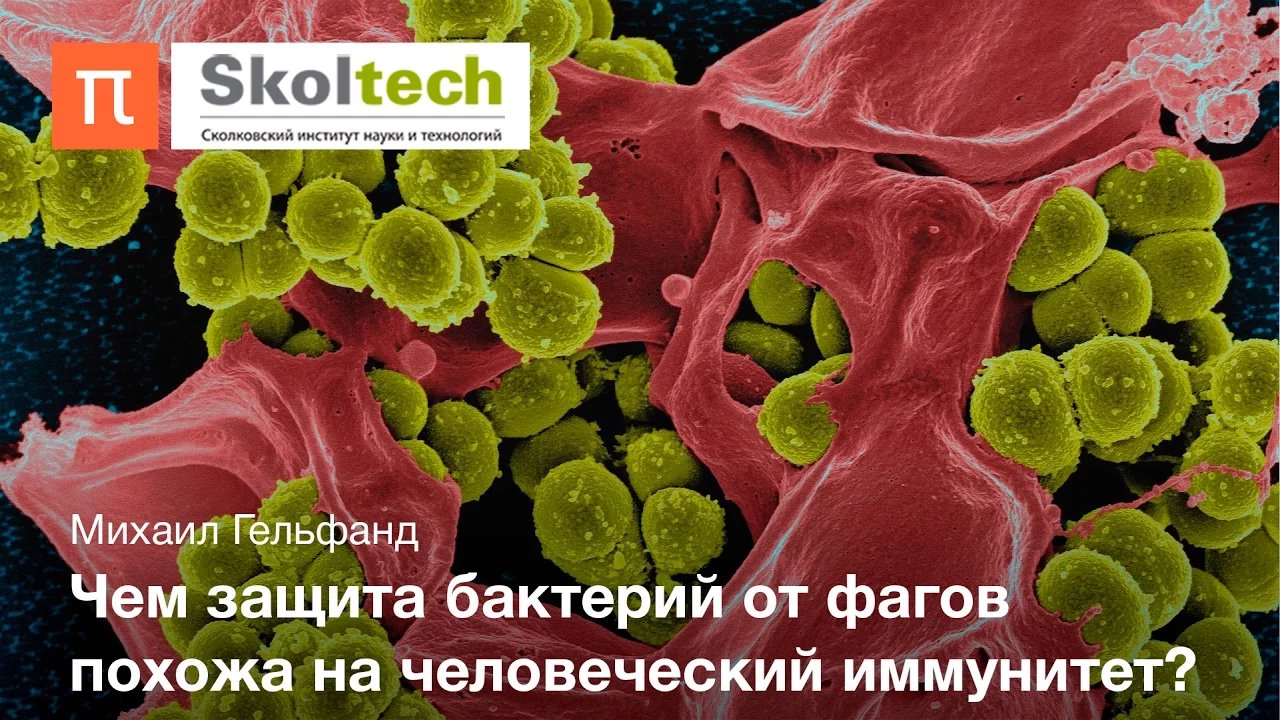
Войны бактерий и фагов — Михаил Гельфанд
19K
Bacteria have evolved multiple sophisticated defense mechanisms against bacteriophage infection, including restriction-modification systems that distinguish and destroy unmodified viral DNA, surface receptor modification to prevent phage recognition, toxin-antitoxin systems that trigger altruistic cell suicide to protect the colony, and CRISPR-Cas adaptive immune systems that store phage DNA fragments for future recognition and destruction. These mechanisms represent an ongoing evolutionary arms race between bacteria and phages, with applications in modern genetic engineering.
Click to watch →

Гонка вооружений. Разнообразие бактериальных систем противовирусного иммунитета. Артём Исаев
1K
Bacteria employ multiple defense strategies against phage infection. Prevention of adsorption includes receptor modification, biofilm formation, capsule secretion, and phase variation. Upon successful infection, bacteria activate restriction-modification systems that cleave foreign DNA, CRISPR-Cas systems that provide adaptive immunity, and abortive immune systems that kill infected cells to prevent phage propagation. These systems work at different stages of infection and provide layered protection. The restriction-modification system is the most common, while CRISPR-Cas provides adaptive immunity that can be inherited by daughter cells.
Click to watch →
IBSG Journal Club 2023 | Tháng Sáu | Tập 27 | Tương tác giữa vi khuẩn, phage, và vật chủ
339
Bacteria have evolved multiple defense mechanisms against phage infection. First, bacteria can modify or reduce surface receptors that phages use for attachment, preventing phage binding. Second, bacteria form biofilms that physically block phage access to surface receptors. Third, bacteria employ restriction-modification systems that recognize and cut foreign DNA while protecting their own genome. Fourth, CRISPR-Cas systems provide adaptive immunity by encoding proteins that recognize and cleave foreign genetic material. These defense mechanisms collectively protect bacteria from phage predation and maintain bacterial population stability.
Click to watch →

RNA Collaborative Seminar: Helmholtz Institute for RNA (HIRI) and Shanghai RNA Club
426
Bacteria have evolved diverse defense systems to protect against viral infections (phages). Restriction-modification systems recognize and cleave foreign DNA, while CRISPR-Cas systems use non-coding RNAs to guide Cas proteins to invading nucleic acids. Over 250 defense systems have been discovered, many with evolutionary origins in human innate immunity. The bacterial ISG15-like system, encoded by a four-gene operon, conjugates a ubiquitin-like protein to phage tail fiber proteins, generating tailless or receptor-binding-defective phages that cannot infect new bacteria. This system is analogous to the human ISG15 innate immune system, demonstrating evolutionary conservation of ubiquitin-like protein conjugation for antiviral defense.
Click to watch →

Rotem Sorek talk at the 22nd HFSP Awardees Meeting
764
Bacteria face constant viral infection pressure from bacteriophages, which are the most abundant entities on Earth (10 times more than bacteria). Bacteria have evolved two main immune arms: CRISPR-Cas provides adaptive immunity by memorizing past infections through stored viral DNA fragments that block future infections; restriction-modification systems provide innate immunity using restriction enzymes that cleave foreign DNA while modification enzymes protect bacterial DNA. These systems represent fundamental strategies that bacteria use to survive in environments teeming with viruses.
Click to watch →
Deep Dive
Genetic engineering of bacteriophages (synthetic biology) to enhance their host range, stability, and efficacy in medical treatments.

【医療者向け・音声のみ】薬剤耐性(AMR)危機を打破せよ:ファージ療法の驚異的な進化と未来~発見の歴史から合成生物学、One Healthへの応用まで Cell
129
To overcome limitations of natural phages, researchers use genetic engineering and synthetic biology. Genome editing tools like CRISPR-Cas systems allow precise modifications to remove harmful genes, enhance killing ability, or convert lysogenic phages to lytic cycles. Synthetic phage genomics involves designing genomes computationally and synthesizing DNA fragments to create entirely artificial phages, free from natural constraints. This enables creation of optimized therapeutic phages with tailored safety and efficacy profiles. Synthetic genomes are expressed using specialized bacterial systems or cell-free translation systems to produce functional phage particles for specific therapeutic applications.
Click to watch →

Episode 45 – Bacteriophages for the treatment of antimicrobial-resistant (AMR) bacterial infections
209
Phage therapy demonstrates favorable safety profiles with properly purified preparations, enabling compassionate use for patients with no alternatives. This safety margin facilitates clinical experience accumulation while working toward formal trials. Tuberculosis represents a promising test case because M. tuberculosis isolates show less genetic variation than related species, potentially allowing standardized phage cocktails. Looking forward, the field aims to move beyond natural phages toward synthetic biology—designing phage-like nanomachines with known beneficial genes, optimized host ranges, and enhanced killing capabilities. Current phages contain ~70% genes with unknown functions, representing potential safety concerns. Combining synthetic biology tools with genetic determinant understanding promises next-generation antimicrobials based on phage principles, representing a paradigm shift from natural products to engineered solutions.
Click to watch →

Obiectul Care Poata Salva Intreaga Omenire!
11.7K
The development of genetic engineering has revolutionized bacteriophage therapy. Pharmaceutical companies like Biosciences have created specialized bacteriophages that exploit bacterial CRISPR immune systems to destroy bacteria. Another approach involves removing genetic interactions that allow bacteriophage multiplication and programming them to destroy bacterial DNA. In the United States, genetically modified viruses have been engineered to cause bacterial self-destruction. A compelling case study involves 15-year-old Isabel Cornel, who suffered from antibiotic-resistant cystic fibrosis. After a lung transplant, her infection intensified. Scientists at the University of Pittsburgh, with one of the world's largest bacteriophage collections, prepared a custom cocktail. After six months of therapy, her infection disappeared without side effects, allowing her to live a normal life.
Click to watch →

Design@Large Series: Bacteriophage Therapeutics for Multidrug Resistant Bacterial Infections
138
Academic institutions are establishing dedicated phage therapy centers to provide treatment for patients with no other options and to advance research. These programs maintain networks of collaborating laboratories specializing in different bacterial species. Modern molecular biology enables engineering of bacteriophages with enhanced therapeutic properties, including modification of phage genomes to broaden host range or improve killing efficiency. For mycobacterial infections, researchers converted lysogenic phages (which integrate into bacterial DNA without killing) into lytic phages by removing repressor and integrase genes. This engineered approach successfully treated a cystic fibrosis patient with multidrug-resistant Mycobacterium abscessus.
Click to watch →

شرح محاضرة ٣ (جزء تاني) الهندسة الوراثية + العلاج الجيني (وراثة عام) | زراعة القاهرة 2026
400
Bacteriophages (bacterial viruses) can serve as vectors for genetic engineering. The lambda phage has a linear DNA molecule with 12-base-pair cohesive ends that allow circularization inside host cells. The central region of the phage genome can be replaced with foreign DNA (10-20 kilobases) during cloning. Phages are particularly useful for cloning larger DNA fragments than plasmids and can infect bacterial cells to deliver genetic material.
Click to watch →
Deep Dive
The concept of Phage-Antibiotic Synergy (PAS) and how combining phages with sub-lethal antibiotic doses can prevent resistance.
CF Live | Discovering more about bacteriophage
158
Combining phages with antibiotics may be more effective than using either alone: (1) When bacteria develop resistance to one treatment, they often experience a fitness cost that makes them susceptible to the other; (2) Phages can sensitize bacteria to antibiotics by breaking down biofilms or altering bacterial physiology; (3) This complementary approach leverages the evolutionary arms race between pathogens and treatments; (4) The strategy recognizes that neither treatment is perfect but together they can achieve better outcomes than either alone.
Click to watch →

Lecture 4: Quantitative viral dynamics
103
Combination therapy using antibiotics and phage exploits complementary mechanisms to prevent resistance evolution. Some bacteria possess antibiotic efflux pumps that expel drugs but allow phage entry. By applying both antibiotics and phage simultaneously, bacteria face dual pressure: those retaining efflux pumps are killed by phage, while those losing efflux pumps become susceptible to antibiotics. This strategy prevents selection for single-resistance mutants and allows using sub-inhibitory antibiotic concentrations, reducing the likelihood of generating antibiotic-resistant strains during treatment.
Click to watch →

World Phage Week Webinar 2025 Day 2
136
Bacteria evolve phage resistance by modifying surface receptors, which causes side effects including reduced antibiotic susceptibility. This creates an evolutionary trap where phage-antibiotic combinations can reduce resistance evolution. The mechanism depends on antibiotic type—chloramphenicol specifically selects against rough LPS mutants while gentamicin does not. Understanding these mechanisms enables design of evolutionarily informed combination therapies.
Click to watch →
TWiV 1121: SARS-CoV2 still didn't come from a lab
20K
Phage Parid dramatically enhances carbapenem antibiotic effectiveness against dormant Pseudomonas aeruginosa. When dormant cells are treated with both phage and antibiotic together, viable cell counts decrease much more than with either treatment alone. This synergy occurs because phage kills some cells, releasing nutrients and signals that reactivate neighboring dormant cells, making them susceptible to antibiotic killing. In mouse models of chronic infection, the combination reduced bacterial load by nearly 100-fold compared to either treatment alone. This approach could treat antibiotic-tolerant infections like those in cystic fibrosis patients or burn wounds.
Click to watch →
Why Infections Are Getting More Dangerous | Virus VS Bacteria | Spark
143.2K
At the burn victims' hospital in Brussels, phage therapy is used in combination with antibiotics. The phage cocktail weakens the bacterium to the point where the antibiotic can gain impact and overcome resistance. This synergistic approach demonstrates that phage therapy may be most effective when combined with existing treatments rather than used as a standalone solution. The combination approach addresses both the immediate infection and the underlying resistance mechanisms.
Click to watch →
Phage Discovery
Killer Looks
Prey Selectivity
Attachment Phase
Injection Action
DNA Delivery
Phage Discovery
0:00- 1
Félix d'Hérrelle identified bacteriophages over a century ago.
- 2
Phages are viruses that specifically target and destroy bacteria.
- 3
They are named for their bacteria-eating capability.
Limitations of Phage Therapy: Resistance, Specificity, and Delivery Barriers
While the nanoscale mechanism of bacteriophage T4 highlights the therapeutic potential of phages, significant hurdles challenge their viability as a universal solution to superbugs. A primary limitation is the rapid evolution of bacterial resistance. Bacteria can quickly mutate surface receptors—such as OmpC or lipopolysaccharides targeted by T4—rendering the phages ineffective. Additionally, phages exhibit extreme host specificity; a treatment tailored for one strain of E. coli may have no effect on another closely related strain, necessitating complex 'phage cocktails' and rapid, precise diagnostics. From a clinical perspective, the human immune system often recognizes phages as foreign proteins, generating neutralizing antibodies that clear them before they can reach the target infection. Finally, the pharmacokinetics of administering live biological viruses are highly unpredictable compared to traditional small-molecule antibiotics. These factors suggest that phage therapy faces major evolutionary, biological, and translational barriers that may limit its widespread clinical efficacy.
Basic anatomy of bacteria and viruses, specifically the structural differences between Gram-negative bacteria (like E. coli) and bacteriophages.
The fundamental stages of the viral lytic cycle, including attachment, penetration, biosynthesis, maturation, and lysis.
Key molecular biology processes, particularly how host cell machinery is hijacked for viral protein synthesis and genome replication.
An understanding of antibiotic resistance and how 'superbugs' evolve to survive traditional antimicrobial treatments.
The clinical applications, history, and current regulatory challenges of Phage Therapy as an alternative to antibiotics.
Bacterial defense mechanisms against phage attack, such as the CRISPR-Cas system and restriction-modification systems.
Genetic engineering of bacteriophages (synthetic biology) to enhance their host range, stability, and efficacy in medical treatments.
The concept of Phage-Antibiotic Synergy (PAS) and how combining phages with sub-lethal antibiotic doses can prevent resistance.
2.9M views42.5Klikes1:02@biolutionOriginal Release: 2019-05-21
More than 100 years ago the French scientist Félix d´Hérrelle identified bacteriophages.
They look like spaceships from another world but are most fearsome killing machines.
Fortunately they leave us humans alone and target exclusively bacteria.
Phages stick to their prey with leg-like fibers, which trigger their shaft to ram a needle into the bacterial hull. This punctures the envelope and injects the genetic material contained within the head, an icosahedral capsid made of proteins
Up Next
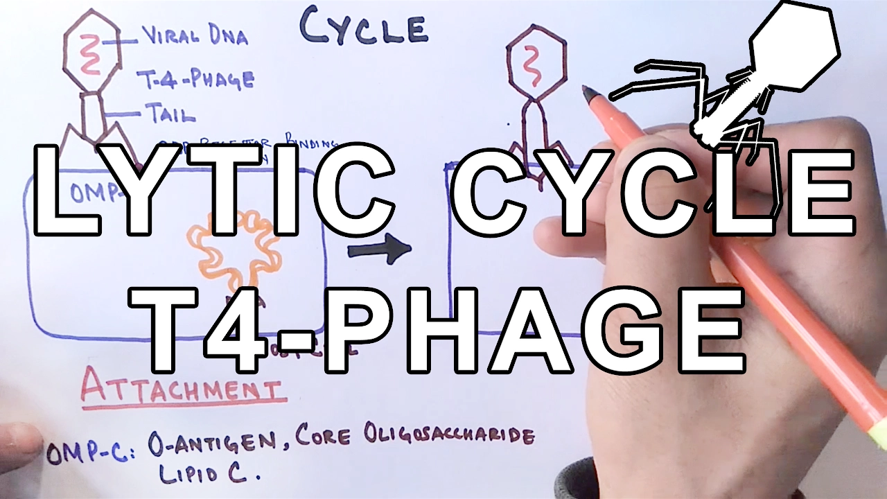
6:17
Lytic Cycle of Bacteriophages: Viral Replication Steps Explained
@hussainbiology
120.6K views•2017-02-20

50:12
Bacterial Communication: Quorum Sensing and Biofilm Formation Explained
@JHUAAP
12.6K views•2011-06-28

5:35
Enteric Nervous System Explained: The Gut's Brain | Neurobiology Lecture
@alumniu6029
438 views•2018-09-12

7:08
Bacteriophages: Earth's Deadliest Killers and Future Antibiotics
@kurzgesagt
34.6M views•2018-05-13
Related Study Plans & Knowledge Roadmaps
Structured learning paths in Biology
biology
RNAi: Biogenesis, RISC & Gene Silencing
01Molecular Biology Foundations: DNA to Protein
02Introduction to RNA Interference (RNAi)
03Molecular Biogenesis of miRNA and siRNA
+ 2 more learning modules
10h5 modules
Explore Roadmap
biology
Mycorrhizae: Signaling, Biophysics & Ecology
01Foundations of Mycorrhizal Symbiosis
02Molecular Signaling and Root Colonization
03Biophysics and Bioenergetics of Nutrient Exchange
+ 1 more learning modules
11h4 modules
Explore Roadmap
biology
Circadian Clocks: TTFL, SCN & Periphery
01Foundations of Molecular Biology & Gene Expression
02Introduction to Chronobiology and Actograms
03The Core TTFL: Molecular Architecture of the Clock
+ 3 more learning modules
14h6 modules
Explore Roadmap
biology
Population Genetics: Math, Drift & Selection
01Genetic & Probability Foundations
02The Hardy-Weinberg Principle and Baseline Models
03Mathematical Modeling of Natural Selection
+ 3 more learning modules
18h6 modules
Explore Roadmap