











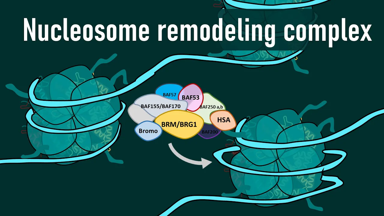
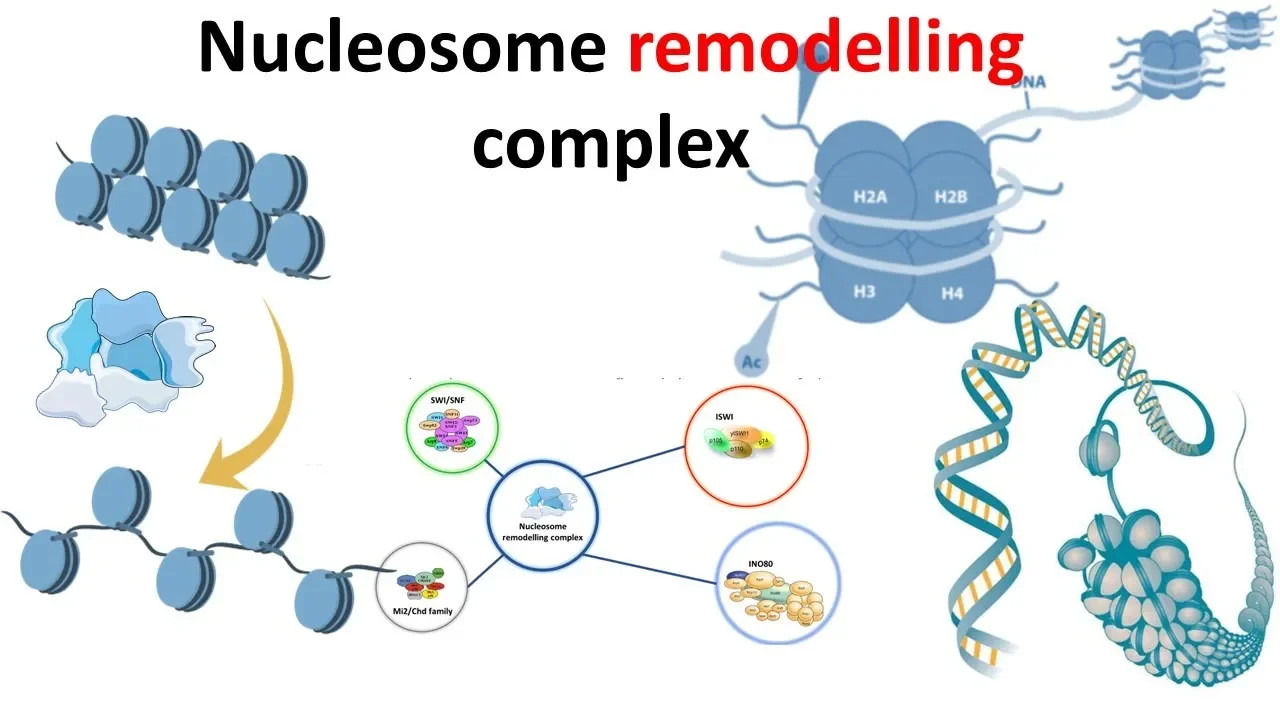
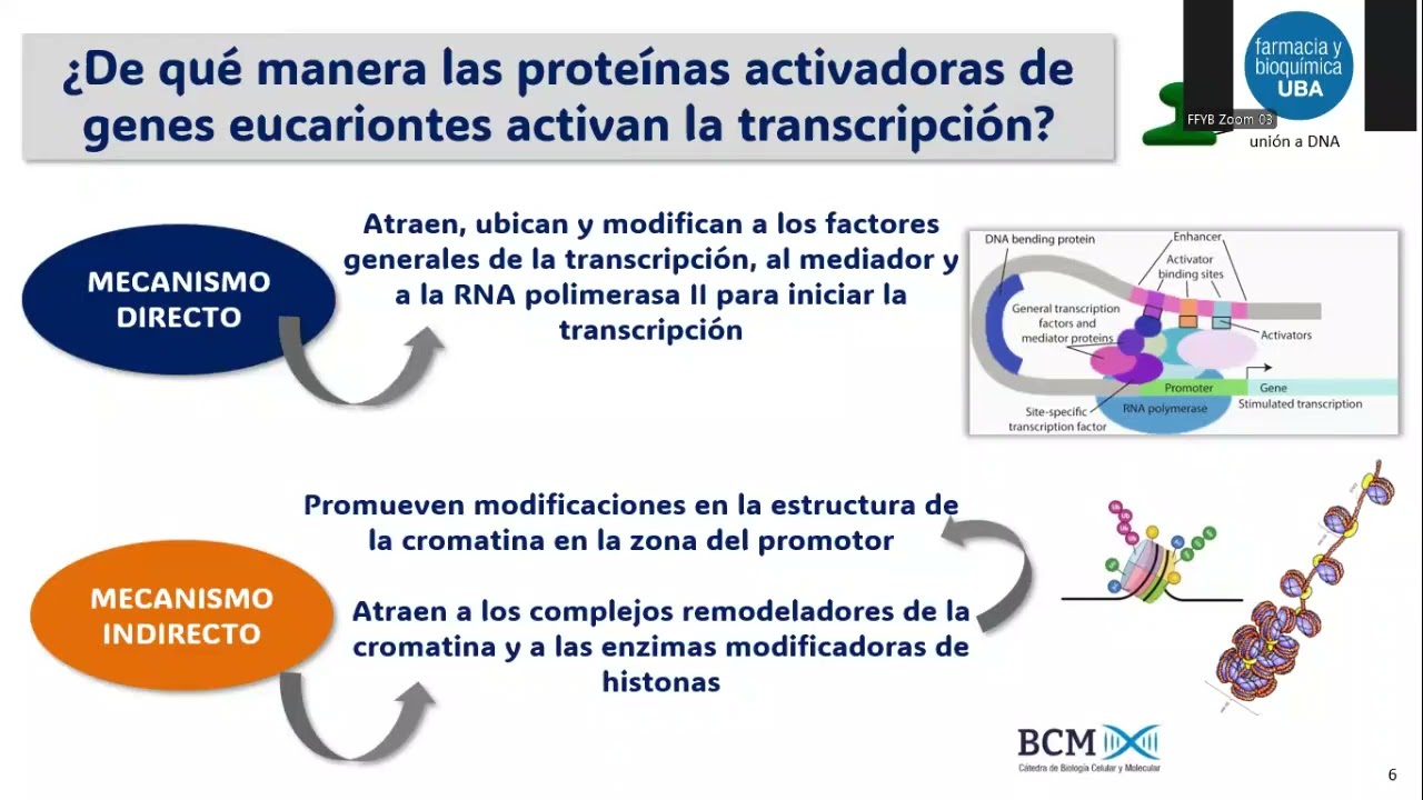





The RSC chromatin remodeling complex relieves nucleosome-mediated gene repression by sliding or removing histone octamers from DNA, with its ATP-dependent activity being stimulated by AT-rich DNA sequences found in nucleosome-free regions (NFRs) at gene promoters, thereby exposing regulatory and transcription start sites for RNA polymerase II transcription.
Nucleosome Dynamics in Chromatin Remodeling: A Yale Lecture
Added:Understanding of eukaryotic DNA packaging, specifically the structural organization of chromatin, histone proteins, and the nucleosome octamer core.
The fundamental mechanism of transcription in eukaryotes, including the roles of RNA Polymerase II, promoters, and general transcription factors.
The concept of gene regulation and how access to genetic information determines cellular function and phenotype.
Basic thermodynamics of DNA-protein interactions, particularly how electrostatic interactions stabilize the DNA-histone complex.
In-depth study of specific ATP-dependent chromatin remodeling families (e.g., SWI/SNF, ISWI, CHD, and INO80) and their distinct mechanisms of action.
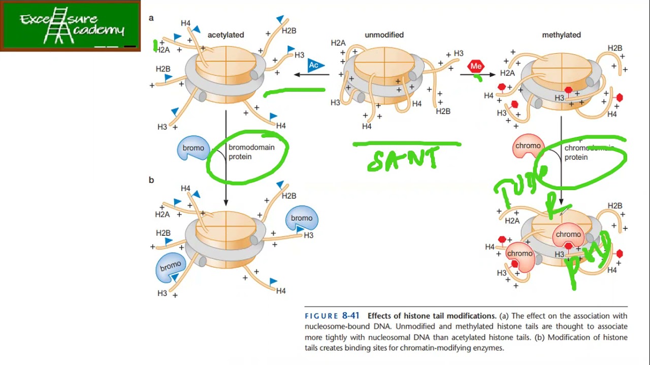
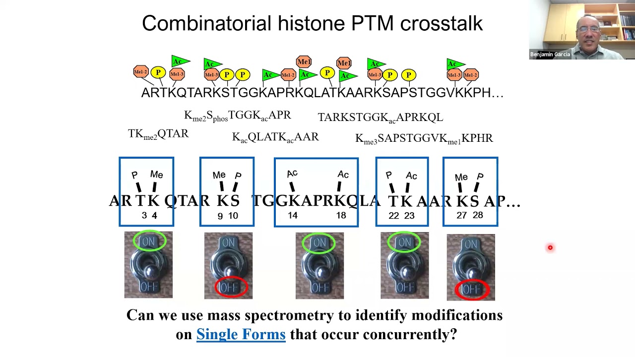
The synergistic relationship between chromatin remodeling and histone post-translational modifications (the 'histone code' hypothesis, including acetylation and methylation).
How pioneer transcription factors initially access condensed heterochromatin to facilitate subsequent remodeling and gene activation.
Clinical applications and pathologies, such as how mutations in chromatin remodeling complexes contribute to oncogenesis, developmental disorders, and potential epigenetic therapies.
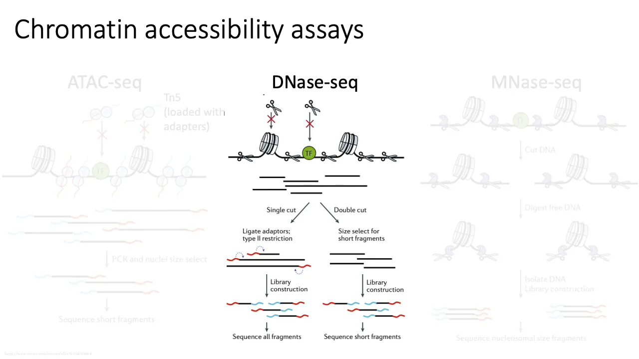
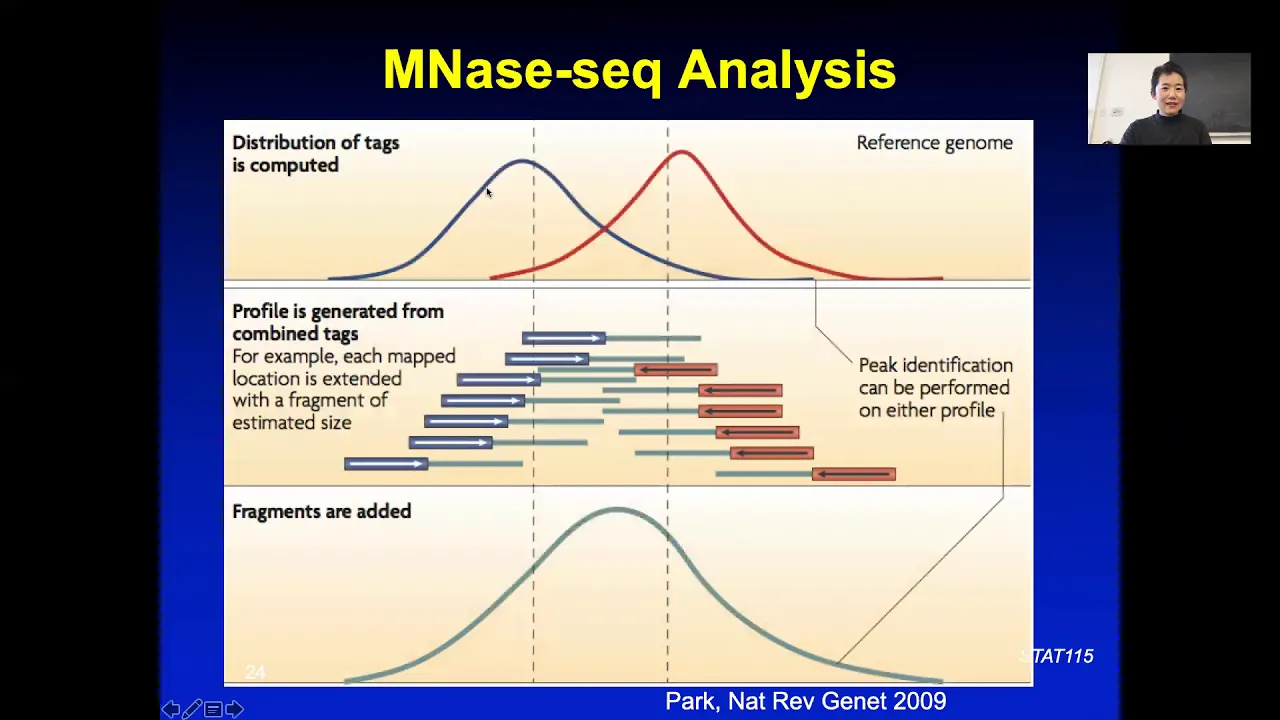
Modern genomic techniques used to map nucleosome positioning and chromatin accessibility, such as ATAC-seq, MNase-seq, and ChIP-seq.
3.5K views41likes35:15Original Release: 2015-05-27
my congratulations to banked and thank you to the organizers for the opportunity of speaking about my work today as the audience is so diverse i will start with basic information about chromosome structure that you need to know in order to understand my lecture the nucleosome is the basic unit of dna coiling in chromosomes the structure may be described as beads on a string in which nucleosomes alternate with linker dna the existence and main features of the nucleosome were proposed by roger kornberg in 1974. the nuclear zone contains a set of eight histone proteins two each of four types termed h2a h2b h3 and h4 147 base pairs of dna are wrapped in 1.6 turns around the octamer the eight histone proteins are arranged as shown here with the h3h4 tetramer in the center of the particle and the h2a h-2b dimers towards the periphery where dna enters and exits the particle double-stranded dna is rigid so bending around the particle with a diameter of only about 100 angstroms imparts considerable strain the energy required for bending is supplied by interactions of negatively charged phosphates in dna with positively charged amino acids of the histones this picture was confirmed by x-ray crystal structure determination by richmond and co-workers in 1997.
here you see two views of the crystal structure on the left is a view from the top of the particle with 1.6 turns of the dna superimposed and on the right you see a view from the side of the particle splitting the particle into between the dna turns and viewing from the top you see one turn of the dna around four histones of the octamer dna enters the particle from the top left and completes one turn at the midpoint of the particle at the top right the dna is bound near the end by an h2a h2b dimer and in the middle of the particle by the first two h3 and h4 molecules of the tetramer interactions of the dna with the histones occur at every point where the dna strands contact the histone surface indicated here by the yellow stars these interactions include charge charge interactions i mentioned before all nucleosomes in all organisms share this basic structure the histone proteins are among the most conserved through evolution for example the amino acid sequence of h4 differs by only 2 out of 102 amino acids between plants and animals the structure of the nucleosome is universal the nucleosome was long regarded as a packaging device important for the compaction of dna in the cell for example human dna more than one meter in length is compacted more than a hundred thousand fold during cell division about 30 years ago we asked whether the nucleosome also performs a functional role we investigated the impact of the nuclear zone upon gene repression expression as most of you know the rna polymerase ii is responsible for all transcription of a messenger rna that called for proteins two other polymerases perform more limited roles transcribing mostly genes for the rna molecules involved in protein synthesis rna polymerase 1 transcribes genes for the rna components of the ribosome rna polymerase 3 transcribes genes for the trna molecules that bring amino acids to the ribosomes and some small untranslated rnas as the first step in the pathway of gene expression rna polymerase transcription is a focal point of regulation we found that the nucleosome performs a critical role in the regulation of rna polymerase ii transcription we assemble the nucleosome on a promoter consisting of a tata sequence and transcription start site the nucleosome prevented the initiation of transcription by rna polymerase ii this was not a general inhibitory effect of the histones upon rna polymerase 2 because a nucleosome downstream of the start site was no impediment to transcription rna polymerase could read right through the nucleosome transcription through a nucleosome displays the histone optima from the dna to summarize the nucleosome is a general gene repressor nucleosomes prevent the initiation of transcription but allow transcript elongation with the displacement of histones soon after this work was published hannah and groonstein showed that nucleosomes interfere with the initiation of transcription in yeast in vivo they depleted nucleosomes by turning off the expression of the histone h4 gene depletion of nucleosomes turned on transcription of a repressed gene in particular the 4-5 gene which i will return to later in the lecture so the nucleosome represses transcription in vitro and in vivo this leads to the question of how is repression by the nucleosome relieved for transcription the answer comes from large assemblies of proteins called chromatin remodeling complexes the first chromatin remodeling complex referred to as switch sniff was discovered by ira hershkovitz by genetic studies in east when we isolated the complex we noticed that one subunit had a homolog in yeast we pursued the homolog and discovered a 1 million molecular weight complex made up of 17 subunits that we termed risk for remodels the structure of chromatin six proteins are homologous or identical between switch sniff and risk and the corresponding complexes in humans the human remodeling proteins are mutated in 20 percent of all cancers second only to p53 which is mutated in about 50 percent of all cancers switch sniff and risk different both importance and abundance whereas switch and sniff genes can be deleted from yeast risk genes are essential for cell viability there are only about 100 molecules of the switch sniff complex per cell but thousands of risk complexes for the rest of the time i will speak about the essential abundant risk complex among the functions of risk subunits listed here i call your attention especially to the catalytic activity of sth-1 the largest subunit of the complex sth1 catalyzes the movement of dna in an atp dependent manner we found that risk has two atp dependent chromatin remodeling activities first risk can slide a histone optimal along dna in the experiment shown here a nucleosome was formed in the middle of a long dna molecule and treated with risk and atp analysis was performed by gel electrophoresis you see the nucleosome moved to the end of the dna during the reaction it therefore migrated faster in the gel forming the lower band after the reaction second this can remove a histone optimal from dna this chromatin remodeling activity requires both atp energy and the histone acceptor which may be another dna molecule or in the experiment shown here a protein that binds histones termed a histone chaperone products of the reaction are again separated by migration in a gym the histone optima is removed piecemeal in the first few minutes the h2a h2b dimers are removed leaving a complex of the h3h4 tetramer and dna finally the tetramer is also removed resulting in naked dna chromatin remodeling by optimal sliding or removal can expose protein binding sites in the dna these may be sites for binding rna polymerase or sites for proteins that regulate rna polymerase sliding of the octamer exposes sites in the linker region between occlosomes while removal of the octamer exposes the entire length of the nucleosomal dna both sliding and removal involve dna translocation the largest subunit of risk draws dna into the nucleosome from one side and expels it from the other dna associated with the nucleosome indicated in green is propelled around the particle and passed into the adjacent linker region the problem is the dna cannot easily slide across the histone surface due to the many interactions with histones symbolized by the yellow stars that i mentioned before sliding would require breaking all the dna histone contacts simultaneously an idea called twist diffusion has been proposed to solve the problem the idea is that the translocase draws a single dna base pair into the nucleosome without altering the rest of the structure this movement involves a twist of 36 degrees one tenth of the total for a full 10 base per turn of the dna double helix the resulting strain is proposed to propagate through the nucleosome carrying one base pair to the opposite side the idea could be tested by the introduction of nics into the nucleosomal dna because nyx would prevent the propagation of twisting strain we found that nyx did not interfere with resectivity so the idea of twist diffusion was eliminated we performed structural and biochemical studies that revealed an altogether different solution to the problem of dna sliding through the nucleosome in collaboration with francisco asturias we obtained cryoam structures of risk alone and a risk nucleosome complex here you see in yellow an electron density map of risk alone at about 25 angstrom resolution notice the large central cavity a difference electron density map between the ris nucleosome complex and risk alone is shown in blue mesh there is little change in wrist structure difference density is almost entirely in the central cavity rotating almost 90 degrees and removing wrist density for an unobstructed view of the density and the central cavity there are three points of contact with risk the histone optima from the x-ray crystal structure of the nucleosome can be docked into the density in the central cavity as before h2a and h2b are red and yellow and h3 and h4 are green and blue here we remove wrist density from both sides viewing only a slab in the middle indicated by the dashed box we rotate again through almost 90 degrees to view from the top down in the lower panel the x-ray structure of the octamer is shown superimposed on the difference density in blue mesh one h2a h2b dimer had to be removed to fit the difference density and the nucleosomal dna was mostly undetectable the dashed line indicates a putative path for dna detached from the surface of the histone optima detachment of dna from the histone surface could also be detected by cleavage of the dna with the enzyme dna s1 here the dna was radio-labeled at one end symbolized by the pink star the fragments resulting from cleavage by dnase were separated on the basis of size by electrophoresis the column labeled dna is the pattern produced by digestion of naked dna cleavage occurs more or less at random with some cleavage sites favored due to the preference of the enzyme for digesting some dna sequences the column labeled nucleosome is the pattern produced by digestion of the dna in the nucleosome the fragments are multiples of 10 bases a straightforward explanation is shown in the diagram each strand of dna is maximally exposed to cleavage by the enzyme with the 10 base pair periodicity of the dna double helix between cleavage sites the dna is protected by the histone octoma binding of risk to the nucleosome enables attack by dnase at points indicated by the red arrows halfway in between those seen with the nucleosome alone indicated by the blue arrows we interpret this picture as a superposition of two patterns one of cleavage with a protection by the histone and the other of cleavage with protection by risk the dna swings from the optimal surface to the wrist surface within the risk cavity this is a plausible idea for two reasons first we know that the risk cavity is highly positively charged because we show that risk binds a naked dna molecule with the same affinity as a nucleosome second we know from other studies that nucleosomal dna is in rapid equilibrium between fully bound and partially unraveled states dna is readily released from the nucleosome due to the strain of wrapping on the small radius of curvature of the octoma interaction with risk provides an alternative surface for interaction of the dna with less bending strain i have also suggested that risk may cause a slight alteration of histone optimal conformation for example contraction by as little as one angstrom which could help release dna from the surface of the octoma the detachment of dna from the surface of the nucleosome in the wrist cavity solves the dna sliding problem whereas positively charged residues on the histone optimal surface are in register with the dna phosphates to form the strong interactions needed for bending the dna we presume the positively charged surface of the wrist cavity allows interaction in any register and therefore enables sliding dna drawn in from one side by the sth-110 slow case using the energy of atp hydrolysis is free to slide to the opposite side no atp energy is required only risk bind energy of course if we add atp then the dna exposed by translocation is cleaved like dnase by dnase like a naked dna dna molecule as shown on the right i have also told you that risk possesses two atp dependent remodeling activities the sliding and removal of nucleosomes i have also told you i have also explained that risk possesses an atp independent activity forming a risk nucleosome complex in which dna is released from the surface of the histone optima i will now turn to the question of how these activities relieve repression by the nucleosome in yeast cells in vivo here you see an example of results obtained by many workers showing the likelihood of occurrence in a nucleosome as a function of distance along a hundred thousand base per region of yeast chromosomal dna about one percent of the yeast genome below are indicated the locations of rna transcripts in black and genes in blue in the region shown a similar picture would be seen for almost any region of any yeast or for that matter mammalian chromosome it is a picture of more or less uniform coverage by nucleosomes punctuated by occasional absences zooming in on the beginning of one rna transcript you will see the virtual absence of a nucleosome in front of the gene a so-called nucleosome-free region or nfr such nfrs are found in front of all genes active in transcription dna sequences that must be recognized and bound by gene activator proteins are exposed in the nfr the tata sequence or in many cases at a tata-like sequence important for the initiation of transcription is also exposed in the nfr transcription start sites however are generally covered by the first or plus-one noclosome how is the virtual exclusion of a nucleosome from the nfr achieved it is widely believed that short stretches of at rich dna in the nfr results in the destabilization and thereby the exclusion of nucleosomes it is well known that long lengths of the adt dna are especially rigid and unable to wrap around the octamer we have tested whether short stretches of the adt dna as commonly occur in nfrs can be responsible for the exclusion of the nucleosomes we inserted stretches of the adt dna of 7 10 and 14 base pairs at various positions from the end of the dna up to the diode at the center of the nucleosome we found at most a two-fold difference in affinity for the histone octamer relative to one of the highest affinity dna sequences known clearly destabilization by short stretches of the adt dna cannot account for the formation of nfrs what then is responsible the answer is risk on the left you see wrist protects at the adt element within a nucleosome from dna's digestion on the right you see a semi-log plot showing the rate of removal of a nucleosome by risk from a natural dna without the adt element in red and the same dna in which we have inserted a seven residue the adt element in blue insertion results in recognition by risk and removal of the nuclear zone this discovery of a stimulatory effect of the adt elements explained the surprising finding we made 25 years ago we found that regulatory sequences that enhance transcription by rna polymerase 1 also stimulate transcription by rna polymerase 2.
this was unexpected because the control of the two rna polymerases was believed to be very different we observed that the rna polymerase 1 enhancer contains binding sites for proteins indicated here by blue boxes and also at the adt element we placed each sequence separately in front of a gene transcribed by rna polymerase ii and found an enormous stimulatory effect of the dadt element we now know the reason that the adt element stimulated the removal of a promoter nucleosome by risk genetic studies in yeast have recently shown that risk is involved in transcription of all rna polymerases 1 2 and 3. finally we made measurements of both affinity for the histone optima and rate of removal of a nucleosome by risk for the ilv1 gene in yeast we assembled nucleosomes on a series of dna segments dna of the nfr and dna of each of the first three nucleosomes of the gene as shown in blue there was virtually no difference in affinity of nfr and gene dna for the histone optima all dna segments bound dna almost as well as a high affinity histone dna binding by contrast as shown in red risk removed a nucleosome for nfr dna at least 10 times more rapidly than from any nucleosome containing gene dna now the experiment was performed on individual nucleosomes assembled in the lab what what what might we find for actual yeast chromosomes assembled in these cells in vivo to this end we isolated a region of a yeast chromosome containing a yeast gene in its native state in the manner shown here we inserted the cognition sites for a recombinized enzyme on both sides of a gene in yeast in this case the fo5 gene which i mentioned before we then induced expression of the recombinized enzyme in the yeast resulting in excision of the gene as a chromatin circle we isolated the 4-5 circle by an affinity technique we purified it 200 000 fold to virtual homogeneity the complete gene circle carried 12 nucleosomes and because it was isolated in the transcriptionally replaced state there was no nfr rather the promoter containing the tata sequence and transcription start site was occupied by three nucleosomes one of these nucleosomes contained the dadt sequence for recognition by risk and also a binding site termed uasp2 for an activator protein a second binding site termed uasp1 for the same activator is always exposed in the adjacent linker region in other experiments we place the recognition site for the recombinase between the promoter and the gene to isolate a promoter circle bearing three nucleosomes or a gene circle bearing nine nucleosomes we applied risk and atp to the chromatin circles and measured the removal of nucleosomes by two different methods topoisomera analysis and limit nucleus digestion two of the twelve nucleosomes on the complete gene plus promoter circle were removed by risk in vitro exactly the same number as are removed upon gene activation in yeast in vivo the nucleosomes removed from the complete circle must have come from the promoter because none were removed from the gene we conclude that risk removes from motor nucleosomes and creates an nfr in an actual yeast chromosome in further work we investigated the possible role of histone modification in the targeting of risk to promoternoclosons vincent alfrey discovered in the 60s that histones are acetylated and methylated and that these modifications correlate with gene activity more recent work has shown that nucleosomes are also extensively modified by phosphorylation ubiquitination and more mostly on the amino terminal tails of the histones protruding from the central core of the particle shown here in various colors all these modifications correlate with structural and functional states of chromatin but it is unknown in most cases how these modifications actually affect chromosome structure and function more than a hundred histone modifications have been described despite the large number of magnifications they occur in relatively few combinations in addition to post translational modifications there are special forms of histones present at a low level some of which are listed here these histone variants are found in nucleosomes in specialized regions of chromosomes such as the beginning of genes regulatory regions centromeres and sites of dna damage returning to the chromatin circles it is known that histones of promoter nucleosomes are acetylated we use the histone diacetylase enzyme to reverse acetylation we found a marked reduction in the removal of promoter nucleosomes by risk at the same time we observed the removal of nucleosomes from the gene region previously unaffected by wrist because of the widespread involvement of risk and transcription and other dna transactions there must be other factors that determine specificity besides the modification of histones the creation of an nfr is only part of the relief of the repression by the nucleosome regulatory and tata sequences are exposed in the nfr but not the transcription start site this point is nicely illustrated by the case of the 4-5 gene i just described upon activation of the gene two nucleosomes are removed indicated by dashed ovals creating an nfr that exposes a regulatory sequence uas p2 a third nucleosome the so-called place one oclosome is not removed but calls to slide exposing the tata sequence but still covering the transcription start site transcription is still repressed recent work suggests that the detachment of dna from the histone optima in the risk cavity that i mentioned before relieves repression by the plus-one nucleosome henikov and co-workers found a partial loss of histone dna interaction in the plus-one nucleosome of active genes in vivo they show that association with risk is responsible for this loss of histone dna interaction in conclusion i told you how we found almost 30 years ago that the nucleosome serves as a general gene repressor it sets a zero baseline for gene expression it performs an essential role in cellular processes such as differentiation and development by preventing transcription of all genes except those activated by a specific regulatory protein ten years later we discovered risk the most abundant and essential chromatin remodeling complex risk relieves repression by the nuclear zone it slides and removes nucleosomes it is stimulated by the adt sequences in front of genes where it creates nfrs exposing regulatory and tata sequences risk also forms a complex with the nucleosome in which dna is released from the histone surface exposing sequences such as transcription start sites of active genes these activities of risk help us understand how repression by the nuclear zone is relieved for transcription risk also plays roles in replication recombination repair and other chromosomal dna transactions doubtless by similar mechanisms i would like to acknowledge the contributions of my colleagues at stanford and at the scripps research institute and thank you all for your attention so thank you and we have time for one or two questions before we move into coffee can i release this so so banked bank's always ready fantastic talk you you have moved the frontiers do you have any comment to and defiles periodic phasing well we only tried using this sequence you know not as a periodic uh insertion but per se walking it into the dna sequence we have never tried to use it as a repetitive sequence we know that you know ats prefer to be at the minor grooves of nucleosomes not when they come in fours when they come in twos and if i should provocative this is a microscopic phasing if you take look out some horizons bigger you have the chromosome banding what's that yeah i mean i'm sure that you know a lot of this will understand much better when we understand higher structures of of chromatin we're at this point just limited to nucleosome and not beyond i'm sure many of these modifications and maybe the 80 rich areas you know have to do with uncovering much larger areas so in 30 years so for a next big celebration bank yes a very big one okay so let's thank the speaker and all the speakers of this session
Up Next
10:07
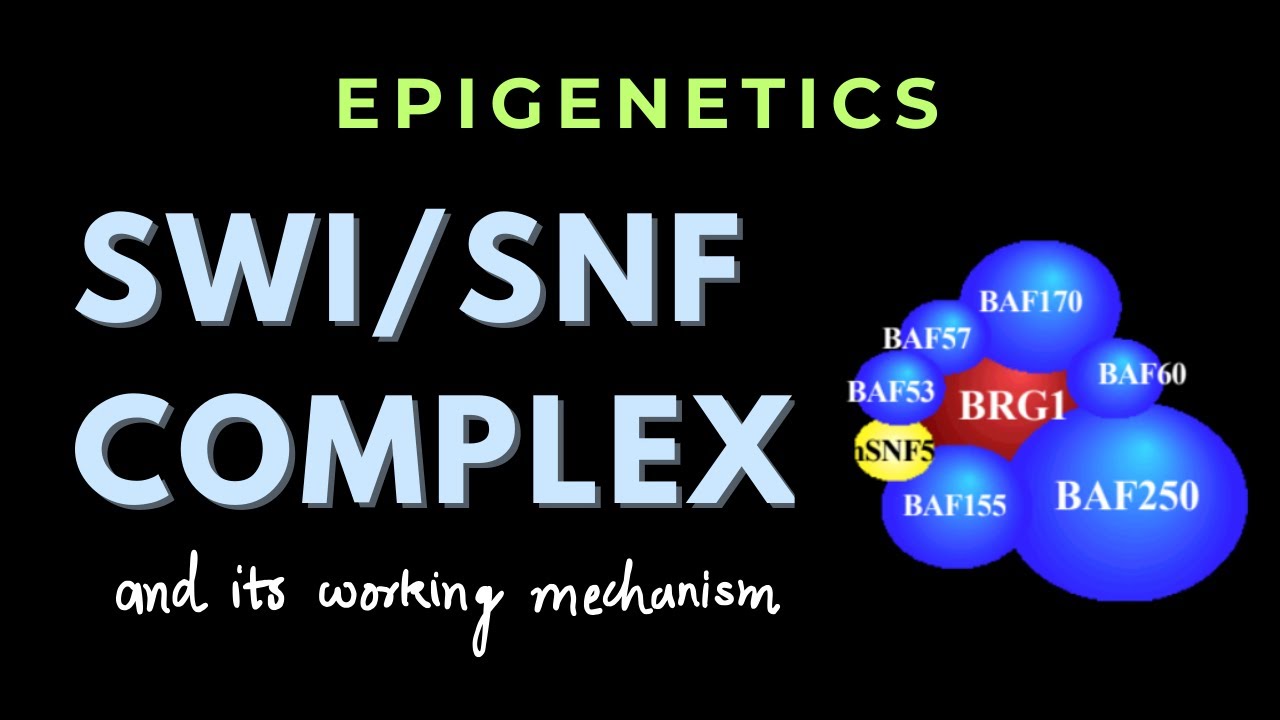
SWI/SNF Chromatin Remodeling Complex: Mechanism & Function
@WindowSeatStudies
592 views•2024-10-25

50:12
Bacterial Communication: Quorum Sensing and Biofilm Formation Explained
@JHUAAP
12.6K views•2011-06-28

5:35
Enteric Nervous System Explained: The Gut's Brain | Neurobiology Lecture
@alumniu6029
438 views•2018-09-12

7:08
Bacteriophages: Earth's Deadliest Killers and Future Antibiotics
@kurzgesagt
34.6M views•2018-05-13
Related Study Plans & Knowledge Roadmaps
Structured learning paths in Biology
biology
RNAi: Biogenesis, RISC & Gene Silencing
01Molecular Biology Foundations: DNA to Protein
02Introduction to RNA Interference (RNAi)
03Molecular Biogenesis of miRNA and siRNA
+ 2 more learning modules
10h5 modules
Explore Roadmap
biology
Mycorrhizae: Signaling, Biophysics & Ecology
01Foundations of Mycorrhizal Symbiosis
02Molecular Signaling and Root Colonization
03Biophysics and Bioenergetics of Nutrient Exchange
+ 1 more learning modules
11h4 modules
Explore Roadmap
biology
Circadian Clocks: TTFL, SCN & Periphery
01Foundations of Molecular Biology & Gene Expression
02Introduction to Chronobiology and Actograms
03The Core TTFL: Molecular Architecture of the Clock
+ 3 more learning modules
14h6 modules
Explore Roadmap
biology
Population Genetics: Math, Drift & Selection
01Genetic & Probability Foundations
02The Hardy-Weinberg Principle and Baseline Models
03Mathematical Modeling of Natural Selection
+ 3 more learning modules
18h6 modules
Explore Roadmap