



















Continuous wave Doppler ultrasound continuously samples returning echoes without receive time, enabling measurement of higher velocities but lacking depth resolution and anatomical context; pulsed wave Doppler uses short pulses with receive time to measure depth and velocity within specific regions, but is limited by the Nyquist limit where maximum detectable velocity equals half the pulse repetition frequency, creating a fundamental trade-off between velocity measurement capability and imaging depth.
Continuous vs Pulsed Wave Doppler Ultrasound: Key Differences Explained
Added:Basic physics of acoustic waves, including frequency, wavelength, and propagation speed in human tissue.
The Doppler Effect physics, specifically how frequency shifts relate to the velocity and direction of moving targets.
Fundamentals of B-mode (2D grayscale) ultrasound imaging and transducer operation.
Basic cardiovascular physiology and hemodynamics, including laminar flow, turbulent flow, and pressure-velocity relationships.
The concept of aliasing and the Nyquist limit in Pulsed Wave Doppler, including methods to resolve it such as adjusting Pulse Repetition Frequency (PRF).
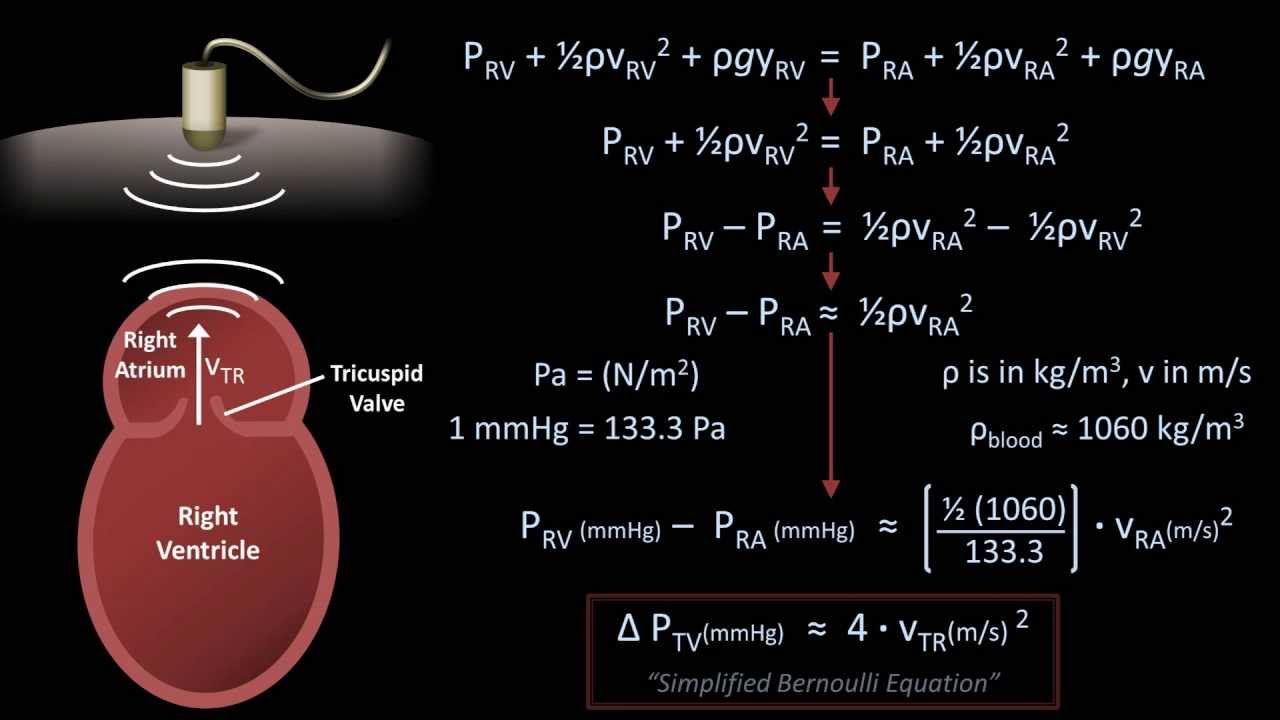
Clinical application of Doppler formulas, such as calculating pressure gradients using the simplified Bernoulli Equation.
Diagnostic protocols for Vascular Duplex ultrasound, including carotid artery stenosis grading and Deep Vein Thrombosis (DVT) evaluation.
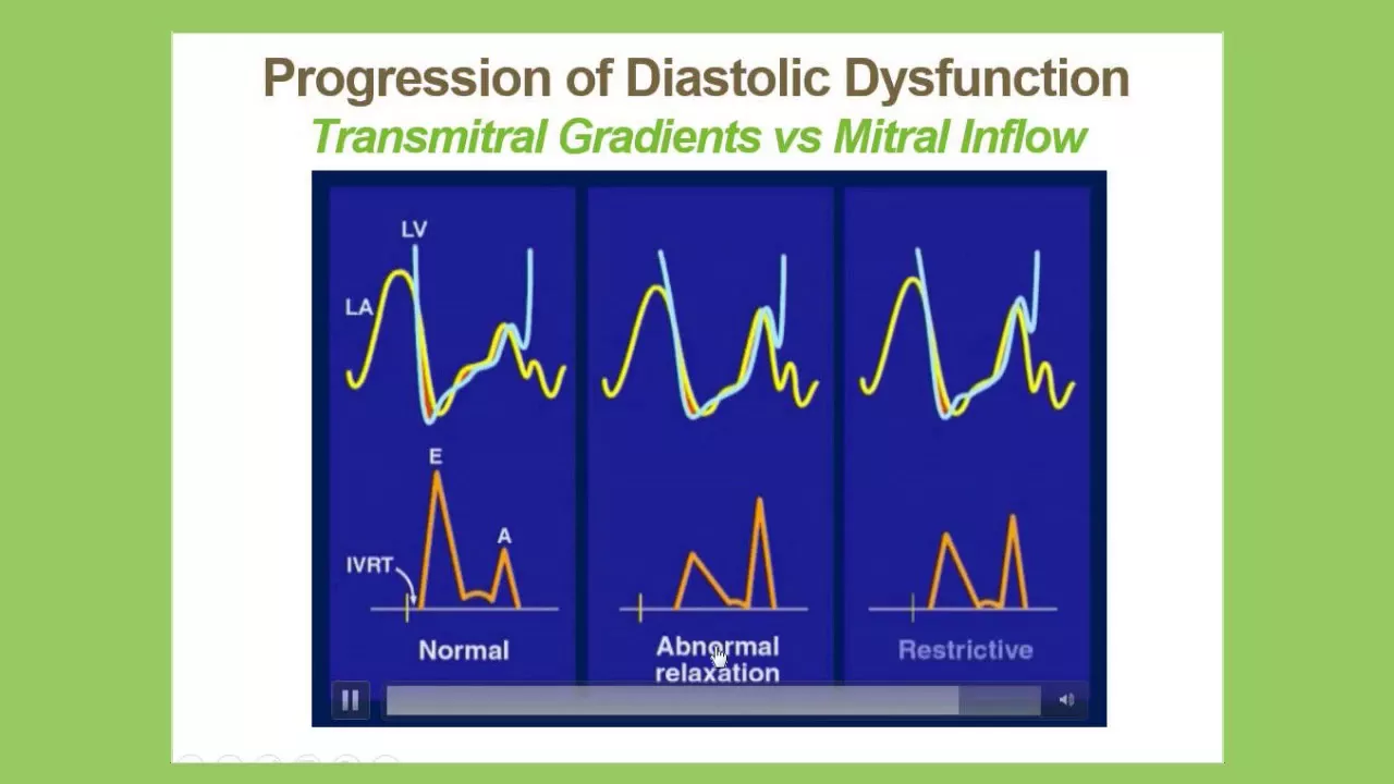
Advanced echocardiography techniques, such as Tissue Doppler Imaging (TDI) to assess myocardial tissue velocity.
60.2K views914likes24:46Original Release: 2023-04-24
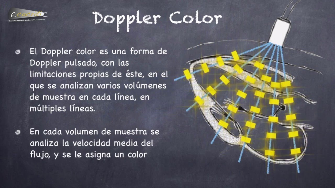
hello and welcome back in today's talk we're going to be looking at the differences between continuous wave Doppler ultrasound and pulse wave Doppler ultrasound imaging now both of these Imaging modalities utilize the underlying principles of Doppler Imaging that we looked at in our previous talk and we saw there that the Doppler shift the shift in frequencies that we measure on our transducer is the difference between the frequency we are receiving within that transducer and the transmitted frequency that we propagated out into the tissues now that Doppler shift can also be calculated using the Doppler equation and the Doppler equation will determine how much Doppler shift we measure on that ultrasound transducer we've seen that the higher the frequency we transmit into the tissues the higher the Doppler shift will be returning back to our transducer also the higher the velocity of the moving object that we're trying to measure the higher the Doppler shift cosine Theta also determines how much Doppler shift we're going to measure on our machine now cosine Theta that value actually increases the smaller that Doppler angle the smaller the Theta angle gets so the smaller our Doppler angle the more Doppler shift we register on our machine now often when we're looking at this equation the unknown variable the thing we're trying to calculate is the velocity of the object moving within our image so we can rearrange this formula to isolate velocity to isolate the unknown value and we can plug in all of these values all of which we will know to calculate the velocity of the object moving within our tissues now way back at the beginning of this course we looked at the principle of dampening and quality Factor and I said that a transducer crystal is much like a symbol on a drum set when you hit that symbol on the drum set it will resonate at a certain frequency and it will resonate for a long period of time now the frequency at which it resonates is due to the diameter of that symbol much like a piezoelectric Crystal within our ultrasound transducer its resonance frequency is determined by the thickness of that piezoelectric material now if we don't dampen that piezoelectric material it will resonate for a long period of time at a set frequency it'll have what's known as a narrow bandwidth the frequencies within this ultrasound wave will be very close to the resonance frequency there'll be a very narrow range between the lowest frequency and the highest frequency it'll all be clustering around that resonance frequency now the problem with this is this long spatial pulse length or this continuous wave allows no time for listening for returning Echoes so we said that this is really good for Doppler Imaging because we get a pure frequency and in Doppler Imaging what we're looking at is the differences in frequencies we're not measuring the strength of The Echoes returning back we're measuring the Doppler shift coming back and using that Doppler shift value in order to calculate the velocity values now when we looked at Pulse Echo ultrasonography we saw that we needed to reduce that spatial pulse length in order to have this receive time in order to register the returning Echoes and plot them at a distance away from our ultrasound transducer and we did this by dampening the piezoelectric material that dampening material is much like a wet rag on top of this symbol it makes that note when we hit the symbol short and have quite a wide range of frequencies it's got a wide bandwidth a low quality Factor but that wide bandwidth allowed us to have a short spatial pulse length a really short sharp pulse that went into the tissues and then we could spend the rest of the time waiting for those Echoes to return and in pulse Echo ultrasonography we weren't really worried about the frequencies of the Waves returning we were worried about the timing of the Waves returning it was the time that we used to determine the depth of the various different reflectors within our tissues and as these two concepts that are really important to keep in the back of your mind when we're comparing continuous Doppler ultrasound imaging and pulse wave Doppler ultrasound imaging so let's start by having a look at continuous ultrasound imaging now in order to create a continuous ultrasound image we need a minimum of two transducer elements one to transmit The Continuous wave into the tissues and one to receive the returning Echoes it's not like pulse Echo ultrasonography where we have a transmit time and that same crystal can then receive the echoes in the receive time this transducer Crystal here is continuously propagating an ultrasound wave into the tissue there is no receive time for that same crystal to analyze the returning ultrasound frequencies we need a separate crystal in order to do that now what happens is we've got this high quality Factor ultrasound continuous wave heading into the tissues that is going to interact with moving objects within the tissues in this example we have a blood vessel here the blood is moving towards the ultrasound transducer now depending on the velocity of that blood we will get ultrasound waves returning back to this ultrasound machine and there'll be no gaps in this ultrasound wave returning it is a continuous wave coming in we're getting continuous Reflections coming back from this moving blood within this vessel now those continuous waves heading back will have varying frequencies depending on the velocity of the blood we know that when blood moves around the body it doesn't move at a constant velocity it moves faster and Sicily it slows down during diastole and in some vessels we can actually get reversal of blood flows for periods of the cardiac cycle so depending on the velocity of their blood and the direction of that blood the frequencies returning to our ultrasound probe will vary now what we can do with these returning frequencies or the returning Doppler shift is use the Doppler equation to calculate the velocity of that blood at every given point Within These returning echoes so continuous wave ultrasound can't create an image by itself we've got no receive time to then time the pulses coming back and then plot those varying distances giving us an image what we can do is continually sample those returning Echoes with no break in between these receiving Echoes that the ultrasound machine is measuring so we've got a continuous stream of data coming back to our ultrasound machine and the data that's coming back is the Doppler shift that is determined by the velocity of the blood within that vessel now the Doppler shift frequency is actually fall within the audible part of the acoustic Spectrum if you cast your mind back to one of the first talks within this course we saw the acoustic spectrum and there was a range of frequencies there that are audible to us these Doppler shift frequencies coming back are within that audible range so although we can't create an image here what we can create is a sound value for the returning Echoes and you may have used a continuous wave ultrasound when looking for viability in a limb when you're worried about blood flow being blocked to the distal part of a limb you may have placed that ultrasound transducer on the radial pulse and heard those Doppler shift frequencies coming back and the higher the pitch of those frequencies the faster the velocity of the blood within this blood vessel now because we're not creating an image here we can't measure this Doppler angle here and we can't get actual velocity values back now if we want to calculate actual velocity values while using continuous wave Doppler Imaging what we need to do is use duplex Doppler Imaging we have an ultrasound machine that is creating a b mode image that is a post Echo ultrasonography that is creating this B mode image then two crystals within that ultrasound transducer can act as continuous wave ultrasound probes the other crystals that lie laterally to this are using pulse Echo ultrasonography these two crystals do exactly what we looked at in the previous slide but now we can calculate our Doppler angle and using that Doppler angle we can get actual velocity values coming back now you can see that the area sampled by The Continuous wave ultrasound transducer elements is a large area here everything within here anything that's moving within this diamond shape will be returned back to our receiving transducer element so when we look at our B mode image and we draw a line down where we want to calculate the velocities we are calculating all the velocities in this area here not just the vessel that we are interested in and that's one of the downsides of continuous wave ultrasound imaging because if we had two vessels within that sensitive area we'll be getting data from both of those vessels coming back and it'll be quite difficult to isolate a single vessel if we were just interested in the red vessel here now blood could be going in One Direction in our blue vessel and a different direction in the red vessel and we would get the summation of those returning frequencies and wouldn't be able to isolate an individual vessel now what happens if we want to isolate a specific vessel well what we need then is our ability to measure depth within a tissue now the only way that we can measure depth in a tissue is to have a receive time to send a pulse out and wait a set period of time that will allow us to listen for those returning Echoes use that round trip time to calculate the distance within the tissue that brings us to pulse Echo ultrasonography and this is the major difference between continuous and pulse Echo ultrasonography so again we've looked at this principle in order to calculate a depth within tissue we need a pause we need a receive time we need to dampen the ultrasound beam that is heading into our tissue in order to create a shorter spatial pulse length and allow for that received Time coming back now in pulse Echo ultrasonography again we create a b mode image that has our vessel of Interest now in order to create this B mode image we have an ultrasound transducer with an array of transducer elements that are sending out pulse Echoes into the tissue a line by a line at a time at a set frame rate which we looked at when we looked at temporal resolution now what we can do is Select an active area within this B mode image now the B mode elements that are responsible for creating the image within this active area are no longer listening for the strength of the returning Echoes the strength of the tissue boundaries and creating a grayscale value what they're listening for is the Doppler shift of the returning echoes and depending on the Doppler shift of those returning Echoes instead of giving it a grayscale value it will give it a color value now if the Doppler shift is a positive value I.E the blood is Flowing towards the transducer it will give a color value in the red region and the higher the Doppler shift the more that value will turn towards this orange Spectrum here so we're getting a directional value here if blood's blowing towards our transducer we get values in the red scale it Bloods flowing away from our transducer we're getting negative Doppler shift value we assign blue values in this active area so there's a directional component here not only is there a directional component there's a magnitude component the higher the velocity the more the Doppler shift the brighter these colors will be closer to Orange than they are to red and in the blue regions the higher the velocity away from the transducer the more light the blue values get if there's no movement no velocity then we get black in the regions of this active area here now we don't have a set angle of insulation here we can't actually calculate the exact velocity values and that's why this is called color Doppler we are assigning a specific color on a magnitude scale now when we are looking at a vessel blood flow in the vessel the center of that vessel generally has faster blood flowing there's laminar flow within the middle of that artery say by the walls of the artery we get slower more turbulent flow and so often in a color Doppler you will see the center of the vessel being more orange and the peripheries of the vessel being more red now what if we want to know the specific velocity in a point on this vessel here then we need to use what is known as spectral pulse wave ultrasonography now on the outside of this active area we are creating the image using these small spatial pulse lines with receive time here we've got a short pulse repetition period we've got a high pulse repetition frequency within the active area in the color Doppler mode area what we need to do is increase our spatial pulse length a bit we need to narrow that bandwidth down in order to get good frequency data coming back now because we've increased our spatial pulse length and we still need to wait for those Echoes to return what we've done is we've increased our pulse repetition period now the pulse repetition period is the same as a time it's taken to create an a-line of data and we've seen that in soft tissue our pulse repetition period or the time taken to create an a line of data is equal to 13 microseconds times the depth that we are trying to image here so this active area has a longer pulse repetition period it takes longer to create that overlaid color Doppler ultrasound image so our temporal resolution in this part of the image is going to be worse than the temporal resolution of the B mode around here and that's based on the differing pulse repetition periods and we've seen that the pulse repetition period is inversely proportional to the pulse repetition frequency the longer the pulse repetition period the period of time from one pulse to the next pulse the lower our pulse repetition frequency the fewer times per second that we can create individual a Lines within the image and this is a really important concept to remember especially when we start trying to calculate actual velocities within the blood vessel so what happens now if we want to actually know the specific velocity of blood within the center of this vessel we can no longer use color pulsed wave Doppler what we now need to use is called spectral pulse wave Doppler now in order to create a spectral pulse wave Doppler image we take one line of ultrasound data within this image we take a Single A Line crossing the vessel now we set this a line to intersect our vessel of Interest the second thing we do is set what's known as a gate now a gate is a region that we want to measure the velocity within this vessel the region between these two lines is where we are measuring the velocity we can increase the size of this gate or narrow the size of the gate the more we increase the more red blood cells flowing through that gate the more velocity changes we'll see heading back to our ultrasound transducer the narrower we make that gate the more specific the velocities will be to that smaller region within our image the narrower our spectral waveform will be now the last thing that we need to set in order to be able to calculate an actual velocity value is the Doppler angle we set this line on our ultrasound machine to match with the direction of the vessel and we've seen that angle correction accurate angle correction is really important for accurate velocity measurements now that we've set these three parameters the a line the gate and our Doppler angle we can calculate the velocity heading through this particular region in our image and because we are sending multiple pulses over a period of time our pulse repetition frequency we can then calculate or plot those velocities over a period of time and this is what gives us a spectral waveform now in the next talk we are going to look specifically at spectral waveforms and break down the various different values here but for now the y-axis on our spectral waveform indicates the velocity of the blood within that vessel the x-axis represents time as time goes what is the changing velocity over time now we can see that as the heart contracts during systole we get an increase of velocity flowing through that vessel we then have blood flowing still in the same direction over diastole until the next contraction of the heartier and as I say we're going to break this down further in the next talk now cast your mind back to when we were looking at the continuous wave Doppler we saw that the returning ultrasound Echoes were continuous there was no break in the sampling of those returning Echoes and we got the exact frequency shift over time because it was continuously returning back now in pulse wave Doppler we are sending short pulses into the tissue and then waiting for those pulses to return back and we're only measuring the frequency shift of those returning pulses there are periods of time when we are not in fact sampling the actual velocity within this vessel and if we want to plot these velocities over a period of time and we want to accurately measure the frequency shift we need to sample this regularly as possible in order to accurately measure now it goes without saying because we are sampling at a set rate there's a maximum velocity that we are actually going to be able to accurately calculate and this is what's known as the maximum Doppler shift within the tissues now if you have a look at this graph here you can see that we've got returning frequencies coming back towards our ultrasound machine now this frequency that is returning is representative of the Doppler shift we are measuring each one of these black lines represents the sampling period of pulse repetition frequency how often are we getting returning Echoes from the gates that we have mapped out now if our post repetition frequencies samples that returning Doppler shift frequently enough we will be able to accurately measure the frequency of that returning Doppler shift now there will come a limit when we can no longer accurately measure the returning Doppler shift frequency and that's what's known as the Nyquist limit now the Nyquist limit states that we need to sample a returning wave a returning dockership frequency at least twice within one wavelength so if we have a look at this wavelength returning and we take two consecutive points on that wavelength we take the peak here and the peak here we can see that we've sampled once sampled twice before the next wavelength starts we will be able to accurately assess the frequency here if we take this peak here and look at the next peak one wavelength in this returning ultrasound we see that we've only sampled this wavelength once and if we only take one point on a wavelength we haven't got two points to join we can no longer accurately represent that frequency returning the frequency that the machine thinks is coming back here will be way lower than the actual frequency coming back and ultimately the velocity that we calculate will be wrong so our pulse repetition frequency determines the maximum Doppler shift that we can detect and we can use this equation the Nyquist limit to determine that maximum Doppler shift that we are able to detect the maximum Doppler shift we can detect is half the pulse repetition frequency we need to repeat that pulse twice within one wavelength here now we can use this equation and plug it into our velocity equation the maximum velocity that we can calculate is determined by the maximum Doppler shift frequency so we can plug that maximum dopplership frequency into our doctor equation here and because we have a prf value we can then substitute the prf value into the Doppler equation now you can see here that the higher the pulse repetition frequency the more we are sampling per second within our tissue the higher the velocity we are able to measure within our tissues we can also see that the frequency of the ultrasound transducer that we're using plays a role in the maximum velocity that we can calculate the lower the frequency of our transducer the lower the Doppler shift that will be returning to the transducer the velocity of the blood in the vessel hasn't changed but the amount of Doppler shift has changed based on the incoming frequency that we've sent from the transducer and then that Doppler shift heading back will be slightly less we then don't need to sample that Doppler frequency as much for the same velocity allowing us with a lower frequency transducer to detect higher velocities within our pulse wave Doppler now we've seen that the pulse repetition frequency is the inverse of our pulse repetition period now we set our pulse repetition period depending on how deep in the tissue we want to image the deeper we're trying to measure within the tissue the longer that receive time has to be therefore the longer our pulse repetition period has to be and the longer that pulse repetition period the fewer times per second we can sample the tissue the lower our pulse repetition frequency so the deeper this vessel is within the tissue the lower our pulse repetition frequency can be the lower the maximum amount of Doppler shift that we can measure and ultimately the maximum velocity of blood that we're able to measure decreases the deeper we go into tissues so you can see that continuous wave ultrasound imaging has various positives it is continuously sampling the frequencies returning so we can pick up much higher velocities although we can't actually create a specific image with A continuous wave Doppler ultrasound image we can't create these color active areas within a b mode image we also have a larger active area within the continuous wave Doppler ultrasound imaging we can't select a specific depth pulse wave Doppler has various advantages we can superimpose color images over a background B mode image we can select specific depths and specific regions on our image to measure specific velocities but because we are waiting for those returning Echoes In order to accurately plot those we can only measure up to a certain maximum velocity and there will be periods of time when we need to change over to continuous wave Doppler in order to measure those higher frequencies now in this talk we've looked at color pulse wave Doppler and we've looked at spectral pulse wave Doppler and I just want to end off by showing you the last type known as power pulse wave Doppler Now power pulse wave Doppler doesn't take into account Direction both spectral and color Doppler take into account which direction the blood is traveling power pulse wave Doppler only takes into account the magnitude of the frequency shift heading back towards our ultrasound probe and depending on the magnitude it will plot a red to orange value here so blood traveling away from the transducer at a specific velocity and blood traveling towards the transduce at a specific velocity will have the same color scale on a power mode Doppler it's insensitive to Direction and because it's insensitive to direction we don't need to set a set Doppler angle and power mode Doppler is really good for picking up low flow within a tissue if we're looking at a specific mass within an ultrasound image and all we want to know is is there flow of blood within that mass we can use power mode Doppler to pick up low flow States as well as deeper structures where we can't measure High velocities within the tissue so we have power doppler color Doppler and spectral Doppler and any time where we use these pulse wave dopplers and superimpose it on top of a b mode image that's what's known as duplex Doppler Imaging now we've looked briefly at spectral Doppler Imaging and there's a lot of information that we can get from these spectral pulses that we plot on this graph here in the next talk we're going to be looking at various different types of spectral waveforms and how we can use them clinically in order to determine blood flow within the vessel after that we're going to look at what happens when we reach that maximum velocity in our pulse Doppler Imaging and we'll get a phenomenon known as aliasing which is an artifact within pulse wave Doppler Imaging and in that talk we'll look at various mechanisms that we can use in order to reduce aliasing within our spectral pulse wave Doppler Imaging so I hope that helps knowing the difference between continuous and pulse wave ultrasound and knowing the benefits and drawbacks of each is really important when it comes to Ultrasound physics exams and if you're studying for a46 exam I've linked a question Bank below where we go through some questions that look at the differences between continuous and pulse wave Imaging so if that's you go check it out in the first line in the description otherwise I'll see you in the next talk goodbye everybody
Up Next

65:15
Exploring the Universe, Time Travel, and God with Dr. Michio Kaku
@VALUETAINMENT
1.2M views•2021-05-07

12:53
21cm Hyperfine Transition in Neutral Hydrogen: Radio Astronomy Basics
@AaronRobertParsons
12.4K views•2011-10-13
38:00
Phase Encoding Gradient MRI | MRI Signal Localisation | MRI Physics Course #9
@radiologytutorials
74K views•2023-07-06

27:14
Entropy and the Second Law of Thermodynamics Explained
@veritasium
27.5M views•2023-07-01
Related Study Plans & Knowledge Roadmaps
Structured learning paths in Physics
physics
Nuclear Physics (Decay, Fission & Reactors)
01Atomic Structure and the Binding Force
02Radioactive Decay and Half-Life
03Nuclear Fission Mechanics and Chain Reactions
+ 2 more learning modules
12h5 modules
Explore Roadmap
physics
Soft Matter (Polymers, Colloids & Rheology)
01Foundations of Soft Matter & Thermal Forces
02Polymer Physics and Macromolecules
03Colloids, Surfactants, and Self-Assembly
+ 2 more learning modules
38h5 modules
Explore Roadmap
physics
Analytical Mechanics: Lagrangian & Hamiltonian
01Mathematical Foundations: Calculus of Variations
02From Newtonian Mechanics to Constraint Dynamics
03The Principle of Least Action & Lagrangian Mechanics
+ 3 more learning modules
28h6 modules
Explore Roadmap
physics
General Relativity (Spacetime & Cosmology)
01Foundations of Relativity: Special Relativity
02The Geometry of Spacetime: Equivalence Principle & Curvature
03Einstein's Field Equations: The Mathematics of Gravity
+ 2 more learning modules
55h5 modules
Explore Roadmap