
![Embryology/Neurology - Neurogenesis [Animation]](https://i.ytimg.com/vi/WmqvOUBh2cc/hqdefault.jpg)

















Cerebral organoids are three-dimensional brain-like structures generated from human pluripotent stem cells that recapitulate early human brain development, including the formation of cortical layers and functional neuronal networks; these organoids enable researchers to model neurodevelopmental disorders like microcephaly by comparing patient-derived organoids with healthy controls, identify disease-causing mutations through genome editing, and study complex processes such as GABAergic interneuron migration from ventral to dorsal brain regions, thereby overcoming limitations of rodent models which cannot fully replicate human brain development.
3D Brain Organoids Model Human Neural Development
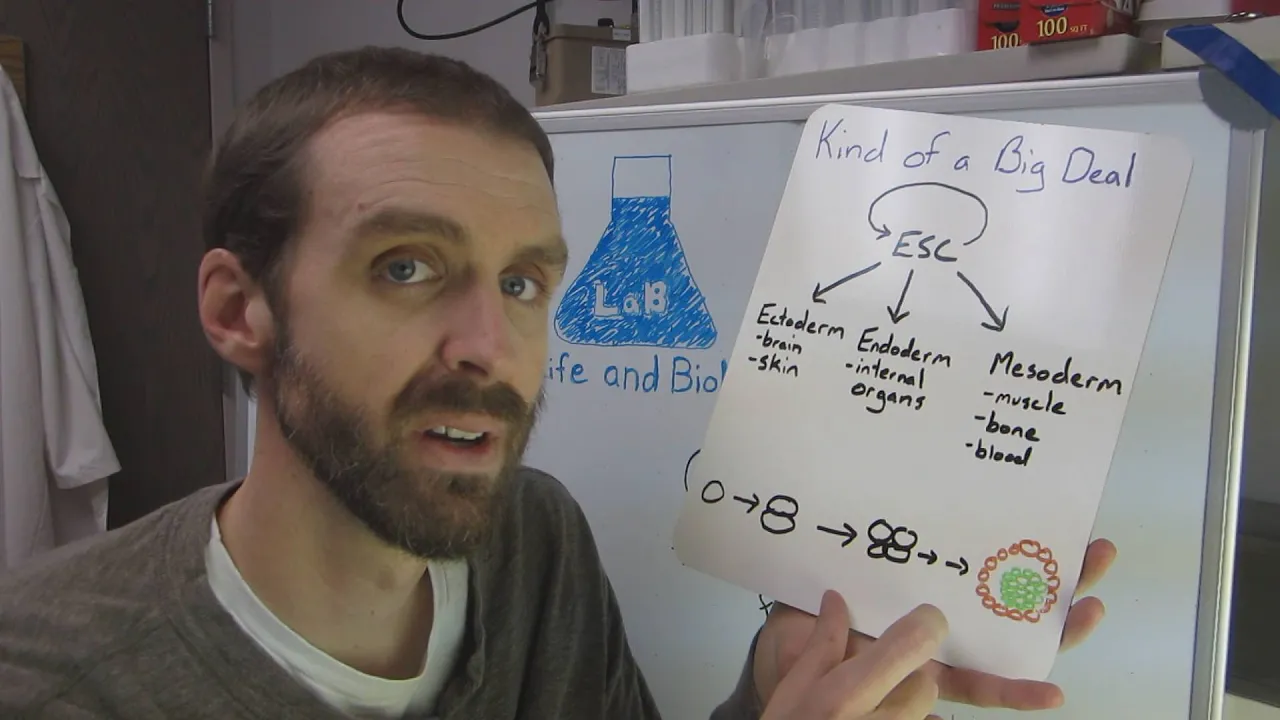
Added:Fundamentals of stem cell biology, specifically the characteristics and pluripotency of embryonic stem cells (ESCs) and induced pluripotent stem cells (iPSCs).
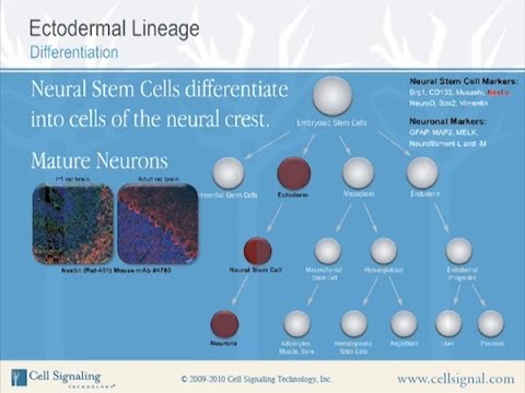
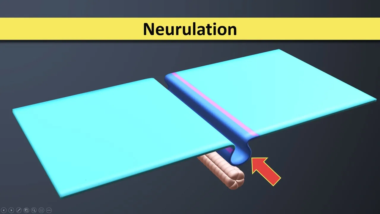
The basic stages of embryonic brain development (neurogenesis), including neural tube formation, progenitor cell proliferation, and neuronal migration.
The distinction between traditional 2D cell culture systems and 3D cell culture systems regarding microenvironments and cell-to-cell interactions.
Basic genetic engineering principles, such as CRISPR-Cas9 gene editing, which are used to introduce or correct disease-associated mutations in stem cells.
Advanced techniques in organoid vascularization and microfluidic integration (organ-on-a-chip) to sustain larger, more mature organoid structures.
The application of brain organoids in high-throughput drug screening and the development of personalized medicine for neurological disorders.
The ethical and bioethical debates surrounding advanced cerebral organoids, including questions of consciousness, sentience, and transplantation.
Methodologies for modeling specific complex neuropsychiatric and neurodegenerative disorders, such as schizophrenia or Alzheimer's disease, using patient-derived organoids.
33.9K views760likes30:00Original Release: 2017-09-20
hello my name is Yuri Knoblauch I am a scientist at the Institute of molecular biotechnology of the Austrian Academy of Sciences in Vienna and in this second part of my lecture I would like to tell you about how we can recapitulate human brain development in the lab starting from pure potent stem cells using three-dimensional culture methods now I'm sure every one of you would agree with me that the human brain is the most complex or also the most fascinating structure that Nature has generated it contains about 87 billion neurons that have to be born at the right time migrate to the right position and wire up in the right way in order to allow us to perform the cognitive processes that we're able to do the despite its complexity the brain develops from a limited number of stem and progenitor cells in a set of predefined rules and essentially everything that we know about those rules comes from experiments that were done in rodent model systems particularly in the mouse so this shows you a very simplified view of Mouse cortical development the mouse neocortex develops from a neural epithelium that lines a liquid filled cavity which is called the lateral ventricle it's a polarized epithelium with basal to the outside apical to the inside and all the different cell types in this epithelium are nicely arranged in a layer type fashion along the apical basal axis of the epithelium so here on the more basal side is the so called cortical plate and this is where the neurons are and where they send out their accidents and here on the more basal side is the so called ventricular zone and this is where the stem and progenitor cells reside and these progenitor cells undergo either one of three different types of division early during development they divide symmetrically and this leads to an initial amplification of the progenitor pool later they divide asymmetrically where one Purge inator generates one progenitor cell in one cell that migrates out to become a neuron or the other cell forms a so-called intermediate progenitor which divides once more into two terminally differentiated neurons and this is called direct or indirect neurogenesis now when we move from rodents to humans there is two characteristic differences first of all the initial set of symmetric divisions lasts a lot longer in humans and in primates this is a rodent context in a rodent brain and the primate brain at about at a comparable stage of development magnified to a comparable size and what you can see is that while the more posterior brain parts are actually very similar the cortex is vastly expanded in the primate brain and this is because these cells had a lot more time to divide symmetrically and to amplify the second big difference is that in addition to the ventricular zone and the cortical plate the primate brain contains an additional layer that is called the outer submarine trigger zone the outer subventricular zone is characteristic for primate brains and it contains a cell type that is called outer radial clear cell or basal radial glia cells these cells are not or in only very small numbers present in rodent brains but in primates they act as a transit amplifying population during neurogenesis so while in the rodents per progenitor division either one neuron or two neurons are generated in the primates the progenitors generate is outer radial glial cells which continue to divide asymmetrically generating hundreds of intermediate progenitors in hundreds of neurons so this is good news because it explains why we have so many more neurons than a mouse but it's also bad news because it means that there are certain brain developmental processes that cannot be modeled in a rodent model system and this is particularly emphasized and emphasized on this slide this shows you an MRI scan of a patient who suffers from a very severe neurodevelopmental disorder that is called microcephaly but you can see is that the patient's brain is much smaller than that of a healthy patient this patient carries a mutation in a gene that is called nd e1 and e1 is conserved all the way through evolution from yeast to humans but when you make a mutation in a mouse in the e1 you see no or only a very weak phenotype so certain neurodevelopmental disorders cannot properly be modeled in rodent models and for this reason there have been many attempts to actually model brain development in a human setting so how can we actually do this the easiest way of course is to use human tissue human cortical tissue can be obtained from aborted fetuses it can be fixed it can be stained the one can even do life imaging and cell tracing on this but of course such experiments plays a huge experimental burden an ethical burden and are very complicated and usually suffer from low and numbers and for this reason people have been trying to model brain development starting from pure potent stem cells the easiest way of doing this is to turn the pluripotent stem cells into neural stem cells which can then be forced to undergo neuronal renunciation and one can study the phenotype in those patient derived neurons but of course neurons like to be in a three-dimensional environment and so there's many limitations to those two-dimensional experiments and so there have been many attempts to actually model the development of human tissues in three-dimensional culture and really the pioneer of this has been a Japanese scientist named Yoshiki society if she kisses I around 2012 was able to recapitulate the development of a human eye in culture and this was clearly a pioneering experiment in the field of generating human tissues in culture now I think Yoshiki society clearly was one of the leading developmental biologists of our times and it's very sad that he's no longer with us and so based on his work but also adding other experimental approaches to it not named Lancaster a couple of years ago in my lab has decided to develop in vitro three-dimensional cultural system that we can use to model the development of a human cortex in culture and here's the method that she came up with we start with pluripotent human stem cells which we dissociate and then rapidly reactivate on the bottom of a 96-well plate we then allow those cells to develop into embroid bodies giving them just enough time to form the three germ layers we then replace the medium with a neural induction medium so that only the neural ectoderm survives in these balls of neural ectoderm are then placed into droplets of Maitre gel which is a collagenous three-dimensional support matrix that supports the development of stem cell derived tissue with an culture these Matrixyl droplets first and floating culture and later either in a spinning bioreactor or more recently in an orbital shaker and over time Marlene saw the development of fairly complex tissues so these are two examples of what we call cerebral organoids here is one example you can see a developing human cortex here here is a lateral ventricle here is another piece of cortical tissue and here is another one this is another example there is cortical tissue here and over here and down here you see the development of a human eye this is a cross-section through a cerebral organoid there's cortical tissue here you see a ventricular zone here here is the lateral ventricle and here are the differentiating new down here is what we call the choroid plexus which is the area that generates the cerebrospinal fluid and here are other brain areas in the absence of markers I do not know what they are but I want you to note that they have a different histology indicating that there are different areas of the developing human brain so cerebral organoids can be used to model the development of various parts of the human brain but for our analysis we focused on the developing cortex which is the most complex but also the most fascinating part of our brain and also the one that is most different between us and rodents this is a cross-section through a cerebral organoid this is the cortical part in red up for generators in green on the differentiating neurons and what you can see is that at this stage the organoid histology is essentially indistinguishable from that of a developing mouse cortex so cerebral organoid can recapitulate the three-dimensional organization of a developing human cortex we also analyzed neuronal differentiation the neurons in the cerebral organoids sent out long axons that contain growth cones they often branch out they can bundle together and the neurons also send out large dendritic trees but most importantly the neurons in our organize are electrically active this is a calcium imaging experiment where you can see that the neurons spike action potentials and they communicate with each other and the pattern of these electrical firings is far from random and and and there are certain correlated neurons and anti-correlated neurons so cerebral organoids recapitulate both the three-dimensional organization of the developing human cortex and proper neuronal differentiation and function now the neurons in the developing cortex actually come in different flavors they are arranged in a layer type fashion and these layers are formed in an inside-out manner where the deep layers are formed first and then newly formed neurons have to migrate through these deep layers adding additional layers to the outside their various markers for these layers suck p2 is a layer for the outlines a macro for the outside neurons see tip 2 for the inside when we stain early organoids essentially all of the neurons are positive for the deep layer marker see tip 2 a bit later suddenly to positive neurons appear initially they are intermingled and then they show some kind of sorting out although we do not really see the formation of proper neuronal layers but the temporal specification of various different neuronal subtypes can be recapitulated in the cerebral organoids so we've generated a three-dimensional cultural system that we can use to recapitulate the development of the human cortex in culture so what can we do with it in my view we are currently experiencing a complete revolution in a way of how we do biomedical research and this is because there are currently four technological developments that are coming together the first one is the availability of more and more complete human genome sequences many of them associated with complete patient records the second one is our ability to generate pluripotent stem cells from each of these patients and the third one is our ability to generate in the lab more and more different tissues as organoids that recapitulate particular organs in those patients and finally we can edit the genome and introduce or remove mutations from any of these pure important stem cells and how we can combine these technologies to analyze human neurodevelopmental disorders will be shown on the next couple of slides for this we teamed up with Andrew Jackson a pediatrician neurologist at the University of Edinburgh who works with a patient who suffers from a severe form of microcephaly this is an MRI scan of the patient you can see the brain is much smaller than that of an age matched healthy patient we obtained a biopsy from the patient to reprogram the cells into IPS cells and then generally organized from them these are healthy control organoids and patient-derived orgonites and what you can see is that the control organize contain a nice differentiated ventricular zone large ventricles and many neurons but the patient arrived organized are much smaller they contain a lower number of neurons and only a very tiny ventricular zone so using our organoid system and patient direct IPS cells we can recapitulate the small brain phenotype of a microcephaly patient and we can now go back in history and ask why are there so few ur neurons in this microcephaly derived organoids this is a control organoid and a patient derived organoid at a much earlier stage of development at this early stage in the control organoid all the progenitor cells are still undergoing the symmetric divisions and neurogenesis has not started this is very different in the patient-derived organoids where we can already see the appearance of individual neurons and we conclude from this experiments and many others that i don't have the time to show you that the cells have switched to an asymmetric division terror in a premature State this leads initially to the formation of too many neurons at a too early stage but at the same time the progenitors are not sufficiently amplified and we believe that this is the reason for why those organoids are so much smaller so premature neurogenesis and incomplete progenitor amplification are correlated with the appearance of a microcephaly phenotype so how can we explain this for this we performed whole exome sequencing on the patient and we found that the patient carries a compound heterozygous mutation in a gene that is called c DK v r up to all of these mutations introduced premature stop codons and consistent with this we do not detect the silica fiber up to protein in the patient derived cells so what is C DK v r up to C D k5r up to is a protein that originally has been identified in the fruit fly Drosophila but it was called centrist almond it's a sin a protein that is absolutely required for the correct orientation of the mitotic spindle and consistent with this while in the control organoids at an early stage all the mitotic spindles are precisely aligned with a ventricular surface in the patient arrived organoids they assume a more or less random orientation and so we believe that during the early stages of cortical development the orientation of the mitotic spindle is very important to ensure the equal distribution of the apical and base a lateral plasma membrane domains and to make sure that both daughter cells can maintain the progenitor fate in the patient arrive organoids however a tilt of the mitotic spindle no longer allows the symmetric inheritance so that some cells start undergoing neuronal differentiation and only a few maintain the progenitor status so a defect in spindle orientation leading to a lineage defect might be responsible for the microcephaly phenotype so this is where a normal human genetics analysis would stop we've recapitulated the disease we've found a plausible gene and a plausible mechanism but now genome editing tools allow us to unambiguously ask was it really this mutation that was responsible for the disease phenotype and for this we use genome editing to repair one of the two premature stop codons this is a control organoid a patient derived organoid and an organ or that's derived from cells where we have repaired one of the two mutations and what you can see is that both the signs the effect as well as the premature neuronal differentiation can be rescued so cerebral organoids can be used to recapitulate neurodevelopmental disorders and they can also be used to unambiguously associate particular mutations with those disorders now microcephaly is a very severe disorder and ultimately we would like to recapitulate more subtle disorders like epilepsy or autism the holy grail of neurodevelopmental disease modeling is a type of neurons that have a very complicated developmental origin and these are the GABAergic inter neurons this is a schematic view of the developing cortex the cortex develops from the dorsal neural epithelium and all the neurons that arise in the dorsal neural epithelium Express an excitatory neurotransmitter glutamate and become excitatory neurons in addition to those neurons there are inhibitory GABAergic inter neurons which arise from the ventral part of the developing cortex they then migrate tangentially to go into the dorsal areas and integrate into the circuits of the dorsal cortex and it is thought that migrating inter neurons is what actually is absolutely essential for creating functional neural circuits and the types of mutations that are associated with diseases like epilepsy or autism suggests that effects in inter neuron formation and migration may be associated with many of these diseases and so we asked can we actually model this very complex developmental event in our cerebral organized first thing we wanted to know is do we actually have ventral and dorsal cortex in the organoids this is shown here the dorsal cortex can be identified because the neurons on the intermediate progenitors express a gene that is called t BR 2 whereas the ventral cortex the lateral ganglionic eminence in this case can be identified by the expression of GS x 2 when we section an organoid we find typically that the organelles contain both t BR to positive dorsal regions and G's X to positive ventral regions so both ventral and dorsal areas are actually present in our cerebral organize so can we use them to model into neuron migration this slide shows you a dorsal cortical area where the intermediate progenitors express the dosa marker TBR to and when which stained the adjacent section with we got an interneuron marker and somatostatin a marker of particular interneuron subtypes we find that interneurons are actually present in the dorsal cortical areas of the organizer so interneuron migration from the ventral into the dorsal part can actually be modeled in cerebral organoids but there's one key problem with this and this is illustrated here in the current protocol organoids are a little bit like a car where the wheels are up the engine is up the seat is in the back the windshield is in the front so all the individual parts are present but they are in a random arrangement and so if we make a section through such an organized we have to be very lucky to hit both a dorsal and ventral part and to be able to model inter neuron migration and so we asked can we actually introduce a polarity axis in organoids for example a dorsal ventral axis and so in order to do this Josh Phegley a postdoc in my lab set out to develop a protocol where he separately generates also organoids and ventral organoids and then he places these two orgonites together into one droplet of Maitre gel one organoid is labeled with rfp expression another organoid is labeled with GXP gfp expression initially when the two organoids are Co embedded they lie next to each other but all the time they fuse with each other and if I would not show you the red and green color you would not be able to distinguish the boundary between the two organized so we have developed a co culture protocol that we can use to generate organoids where one part is dorsal and the other part is ventral so can we use this to model interneuron migration this slide shows you a typical dorsoventral fusion organoid and what you can see is that within the dorsal area you see these green spots when we focus in on this we that these green areas are actually full of cells that have the migratory appearance of interneurons when we fused also with dorsal orgonites we no longer see this and so from this experiment and many others that I don't have the time to show you we conclude that when we fuse ventral with dorsal organoids there are cells that are migrating from the ventral into the dorsal part of the orgonite so what are those cells this slide actually summarizes a very large number of experiments and i only show you the key experiments here i show you that the migratory cells Express GFP and all of them also Express GAD 1 which is a marker for developing inter neurons this is a high magnification view and you can actually see the elaborate cell shapes that these develop these migrating inter neurons actually have we also use many other markers of migrating inter neurons and various interneuron subtypes like parvalbumin somatostatin neuropeptide Y and also called Benjamin Cal retinal and we find all of these subtypes in our migrating fused organoids so taken together this tells us that we can use fused organoids where we have regenerated a dorsal ventral axis to model the long-range interactions of various parts of the human brain in particular the migration of cells from one part of the brain into the other now of course ideally we would like to image this process of inter neuron migration in real time and in order to do so Josh developed a method that we can use to do long-term imaging of migrating inter neurons and this is shown here for this Josh makes very thick slices from fused organoids and those sliced slices are then imaged on a spinning disk time-lapse microscope for about three days this is a still of one of these migratory movies and what you can see is that we can actually see migrating cells that go from one part of the organ or it into the other this slide shows you a movie of migrating interneuron the interneuron can be seen here here is the cell body and here are two processes and would like you to focus on this one cell and you will see that it migrates across the entire field of the movie and interneuron migration is one of the most complex but in my view also both beautiful processes that occur during development you can see the cell extends many processes then some of them are retracted and it migrates along other processes the second movie shows you a more realistic view it shows you how crowded the field is there is many different migrating interneurons I want you to focus on this one and you will be able to see that this cell actually migrates across the entire field of the image field so here you can see that the cell extends different processes some of the processes are then retracted it makes decisions to actually follow processes which initially are weaker and so with this we can now use organoids to model long-range interneuron migration and we're in the process of deriving IPS cells from patients that actually suffer from neurodevelopmental disorders to see whether internal migration is affected now it is known that the migration of interneurons is in response to chemokine that is called CX CL 12 and the receptor for this key note first chemo kind is cxcr4 and there is a drug called AMT 3100 or plural safor which actually is an inhibitor of this chemokine receptor and so we asked is inter neuron migration that we actually see in our cultures dependent on this receptor this is a normal organ or where you can see a lot of inter neurons migrating from one into the part and this is an organelle that has been cultured in the presence of the cxcr4 inhibitor and what you can see is that the in migration of the inter neurons is actually almost completely suppressed I want you to note that the growth of axons from one part of the organ or it into the other is actually not effective indicating that the other part of the organ is actually still alive so we can use fused organoids to model inter neuron migration and also to test chemical compounds for their effect on interneuron migration and we are currently generating various models for neurodevelopmental disorders that we can use to then test where the inter neuron migration is effected so in the end I would like to summarize what I've told you I've told you that we've generated a three-dimensional cultural system that we can use to recapitulate early human brain development in culture our cerebral organoids generate various parts of the human brain that can actually functionally interact with each other we can use the organoids to model neurodevelopmental disorders and we can generate various parts of the human brain separately in culture then fuse them together and the interactions between these various parts are maintained and particularly last part of this course has huge potential we will be able to generate any pairs of brain regions and watch the migration of cells from one into the other or the migration of axons and hopefully eventually even be able to visualize the formation of some of the major exon tracts in the end I would like to acknowledge people who actually contributed to this work the hero of the organoids is Madeleine Lancaster she was a postdoc in my lab now it's her own lab at the lnb in Cambridge for looking at also ventral patterning she worked together with a grad student Magdalena Elena the fusion organoid system was generated by josh bailey another postdoc in my lab these are the names of people who work together in this various projects the reprogramming of the cells was a collaboration with the lab of Joseph pending her I would particularly like to thank the patient and their family for allows for allowing us to use the cells in our experiments and our collaborator at the University of Edinburgh is Andrew Jackson I would like to thank the Austrian Academy of Sciences for funding our work in addition we obtained funding from the European Research Council the Austrian Science Forum and from embo and I would like to thank my entire lab for being such a fantastic group you
Up Next
11:49
Neural Proliferation Migration & Aggregation | Brain Development
@professorgalvan4469
20K views•2018-01-30

21:55
Deciphering Neural Circuits Controlling Anorexia in Mice | Richard Palmiter
@AllenInstitute
2.3K views•2011-10-10

38:06
Neuromuscular Connectomics & Synapse Elimination | Jeff Lichtman
@scicommlab
20.9K views•2014-03-31

13:03
How Exercise Benefits Your Brain: Science Explained
@TED
11.4M views•2018-03-21
Related Study Plans & Knowledge Roadmaps
Structured learning paths in Neuroscience
neuroscience
Appetite Regulation: Brain Circuits & Hormones
01Foundations of Endocrinology and Brain Anatomy
02Introduction to Energy Homeostasis
03Peripheral Signals: Leptin, Ghrelin, and Insulin
+ 2 more learning modules
14h5 modules
Explore Roadmap
neuroscience
Neuroeconomics: Value, Risk & Brain Mechanisms
01Foundations of Neuroscience and Brain Anatomy
02Introduction to Neuroeconomics and Subjective Value
03Dopaminergic Prediction Errors & Reinforcement Learning
+ 3 more learning modules
18h6 modules
Explore Roadmap
neuroscience
Spatial Navigation: Cells, Grids & Brain Maps
01Foundations of Brain Anatomy and Memory
02Place Cells and Head-Direction Cells
03Grid Cells: The Brain's Coordinate System
+ 2 more learning modules
12h5 modules
Explore Roadmap
neuroscience
BCIs: Neural Decoding & Prosthetic Control
01Neuroscience Foundations: How the Brain Signals Intent
02Signal Acquisition: Recording the Brain's Activity
03Neural Decoding: Translating Brain Waves into Data
+ 2 more learning modules
25h5 modules
Explore Roadmap