






















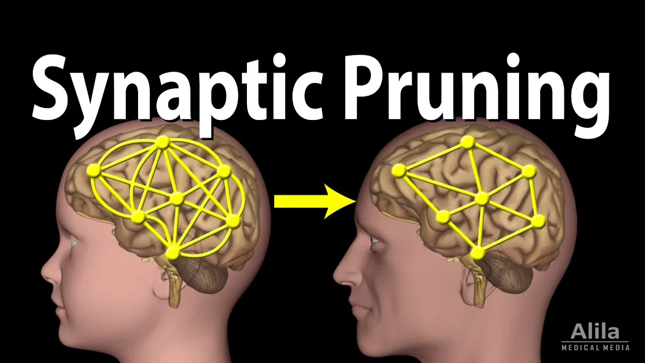


During early development, the nervous system undergoes massive pruning where most synaptic connections are eliminated, transforming initially highly redundant and variable neural circuits into specialized, efficient wiring diagrams that reflect accumulated experience.
Neuromuscular Connectomics & Synapse Elimination | Jeff Lichtman
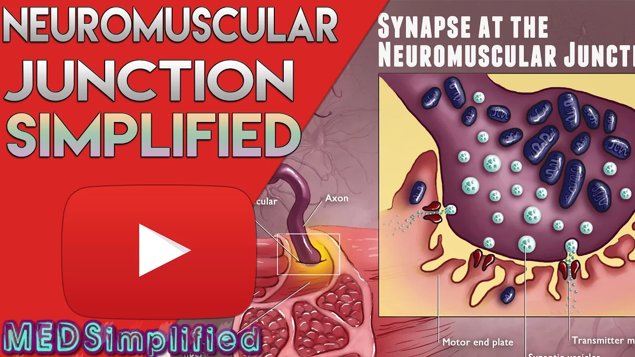
Added:Structure and function of the Neuromuscular Junction (NMJ), including the role of acetylcholine and postsynaptic receptors.
Basic principles of neurodevelopment, specifically axon guidance, target selection, and growth cone dynamics.
The concept of synaptic plasticity and how neuronal activity can influence synaptic strength.
An introductory understanding of imaging methodologies in neuroscience, such as light and electron microscopy.
Advanced computational connectomics, focusing on reconstruction algorithms and handling petabyte-scale electron microscopy data.
The molecular mechanisms of synapse elimination, including the roles of glial cells (astrocytes and microglia) in pruning.
Pathology of synaptic pruning, examining links to neurodevelopmental disorders like schizophrenia and autism, or degenerative diseases like ALS.
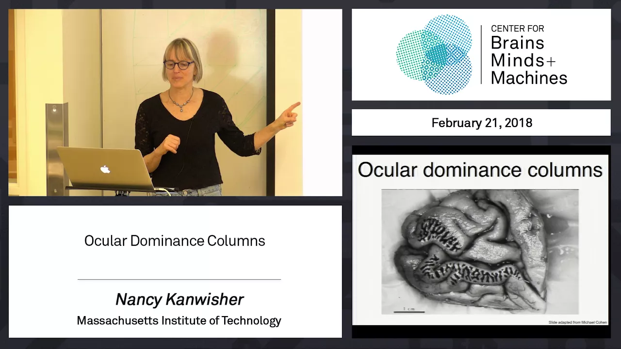
Activity-dependent refinement in the central nervous system, comparing NMJ elimination to visual system refinement (e.g., ocular dominance columns).
20.9K views403likes38:06Original Release: 2014-03-31
Hi, I'm Jeff Lickman and I'd like to uh talk about some uh attempts we've made to try to understand the relation between the wiring diagram of the nervous system and its function. And I'm going to focus mainly on the most accessible or perhaps one of the most accessible parts of the nervous system which is the connections between cells in the brain stem and spinal cord and muscle fibers. These are the neuromuscular junctions and it's the final output for most of the behaviors we have as we move our muscles through all the actions we do. And that part of the nervous system uh is simple because it's the a dead end. you know, the axons go into muscle and they make synapses and that's the end of the road and there's not much else going on except this one-way street. Uh, and we've used techniques that allow us to see those wires between the neurons and muscle very clearly and uh, in this system perhaps one can get an inkling of what way of some of the ways in which perhaps experience are instantiated in wiring diagrams. I'm going to talk about peripheral muscles in mice. Largely I talk about mice because these are animals where we have transgenic tools uh like fluorescent protein expression uh in nerve cells that highlight these cells. So you can see them without a great deal of effort just using fluorescent microscopy. And some of the tools I'll talk about are brain. I've already talked about in a previous lecture that technique. Uh I'm going to try to show you how we take advantage of that technique to understand something about the connections and the omix of connections full wiring diagrams. So uh let me begin by just giving you a uh a view of what neuromuscular connections look like by uh viewing um the neck of a mouse. This is a a anesthetized animal that's being intubated. And uh what's exposed on this side is a muscle that sits right underneath the salivary glands. This is the submandibular and sublingual salivary glands that have been retracted to expose this muscle.
It's called the sterno mastoid muscle.
Humans have this muscle. It's this big V-shaped muscle you have, if you have a neck, unlike me, uh a long neck, you can see it. And if you don't have as much fat on your bones as I do, you have this nice V here. That's the sternoccllyommastoid in humans. In in mice, the sterno mmastoid is separate from the kyom mastoid. So it goes from the sternum at the notch of where your collar bone enters uh your rib cage all the way back to the mastoid process on your jawbone. That's why it's called sterno mastoid. And the nerves that activate this muscle that make it twitch uh converge on one little part of the muscle called the end plate zone. The endplate zone. The word end plate is an old term uh that was invented when hisystologists first noticed the sites where nerves touched muscle fibers and they're called end plates. They're ends because they're terminations of axons and plates because they sit on a little plaque shaped region in the muscle. So that's the end plate zone. And in these transgenic mice where for example a mouse that might have under the control of the thigh 1 regulatory element the expression of a single fluorescent protein let's say yellow fluorescent protein in all nerve cells. If you look at a mouse like that at that region with a low magnification fluoresence microscope like a dissecting fluoresence microscope this is what you see. see this kind of treeshaped structure in a line of mice. In this case, it's called the YFP16 line that Goping Flang Josh Sainz and I uh developed a number of years ago. Uh that is very nice because without doing anything more than just looking you can see these big nerves that branch into smaller and smaller branchlets and at the end are these little reddish things which are not yellow fluorescent protein but that is a stain of the acetylcholine receptors on the muscle fiber at the site where the nerve releases acetil choline. So these are the synapses of individual axons. Uh we can zoom up with a higher resolution microscope and look with conf focal microscopy at a small region of this muscle. And you see that those uh big fat uh yellow branches are actually composed of many single axons that are branching to these neuromuscular junctions. And in the adult you see that each of these neuromuscular junctions has one axon uh coming up to it. If you counted the number of axons in this whole branch here, you would see there are fewer axons there than the number of neuromuscular junctions. And that's because many of these axons branch to more than one junction. You can see it's a little hard to see where those branches are, but I'm going to show you uh how we ultimately got to trace out all these branches. The other thing uh worth noting is this kind of haphazard appearance here. And the question is, is this regulated in some way or is it variable from one animal to the next?
I'd like to zoom up even further to single neuromuscular junctions on two adjacent muscle fibers to give you a sense of the exquisite alignment between where the nerve is releasing its neurotransmitter and where the target cell in this case a muscle fiber has the receptors to respond to the chemical being released by the nerve. So these are two adjacent muscle fibers. Uh, and if you pour onto a muscle a fluorescent, in this case a red fluorescent version of a snake toxin called bungrotoxin from cobra type snakes that inactivate animals by paralyzing them by blocking the ability of the receptors to respond to acetylcholine. If you make a fluorescent version of that toxin, it binds very tightly as you would expect to the acettooline receptor which is its normal target. And there are the acettooline receptors on two adjacent muscle fibers. Each one is a unique shape. Uh so they're easily recognizable. That is every one looks different from every other one like like faces. And here's the overlying nerve terminal. And if I just blink on and off the yellow fluorescent protein on top, you see how beautifully the aocoline receptors are covered exactly by the nerve. And it's an interesting question.
What gives rise to this perfect alignment? Are the axons particularly attracted to grow on top of receptors?
It's no there's no doubt that that is the case because in these animals which you can stain like this in a living animal, if you crush a peripheral nerve, the axons will grow back and when they get back to those receptors as shown in this time-lapse diagram uh movie here, over several hours, they grow right back on top of the acylcoline receptors very quickly. this case it's a blue fluorescent protein but the same idea again this is work of Hono Kang so we would like to use this technique um of labeling individual axons to trace out all the axons in a muscle and we did that this was work that Julu did he worked on a muscle that is very small also in the neck but in this case the back of the neck called the intercodilaris muscle it's an extremely small muscle it only has about 15 axons coming in. These are the individual neuromuscular junctions.
That's just a a joke up there, but it looks a little bit like a tree. And here is uh an example of tracing out every single axon in data sets that were about 100 gigabytes large with a very fine resolution necessary to trace every axon in them. So this is the tree now with every axon labeled a different color and it is a complicated tree. Uh you see that many different colors are in every part of the muscle. Some of the axons have a large amount of territory. Other axons have very small amount of territory but every axon branches uh to more than one neuromuscular junction.
And this provided us with an opportunity to ask a a fundamental question that uh depending on your point of view uh would either be obvious what the answer is or surprising. And the question is if you look at this wiring diagram let's say on the left side of a mouse and you look at the same muscle on the other side are the wiring diagrams going to be mirrory symmetrical sort of like your hands are mirrory symmetrical or the rest of your body is certainly the axons come into these muscles in a symmetrical way and also I should tell you that in insects and in uh other invertebrates like worms axons often have highly stereotype branching patterns that are genetically based. So if the genes have something to do with this wiring diagram, one would expect that the left and right side would either be the same or if they were different, if you compared the left of one animal with the left of another animal, they would be similar and the right of one animal would be similar to the right of the other or as I said, the left and right should be mirror symmetry of each other. Turned out that neither of those were the case. Every single instantiation of this wiring diagram was unique. every single one was different from every other one. This is a cautionary tale perhaps for what to expect out of mamalian wiring diagrams in terms of stereotyped organization.
Everyone had its own particular features. And when I say unique, I mean exactly where axons went, exactly what the branching pattern was, was different from one animal to another. Even though the nerve always came in in the same place, once it got to the muscle, it made its own unique branching pattern uh even different from the left and right side of each animal. Perhaps more surprising is that the wiring diagram was not that pretty.
It had lots of suboptimal things about it. It had places where the wires would make useless loops that didn't have any purpose as far as we could tell. places where branching was premature, that is, branches extended farther than they had to, the branches happened early. I'm just going to give you an example of this in the next slide. So, this is a uh one of Julu's reconstructions. In this case, all the axons were labeled, but only one is now highlighted for you in red. And and I'm this is just a zoom up of of the region in the box there. uh and I just want to show you why this axon was chosen for demonstration here. All the axons uh except for this red axon uh go where this black arrow says they go up this large trunk right here. This red axon and one blue axon however bifurcate to the left. The one that is to the the blue axon that is going up here is actually a bifurcation of an axon. It goes both ways. But this red axon is the only axon in this particular muscle that chose only to go left. And who am I to say that was a bad idea. Maybe there's a good reason for that. But if you follow the yellow arrows, you see that when the axon gets up to here, it then makes a hairpin turn and goes back down. Keeps going down, down, down. And if it keeps going any further than this, it will leave the muscle and then makes a hairpin turn again and comes back up that branch there. give you a sense of how wasteful that is. This little branch down here is this branch to this little neuromuscular junction there. There's an easy way to get there. You just go up this way and go right down there. But no, it takes this very route. Another example is this bifurcation here generates the branch that goes here. That seems fine. But that bifurcation occurs and then the bifurcated axon instead of just going straight over there follows the white arrows. It just co-aciculates with the other branch of the same axon goes all the way up here comes back down here then peels off crosses over itself to make that neuromuscular junction. In every single animal we found examples like this of wiring that wasn't optimal. And this raised many questions about why uh this might occur. And what I'd like to tell you about is that we think one of the reasons this occurs is that the wiring diagram one ends up with in a muscle is not the wiring diagram that the animal is born with. The animal is born with a very different kind of wiring diagram.
And what you're looking at is what is selected the subset of wires that remain after a period of massive branch pruning in early life.
So I have to first digress and tell you that what I'm going to try to argue is that the changes that are occurring in development are a way in which experience may alter the connectivity the connections in the nervous system.
But it's not the only way nervous systems can be modified by activity. In fact, there are two general categories.
The first is you could have activity that is changed in a way that synapses get stronger or weaker but they are still present. Uh these would be changes in synaptic efficacy. You could have synapses that facilitate or potentiate or depress. These are well-known phenomena that take place at synapses.
They're quite common and they're found as far as I know in all animal species.
And animals can jazz up or lower the action, the behavioral state of an animal by virtue of of these kinds of effects. Uh, and they may be reversible.
When you're awake, some of your synapses are jazzed up and then maybe they u depress a little when you go to sleep, for example. Or when you think of something and at first it's a foggy thought and then it gets more and more intense as you think more and more about it. That could be a form of potentiation. There is a second kind of change experience might do which is one where you don't change the strength of synapses but you actually change which synapses are present in the nervous system and which ones are absent. You change the actual wiring diagram. You might add new synaptic connections or you might completely eliminate connections and these might last forever. And in fact, my bias is to think that these long-term uh changes in the nervous system that come about through experience, the indelible changes of memory that are hard to wipe out are likely to be changes in the physical connections between nerve cells rather than changes just in the strength of synapses. And I'm going to suggest that this second idea is important by showing you that during development when mammals are doing a lot of their learning, there's a huge change in which connections are present and which ones disappear. And one way to see this is to look in the lowly muscle. Look at the neuromuscular junctions in a young animal. So if you take a baby mouse at the age of a week and you uh again expose the muscle fibers to bungtoxin to see the receptors and you use a line of mice where all the nerve cells are labeled with yellow fluorescent protein, the neuromuscular junctions in baby mice are quite different from adults. Instead of having that one axon there, there's often more than one. And so in this picture that Thomas Mskell took, there's unambiguously two axons converging on the same little neuromuscular junction site.
What happens over the first couple of weeks of life is all this multiple intervation, all these extra inputs disappear. And I'll show you a time lapse of looking at a neuromuscular junction on three successive days where you can see what actually happens. So this is work that Mark Walsh did in a paper from Cindy Keller Pek where we first took a picture of two neuromuscular junctions at post-natal day seven.
And you see that the one on the left has two axons interervating it and the one on the right has one axon and that axon is actually a branch of an axon that goes to both junctions. Come back. Take that picture.
Sew up the wound. Allow the animal to recover. Reanesthetize the mouse. Expose the neck again. find the same two neuromuscular junctions on the next day of life in the same animal. And you find that again the one the neuromuscular junction on the left has two inputs and the one on the right has one. But notice that one of the inputs to the junction on the left now is quite thin all the way back to its branch point compared to how thick it was the first day. So there is some kind of atrophy that has taken place in that axon. And then when Mark Walsh came back a day later, he found that this junction is now singly innervated. And the other axon is now just a bulb that's no longer connected to the muscle all the way back to this branch point. And not shown here is eventually that little branch withers away entirely and disappears. And this is how the muscle ends up with every muscle fiber choosing one axon.
Importantly to realize it's important to realize that the branch that got eliminated didn't eliminate that axon in the muscle. That axon is still making a good connection here, but it's not making a connection with this muscle fiber. So this muscle fiber has chosen one input to keep and one uh to disappear. So there is this retraction step. So from this I I want to give you a sense of what this is part of. It's part of the fact that axons start out branching to many muscle fibers and end up branching to fewer. So here's a cartoon uh that shows this. You have a bunch of motor neurons in the spinal cord. I've only drawn four here. And in early postnatal life, many of them send multiple branches such that individual neuromuscular junctions are multiply innervated as it's called. And then over the first couple of postnatal weeks in a mouse, it may take much longer in a human being. Each neuromuscular junction becomes singly innervated.
This phenomenon of going from multiple to single intervation requires one to think about what actually happens at the neuromuscular junction. For example, do the sites that are eliminated from one axon uh do those sites get taken over? Is that territory invaded by the other axon? And one way to study that is to take advantage of multiple colors. So for example, if you have a blue fluorescent protein axon and a yellow fluorescent protein axon at the same neuromuscular junction and then you watch them over time, you can learn something about how synapse elimination, as it's called, occurs. And that's what I'll show you on this slide. So this is a neuromuscular junction at postnatal day 11 that Mark Walsh took a picture of. It has a yellow axon and a blue axon sitting at the same neuromuscular junction on the red acetylcholine receptor sites. And then after taking this picture, he sewed up the wound and he waited a day and found the same neuromuscular junction a day later and got this picture. I'll just go back and forth for a second. See that in this picture, the yellow axon seems to have added some territory and the blue axon has withdrawn a little bit and the boundary between them is at that yellow arrow. He came back a day later at postnatal day 13 and now the yellow axon has taken over more of the territory and the blue has withdrawn further. And at postnatal day 14 now it is a singly innervated junction. The yellow has all the territory and the blue has retracted and postnatal day 15 the yellow axon has kind of plumped up a little bit. The blue is going away and eventually that branch of that blue axon will be entirely gone. When you look at this process, you can't help but think it looks sort of like an invasion of territory uh by one axon that is competing with the other for the same post synaptic sites as if the receptors are territory you want to have and you take it over if the other one goes away.
It implies competition but it doesn't doesn't prove competition. But we realized that once we could watch the process like this, maybe we could see whether they are actually competing. And one way to ask about competition is to say if the invader suddenly disappears, does the axon that's lost territory suddenly say, well, now I have all this territory I'm going to grow back in. If it does, then you can say the reason this axon left is because of competition with the other axon. And that's exactly what we tried to do. Let me just show you the result on the following slide. This is a neuromuscular junction that has a big fat axon and a very thin axon at the same junction. And we can use a fine laser spot using a two photon laser which allows very little spread of the damage beyond a very small region to completely destroy one axon and leave the other intact. So if you damage one axon and we slowly damage it, we don't make a huge bubble in the muscle. Bo, we don't boil it. We just damage it enough for all the fluorescent protein to leak out of one of those axons. And we just come back an hour later. All you see is the uh axon that is remaining, the undamaged one. And you see it has a very small amount of the territory of this multiply innervated junction in development. But if we come back now a day later, we see that that little axon has now taken over the entire synaptic site. We did this many times and always we got the same result suggesting that the losing axon is losing reluctantly. If you get rid of the big axon, the little one constantly will just change its mind and grow back suggesting that there is actual competition between axons in development. And this raises the question of what are they competing for and how does one modify this competition? Does experience have anything to do or neural activity have anything to do with who wins and who loses? The connectivity uh that I've been emphasizing so far is the change in connections between two axons at one junction. I want to remind you by going back to this cartoon that there's another side to this ch transition.
Besides going from multiple to single intervation, axons initially start out with many branches in the muscle and have what are known as large motor units. That's the name of an axon and all of its branches. And they end up with smaller motor units. And that transition from large to small motor units is due to the loss of multiple intervation at single junctions. But the change is now not a change in the convergence on single muscle fibers. But the other side of the coin is that each axon starts out diverging to many muscle fibers. It ends up connected to a smaller number. One question we were interested in is how big a change is this actually?
Uh everything I've shown you so far uh shows two inputs going to one input. Uh but truth be told, all of these experiments are begun somewhere around one week of age. We were interested to know whether this transition from multiple to single intervation was even more profound than two inputs to one input by asking the question what kinds of motor units do we see if we look not in young animals at a week of age but look back at birth or even before birth.
And again, we took advantage of transgenic mice where single axons could be labeled in a single color and we could look at one axon and all of its branches. I'll just show you a diagram of tracings of three single axons from three different ages in a young animal.
And you can see there's no doubt of a profound change in the organization of these axons. So in embryos, this is embryionic day 16, axons have rather modest branching, but in fact the number of receptor sites in gray here is much smaller than at birth. That is muscles are still building their muscles and so axons have small branch numbers of branches, but so do the muscles. If we go to postnatal day one, however, just right after birth and look at an axon's arbor, it occupies maybe 80 sometimes even 90% of all the receptors on every muscle fiber, every receptor site has a piece of this axon. This axon has kind of branched to everybody. There's just very dense uh coverage of all the receptor sites. Whereas just two weeks later, postnail day 13, you can see how sparse an axon's arbor is. So this implies that an axon is touching most of the neuromuscular junctions at P1. But this is one axon. If all the axons are doing this, then there should be lots of axons converging at each of these neuromuscular junctions because there isn't room for all the others. If if each one does 80% and there are 10 axons, there should be a lot of axons at one junction.
In order to see if that was the case, that there were many axons converging, more than two, we looked at a single neuromuscular junction at post-natal day one, and reconstructed the wiring using highresolution techniques using electron microscopes. I'll just show you a piece of work that was done by Juan Carlos Tapia uh by showing you a block of muscle uh from postnatal day one where we have isolated and identified all the axons converging on one neuromuscular junction. So this is the block of tissue and I'm just going to cut through the block. Each image is from a different uh depth. And then what Juan Carlos did was he colored in the muscle fiber green and the axons converging on that site each a different color. And you can see that there are lots of axons converging at that one neuromuscular junction at birth. In fact, they look like they're in some kind of orgy of happiness. You know, they're just glombmed onto each other.
They're all they're not segregated from each other in any way as we see when they're you're down to two inputs.
They're often on opposite sides of the junction. Here there's 1 2 3 4 5 6 7 8 9 10 and and one axon that's already looking like it's leaving 11 axons converging at that neuromuscular junction at birth. And this was typical.
Every muscle fiber we reconstructed at birth uh had large numbers of axons converging. So uh the picture one might want to keep in one's mind is not going from two to one but going from a situation where virtually every axon samples every neuromuscular junction to this very sparse labeling later. To put this another way, this wiring diagram would allow for any possible outcome because every axon is possibly the remaining axon at every one of these muscle fibers. And that what you end up with is a minuscule possibility out of an infinitude of ways this muscle could have wired up. And I'd like to make this statement which is quite tentative but to say this may be a metaphor for what's happening in other parts of the brain as well that in young humans before they know anything it's not that they don't have any wires. It may be that they have wires for every possibility and then through the experience of their parents, television, teachers out in the world, a large portion of what we could have been gets eliminated and we end up being those narrow-minded adults. Uh we or at least I have become at least that's what my children tell me. So in development uh in muscle at least greater than 90% of the synaptic branches are eliminated during early life and I suggest that maybe this is occurring elsewhere as well. This raises an interesting question. Why is this happening and who determines who stays and who goes from each muscle fiber? And I'm going to give you just some tenative hints about what we think might be going on. Taking advantage of mice where every axon is labeled a different color using these brain mice. This is work that Ryan Draft has been doing. This is a muscle from about a week of age, a small neck muscle called the omohyoid where each axon is labeled a different color. If we zoom in on a little part of this muscle, I think you can see this a little more clearly that each of the neuromuscular junctions uh is contacted by accents, but there's a lot of color in this muscle. And if you look at the where these arrows are, you can see that many of these neuromuscular junctions at one week of age are multiply invated. There's more than one color axon at each of those sites. Uh in distinction to the adult where they're all singly innervated. And what Ryan did was he went through uh painstakingly looked at each neuromuscular junction in this muscle and itemized exactly which axon was in each muscle fibers neuromuscular junction site. And he used conff focal microscopy to do this. So by sectioning through the muscle with a conffocal microscope you could see every single neuromuscular junction quite clearly and you could identify exactly which colors were at each muscle at each neuromuscular junction. And this is a muscle where the colors of the axons were different enough that each color represented one neuron. There was no duplications which made it easy to map out the conneto of this developing muscle. One of the things he saw very early was that when you look at muscles like this uh there is no tendency for let's say the blue and the red axon to go to one part of the muscle and the green and the purple axon to go to another part of the muscle. Every axon more or less went everywhere. So what I'm going to show you next cannot be explained by local topography in the muscle. Another thing he found quite early is that certain combinations of colors were very common and other combinations of neurons were very rare.
So let me give you an example by looking at the one of these omohyoid muscles that have 411 muscle fibers 411 neuromuscular junction from postnatal day six. These are the colors of all the neurons that intervate this muscle that contact the muscle. And the most common thing he saw in this particular muscle was that the red neuron and the blue neuron converged on 66 muscle fibers.
That was the most common thing he saw.
The next most common thing he saw in this particular muscle was 61 muscle fibers that shared those two colors. The next most common thing he saw were 60 muscle fibers that shared those two colors. The next most common thing was 54 sharing those two colors and 50 sharing those two colors. So I'm going from 66 down to 50. So uh a good mental exercise is how many would you think are shared between the red and the one at the end? It might be slightly less than 50. But this was the weird thing about the way these results came out. There was only one muscle fiber shared by those two. And that was not the next most common connectivity in this muscle.
It was not a ring structure. The next most common was 29 muscle fibers shared by those two, which in this particular order skips one. And the next most common was 22 muscle fibers shared by those two. Again, in this weird order, skipping one. And 21 shared by those two, skipping one. And 18 shared by those two, skipping one. 16 muscle fibers were shared by those two axons.
And that skips two in this order. Seven by those two. Again, skipping two. If we go around clockwise, six between those two again, skipping two. Four between those two and that skips three in this order. And then finally, there's that one. So, they're not near each other.
You actually have to go all the way around the circle to get to them. So, this shouldn't we shouldn't have drawn it this way because you drew draw it this way, it just looks like a big mess.
And for a long time, we were just thinking this was a big mess because every muscle we looked at had the same kind of weirdness. We couldn't make much heads or tails of it. until we started looking at axons individually. So if we just look at the red neurons a uh connectivity it has a lot in common with this guy 66 29 with this color 16 with this neuron four with this neuron and only one with that one. We shouldn't draw this as a circle but we should draw this as a distance. The nearest neighbor the strongest relations and the distant neighbor the least. So this is the same data now graphed another way where the 66 neighbor is closest and the one is the furthest away. So that's the particular order. And now I'm just going to show you the same data set. Now not looking at the red axon's connectivity but the blue axon right next to its connectivity. And I'm not going to change the order of the neurons. this order, whatever it is, I don't know what it is, but whatever it is, I'm not going to change it. But if I don't change it, the rule still exists. Its nearest neighbors, it's best connected with.
It's less connected to one further away, weaker here, and it's not connected at all to this one who is its furthest neighbor. When I do the same for this neuron's connectivity, it's the same thing again.
The nearest neighbors are its strongest connections and the weaker the farther you go out. The same is true for this neuron. The same is true for this neuron and the same is true for this neuron with one exception that there's no connection here and one connection all the way to the red one. And that's probably within this the noise of the measurement. It's a kind of an amazing connectional matrix. It was not something we were looking for. And therefore, I couldn't just tell you, oh yeah, that makes sense. We didn't know what it meant, but it was quite striking. And we called these a linear order connectional matrix because it wasn't a ring. It was like this neuron was at one end of some spectrum and this neuron was at the other end of something, but there was no topography in the muscle to explain this. So, we kept thinking, well, where are there is there anything ordered in muscle? And one of the things that's ordered in muscle is the order in which nerve cells are activated when you use muscles to do a task. This is known as the size principle of henaman where you always recruit a particular neuron first and if that is not sufficient to generate enough force to do the action, you always recruit a second neuron next and a particular neuron third and so forth.
And so what we've been trying to do is see whether this order has anything to do with that order because that is an order that muscles hold on to into adulthood. And to make a long story short, we've done a number of tests. Um most of them are only focused so far on the neuron that's recruited first. We're trying to do other tests, but we're still at a preliminary stage here.
But the suggestion is that the neuron that is recruited first is always at one end of this connectional matrix. And we know that because the neuron recruited first activates muscle fibers that have slow measin and we stain muscle with slow measin. We see that the axon that most often intervates the slow measin muscle fibers is at one end of the linear order connectional matrix. Why am I making such a big point of this uh correlation between the activity pattern and the wiring diagram? Because the neonatal wiring diagram then perhaps reveals per se the experience that set it up. This may be actually an example where neurons that are firing together because they're recruited right after each other in succession are remaining connected together more strongly than neurons whose firing patterns are very different. And therefore once you have decoded this wiring diagram and related to function then presumably in every muscle where you see this you don't have to actually go through and figure out what the firing pattern is. The physical wiring diagram is an instantiation of the experience that is wiring this up.
So that may be an example of engrammatization. The idea that there's a process that takes experience, turns it into wiring diagrams and once it's in the wiring diagram, you can see it. I think I will end uh by just pointing out that this is a basically a toy problem in muscle. one would like to study uh these same kinds of things in the central nervous system but that requires a whole different set of techniques and I'll talk about that in another lecture. Thank you.
Up Next

38:05
Methylene Blue & Near-Infrared Light: Neuroprotective Mechanisms
@LongStoryShortWithDrBeen
128.6K views•2022-11-24

21:55
Deciphering Neural Circuits Controlling Anorexia in Mice | Richard Palmiter
@AllenInstitute
2.3K views•2011-10-10
30:00
3D Brain Organoids Model Human Neural Development
@scicommlab
33.9K views•2017-09-20

13:03
How Exercise Benefits Your Brain: Science Explained
@TED
11.4M views•2018-03-21
Related Study Plans & Knowledge Roadmaps
Structured learning paths in Neuroscience
neuroscience
Appetite Regulation: Brain Circuits & Hormones
01Foundations of Endocrinology and Brain Anatomy
02Introduction to Energy Homeostasis
03Peripheral Signals: Leptin, Ghrelin, and Insulin
+ 2 more learning modules
14h5 modules
Explore Roadmap
neuroscience
Neuroeconomics: Value, Risk & Brain Mechanisms
01Foundations of Neuroscience and Brain Anatomy
02Introduction to Neuroeconomics and Subjective Value
03Dopaminergic Prediction Errors & Reinforcement Learning
+ 3 more learning modules
18h6 modules
Explore Roadmap
neuroscience
Spatial Navigation: Cells, Grids & Brain Maps
01Foundations of Brain Anatomy and Memory
02Place Cells and Head-Direction Cells
03Grid Cells: The Brain's Coordinate System
+ 2 more learning modules
12h5 modules
Explore Roadmap
neuroscience
BCIs: Neural Decoding & Prosthetic Control
01Neuroscience Foundations: How the Brain Signals Intent
02Signal Acquisition: Recording the Brain's Activity
03Neural Decoding: Translating Brain Waves into Data
+ 2 more learning modules
25h5 modules
Explore Roadmap