















![[LIVE] Lets Talk About NEURONS! Brainbow, MEAs, and More! Ft. Gabriel Licina - Biohack Chat #12](https://i.ytimg.com/vi/zQ46sBiv5zM/maxresdefault.jpg)


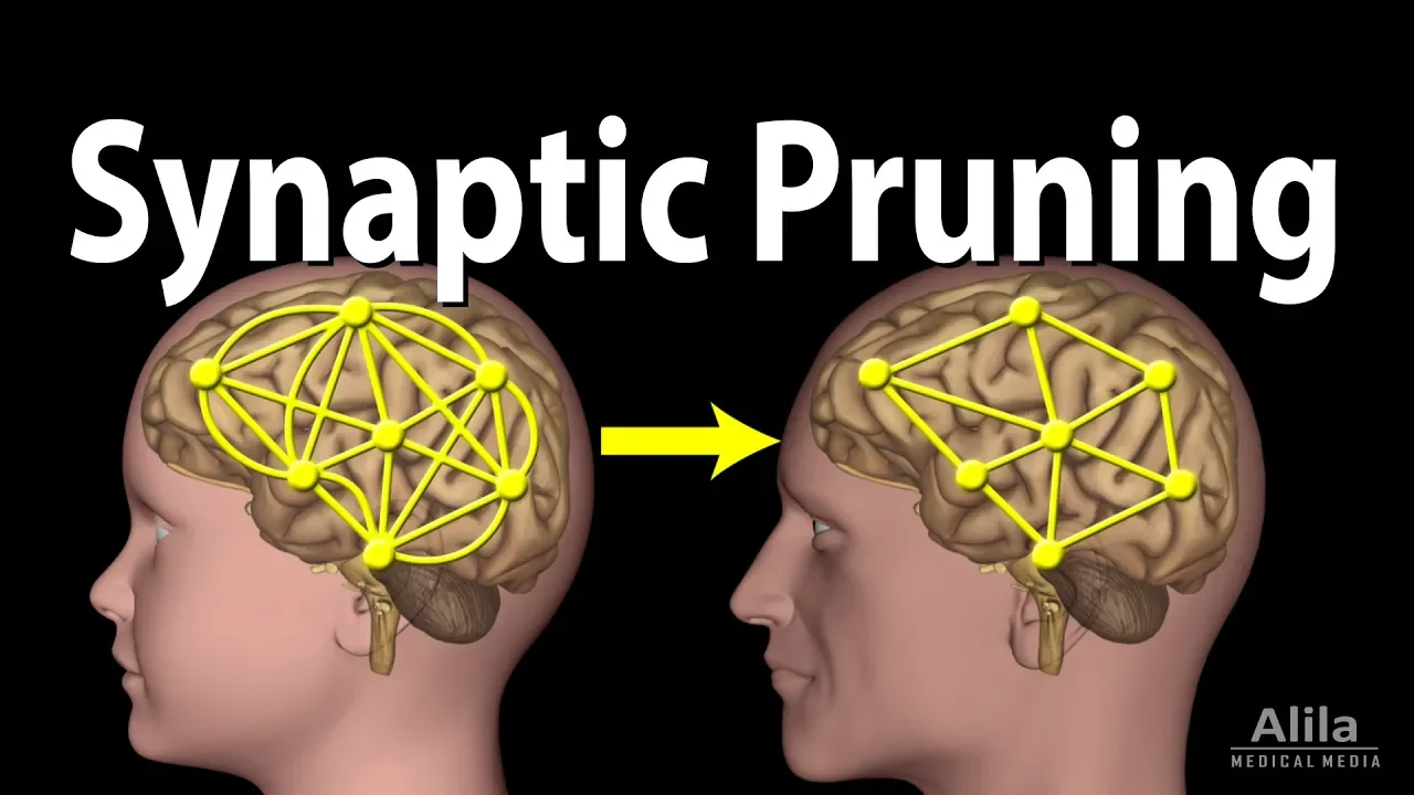
Microglia, the brain's resident immune cells, actively participate in sculpting neural circuits during development by engulfing and eliminating excess synapses through a complement-dependent pathway involving C1q and C3 proteins; this same pathway may become dysregulated in neurodevelopmental disorders like schizophrenia and autism, where genetic variants affecting complement components (particularly C4A) are associated with altered synaptic pruning and connectivity defects.
How Immune Cells Help Wire the Developing Brain | Beth Stevens
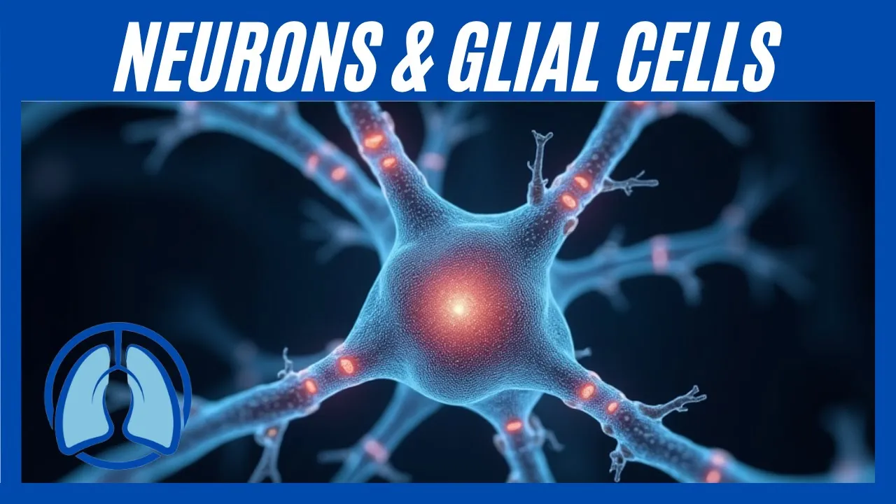
Added:Basic neuroanatomy and brain cell types, specifically distinguishing between signaling neurons and support glial cells.
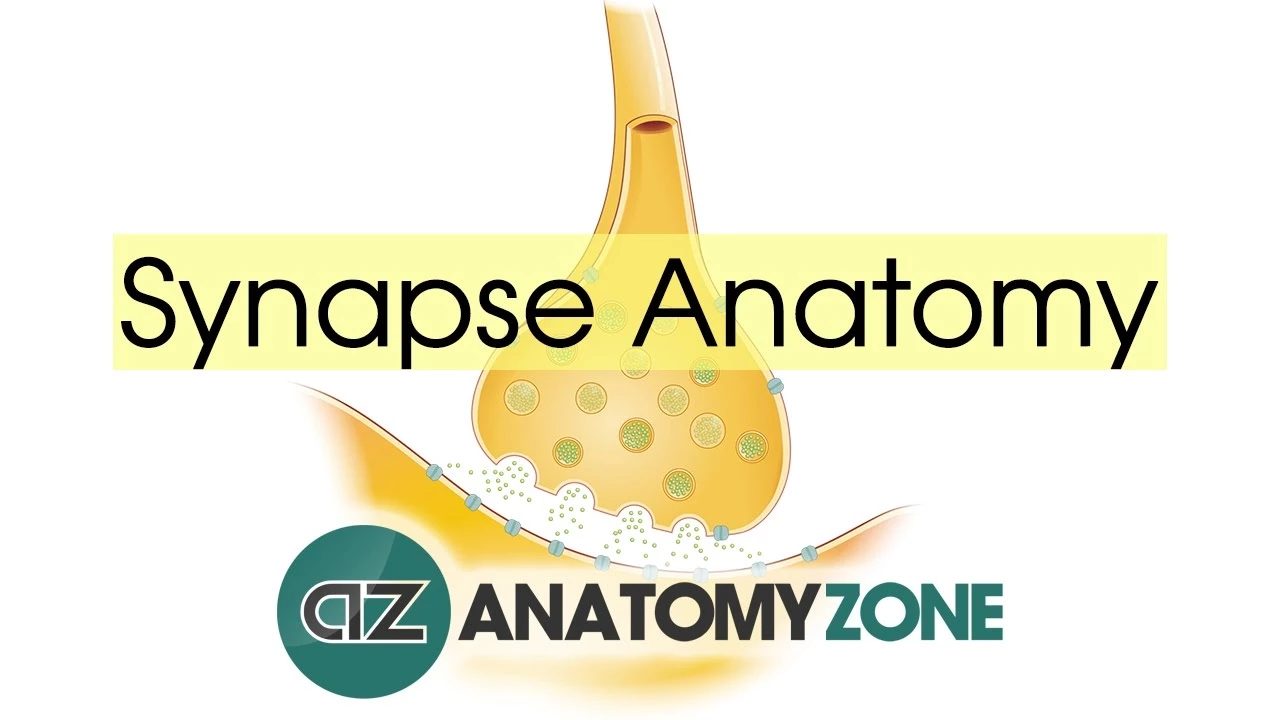
The structure and function of a synapse, including how neural connections are formed, maintained, and strengthened.
Fundamentals of the immune system, particularly how the complement cascade tags foreign pathogens for destruction.
The general concept of neurodevelopment, including the initial overproduction of synaptic connections during early life.
The link between aberrant or excessive synaptic pruning and psychiatric disorders like schizophrenia and autism spectrum disorders.
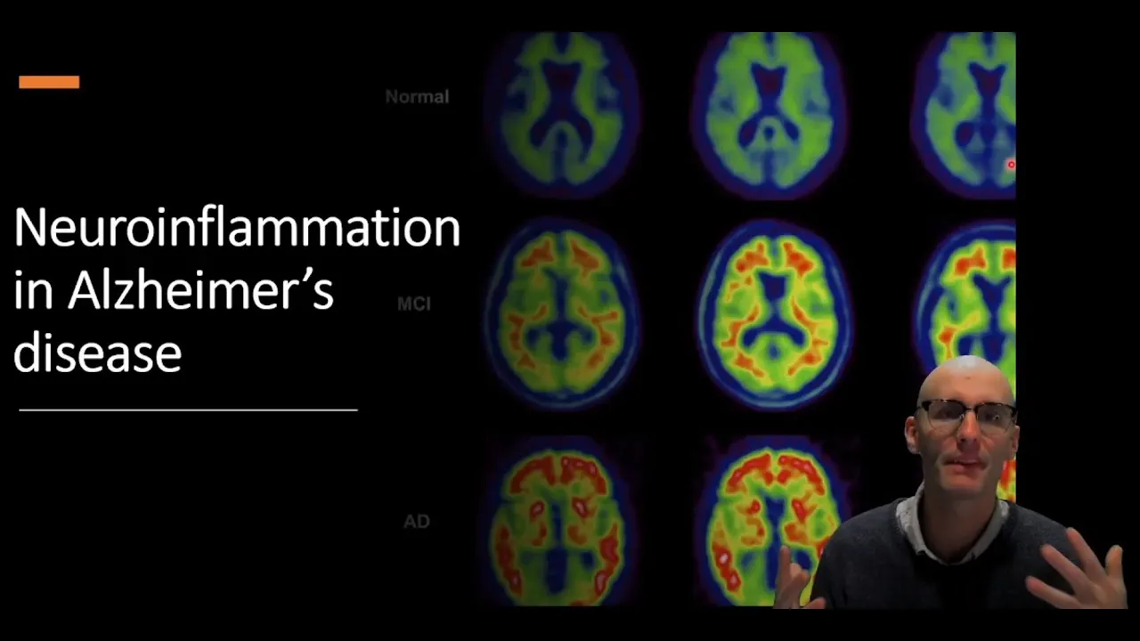
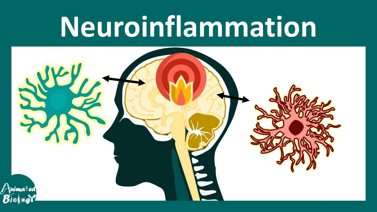
The role of chronic microglial activation and neuroinflammation in late-onset neurodegenerative diseases like Alzheimer's.
Current pharmacological research aimed at targeting immune receptors in the brain to treat cognitive decline.
Advanced neuroimaging and molecular biology techniques used to study live glial-neuronal interactions in model organisms.
174 views0likes1:02:47Original Release: 2024-07-10
okay thanks so much Julia and uh to Simons foundation and Safari for the opportunity and the invitation to be here tonight to tell you about um just our some of our work so you know it's been long known that the nervous system and the immune system interact um in multiple levels mostly in the context of disease or injury when the blood brain barrier is breached you know that you can have interactions between these two systems but I think over the last decade or so it's become increasingly clear that there's actually a lot of interactions between the immune system and the nervous system even in the healthy brain and we now appreciate that a number of molecules traditionally associated with the Adaptive and the inative immune system are actually expressed in the healthy brain by neurons by gal cells all the time and in particular they play um they're more robustly expressed uh in some of these at least during development when the brain is wired up um so today I want to tell you a bit about um sort of how we got into all this and I'm going to focus um my attention uh on this cell here microa our resident immune cells and until recently much of what we knew about these cells was in the context of disease and injury um and and what I'm going to tell you about today is that these cells um play a really important role during development they help sculpt developing neural circuits and synaptic circuits and I'd like to tell you a bit about what we know about them how how they do this in the normal development of a mouse visual system which is the model that we've been using and then in part two I want to segue into new research that suggests that this immune related pruning pathway we've been studying could become aberrantly activated or disregulated in the developing brain and this may be important and and play a role in neuros psychiatric disorders like schizophrenia and and potentially autism okay so let's just start with with this image which I always like to start with kahal right so the wiring diagram the brain is remarkably complex as illustrated by this beautiful image um the human brain contains 100 billion neurons trillions of synapses yet somehow each neuron manages to find the right connection with the right cell they re receive thousands of these connections to form functional circuits that control specific behaviors it's really mind-boggling right however uh despite how beautifully precise the circuit looks we're not born with such Precision actually we start out with a bit Messier uh circuitry so neural circuits underglow tremendous degree of remodeling during development um synaptic connections the connections between neurons constantly form and break um and actually it's this refinement or sculpting process that I'm going to be talking about today it's why a child's brain is so plastic why my daughter for example who's who's six years old can learn French seamlessly where I as an adult cannot um there's a lot of um uh really different features about the developing brain and this idea of plasticity how is that how does that come about well we know if you look at this diagram that initially um there's an excess of synaptic connections this is a a diagram taken from huon looker's uh work that just illustrates that if you were to look through development at different times and places in the brain you see that initially this um this wiring diagram is such that there's an excess of synaptic connections and then through a process called developmental pruning or synapse elimination a large number of these extra connections get permanently removed through a process called pruning now it's known that this process is a good thing during development you want to prune some of your synopses right this is the way that um we sort of the idea of use it or lose it connections that are meaningful to us get strengthened and maintained and those that are less meaningful get eliminated and so we also know that it's necessary for precise brain wiring and connectivity and it's thought that defects in this pruning process or refinement process could underly neurodevelopmental and neuros psychiatric disorders so one of the major questions that we've been focused on for the last many years in my lab is this question of how is it that synapses get eliminated because what we now appreciate mostly from the work in animal models is that this is an incredibly precise process not all synapses are randomly pruned specific synapses get pruned and so illustrated by this cartoon you can see this neuron this post synaptic neuron is inated by red and blue inputs these are the axons coming in and making synopses onto those cells cells and initially you'll see lots of inputs and over the course of development you can start to see that some of these inputs like the blue ones get removed but the red ones remain some of them and some of them even get stronger so the question is how does this work one of the things we know that regulates this process of pruning is neuronal activity it's known that activity sets up a local competition in such a way that the input in this case the red one that's able to more efficiently fire the post synaptic cell wins this local competition at the expense of its nearby neighbor in this case the blue one so we've known this for many years this is true in the peripheral nervous system which has been beautifully studied in our muscular Junction we know it's true in many parts of the brain especially sensory systems like the visual system where it's been well established but the question we've been thinking a lot about is what are the molecules or what are the mechanisms that distinguish the red from the blue input could there be molecular tags if you will that say Okay eliminate the blue one but not the red one could there be protective signals that protect certain inputs from being removed and and also where do they go do the neurons retract them and take them in or are there other ways by which these synaptic connections get pruned now most of the field with good reason has focused on mechanisms regulated by neurons themselves intrinsic mechanisms or signals that are regulated in neurons and not surprisingly these are the cells that are getting prune that makes sense but emerging evidence implicate gal cells in this process of pruning now Gia as as you just heard from Julia they make up the other half of the brain roughly um they are a number of different cells oligodendrocytes asites and and microglia in the central nervous system make up over half the cells of our brain and we now know from the work of many in the field including work from bun baris's lab my my my mentor and many many others now that the gal cells are actively communicating with neurons and synopses and one of the jobs they do during development one of the many jobs they do is they help to sculpt synapses and help to regulate their formation their plasticity and and their elimination today I want to focus on although asites have been quite quite well studied in this context of of synapse formation I'm going to focus on this guy over here microa and I just say that as a Developmental neurobiologist and a hard carrying gal biologist I've completely ignored these cells until about the last 10 years or so and that's in large part because as a developmental neurobiologist they really weren't thought to sort of be there early enough to be playing such a big important role in development but we now appreciate that these cells actually enter the brain extremely early in development embryonic development and I'm going to tell you a bit more about microa to sort of set this set the tone here so first of all it was drawn in this diagram as a sort of immune looking cell and of course they are our resident immune cells but they're actually quite beautiful they have lots of processes as you can see here they tile the brain they make up about 7 to 10% of the cells of our brain give or take and one of the things I'd say the first game changer for me was um was a was a really beautiful and important fate mapping study done by Mar mad's lab and floron Janu where they basically asked the question where do these cells come from and where do they how do they get into the brain and when did they get in the brain and this was a paper that basically showed that it despite the way it's always been thought the Dogma was that they come in through the peripheral nervous system through the peripheral immune system the circulating macras sneak in after birth and they basically become microa so that's suggest a sort of later step we now know that they come from the Yol saac they're a special progenitor of tissue derived macras that go into the brain in a mouse as early as embryonic day 8 right so that says that these are the first gal cells in the brain they're there before any of the other cell types in fact they're there when tons of major developmental Milestones are happening neurogenesis migration pathf finding when all of the layers are being formed microglia are there and I would say that nothing's still known about how they potentially regulate any of that so they're there early now we do know a lot about these cells because they are well known for their roles in injury and disease so there are resident immune cells meaning they do have similarities to their counterparts in the immune system macrofagos um we know that they have both good bad good and bad roles in the brain in the context of disease we know that they um undergo dramatic changes in morphology they go from this sort of fine sort of procy uh sort of uh very beautiful looking cell to this what I would call an angry looking cell they pull their processes in they become more fago cdic meaning that they are capable of engulfing things more they express different markers we know that they're good guys and bad guys meaning that they can certainly promote and and regulate neuroinflammation in the brain they release a lot of things like cyto kindes and inflammatory molecules that could be harmful they also can do a lot of good things um this are sort of one of their homeostatic roles they can clear debris apoptotic cells pathogens like bacteria and they're really good at removing toxic proteins and this has been really well shown in in Alzheimer's disease for example where they're really good at clearing amalo beta plaques so this is just to give you a picture of their sort of good roles and bad Ro but again in the context of disease one of the things unlike any other cell in the brain that they're incredibly good at doing and it's not to say no other cell can be fosic but they're incredibly good at eating things engulfing things and this is illustrating here by a movie that was taken by my colleague Axel nimron uh where in Vivo Imaging studies reveal that if you look in the cerebellum of a mouse this is a pingi cell shown here in red this is a micral cell hanging out next to it associating with it and what Axel did in this experiment is he damaged the pingi cell purposely and then asked what did the microa do in response to that injury and what you could hopefully appreciate is them literally pulling up and eating bits of that neuron right and actually we now know from others that they can even engulf things that aren't undergoing apoptosis like during early development they can actually eat a a live cell a live neuron right so they they are really good at eating cells and debris so this we knew but one of the things that we can now appreciate because of tools that are now available to us in the field is that we have these reporter lines right these microa reporter lines using this fractaline receptor egfp Mouse so all of the microglia are labeled green fluorescent um this was a mouse developed by stefany and using this mouse it enabled scientists to look into the brains of mice and ask what are these cells doing in different contexts and these are some Pioneers uh some of some of the these folks including wow and Demetrius were here at NYU and made these discoveries Axel nimron was the other and what they showed is when you actually looked in the brains of a mouse and said okay what are these microa doing they demonstrated that they're incredibly dynamic cells all right they can respond dramatically to injury not just by eating things but their processes move they can chemo attract towards the sights of injury this is Illustrated in this movie where again using a mic putting Mouse under the scope watching microa that was an example of a local injury and literally the microglia processes recruit towards the sight of injury very very uh robustly now that's in the damag uh case and we've a lot of people been been focused on that but one of the things that I became very interested in is this other observation which is even without injury if you just watch them what we now appreciate is that they're always Dynamic their processes are always moving they're constantly surveying the brain perena all the time and it's one of the unique features of these cells no other cell that I know of does this and this raises a number of questions that became of interest to me which is what is it that they're surveying and in particular since much of the work had been done in the adult brain the question is what they might be doing during development right so one of the things we now appreciate um that they're surveying are synapses so if we overlay neurons in this case a neuron this particular neuron was labeled with a th1 RFP so you can see the red spines these are the synapses here overlaid touching the microglia this is a movie taken by my graduate student Janelle in Vivo Imaging an a wake behaving Mouse actually in this case now you can see that one of the things they're constantly sampling and Sur are the synopses the connections between two neurons right they're doing this all the time very dynamically and we also appreciate that they can respond to local changes even changes in the in the context of firing so if a neuron is firing more the microa no they can recruit themselves and touch synapses differently depending upon whether they're more active or not this raises all kinds of possibilities for what these cells might be doing in the context of normal development and that led us to wonder could they be remodeling or pruning synapses during development so what I want to do is tell you a bit about uh how we uh how we learned about this and and the kinds of experiments we set up to test the hypothesis that microa are sculpting circuits sculpting synapses and how they're doing it and then I'm going to segue into implications for for disease okay so one of the things we appreciated when we looked during development now from the time the animal was born until about the second or third postnatal week uh is that the microa are undergoing dramatic changes if you just stain the brain with microa markers like this or use that reporter line you can hopefully appreciate that they are dramatically changing their shape and their morphology and if you then overlay um markers like L cd68 which which actually uh looks at their aiic capacity their lysomal activity what we also appreciated was that they were more fago ciic during this early phase in the development in times and places that were um happening during PR in so in Windows of pruning different parts of the brain prune at different times and in parts of the brain like the visual system that were undergoing robust remodeling microa were particularly fosic and that led us to ask well are they actually sculpting or engulfing these synopses so to address this we move to the mouse visual system which is a really a really terrific model for studying synaptic pruning and synapse elimination um one can look at the synapse between the retina the retinal gangan cells and the retina that project to the visual ual Thalamus the dorsal lateral geniculate nucleus the relay the relay neurons in the visual Thalamus this just shows that if you put dyes in the m and The Eyes Of Mice red and and blue you can watch where their projections go and that uh image I showed you earlier was a zoom in of one nuron that happens to be right there and that shows that early on a postseptic cell is interated by axons from both eyes shown here but by maturity you can see that there's a clear elimination of one eye input and a strengthening of the Curve eye input so that's actually pruning of eye specific inputs and we knew when and where this happened thanks to the work of Carla Mason and Char K shats and many others but now we wanted to Overlay that with our tools to study microa and so that's where Dory came in my first postto fortunately for me I recruited her to my lab she very clever and she came up with a way to test the idea we knew that the microa here in green were interacting and touching the inputs from the two eyes right so they're touching all these little puncta these are the endings of the red gangan cell inputs and what Dory asked is well okay they're touching them but are they actually eating them are they engulfing them and so to get at that question she labeled the inputs like like I just showed you and then she used that reporter line and said well if they're engulfing them we should see bits of those red and blue puncta within the lomes within their bellies and processes and pretty much what she showed is almost every microgo we surveyed throughout the visual Thalamus was full of synaptic inputs from both eyes and this was quite robust and reproducible but importantly um what they seem to be nibbling by the way is the pre synaptic terminals um they're like basically plucking off the endings of the of the axons where the preseptic elements are but importantly and quite relevant for what I'll talk about in the second part which is relevance to disease is that this is tightly regulated process it's not happening all the time it's happening in these windows of development and in particular in this uh part of the brain this eye specific segregation or prunings happening between about postal day 5 and 10 we see a lot of microG engulfment then and then they really diminish and decrease their capacity to do this after that we've since gone on and identified a second window of pruning that happens when the eyes open and when the feedback from the cortex is coming in and we actually think from the work of our lab and now many others that there are multiple windows of microG Gia pruning that are happening in different parts of the brain and there are windows in critical periods they don't happen they're happening in very precise way raising many questions about the mechanisms now around the same time we uh uh made this discovery another group um from Cornelius gross's lab made a very similar observation in the hippocampus so this says that it's not just the visual system this is the model that we use but it seems to be happening in other brain regions they use a different Mouse this fractaline receptor knockout Mouse which is a receptor that's only on microa and when they looked in mice that don't have this receptor they noticed two interesting things there are about half as many microG GLA in the brain for reasons unknown actually during development and one consequence of that is that you saw um too many sort of a lack of pruning uh so you had you had a decrease or you had a difference in this in the number of spines and synapses their maturation defects were happening and importantly and quite interestingly when they looked at another study there was actually evidence of a a weaker functional connectivity not just in the hippocampus but in the connections between the hippocampus and the prefrontal cortex they actually were um essentially you know hypo wired or less functionally connected now this is based on sort of fmri bold Imaging which is I think intriguing we don't understand it yet but it's evidence that if you mess around with something in the microa and their ability to interact with synapses one of the consequences of that is that the functional connectivity in the brain at least in a mouse is different and I think that has some relevance and significance for for way thinking about this in terms of um their contribution to brain to brain wiring now what are the mechanisms so it's one thing to say that they're there at the right time in the right place we have evidence that they're engulfing synapses how are they doing this and this brings us back to the same question I started with at the beginning was we really want to know what makes the red synapse maybe different molecularly from the blue one could there be molecules or cues that tell the microa engulf me the red one the blue one but do not engulf me the red one so this brought to bear a work I did as a postto in Ben baris's lab Where We unexpectedly in through an unbiased screen initially we identified um a role for a group of molecules called complement which are molecules traditionally associated with the um with the innate immune system and this was a surprising finding back then because these molecules really weren't thought to be playing a role in the healthy developing brain but lo and behold we found that the molecule called c1q which I'm going to tell you more about in a minute this is the initiating protein of the classical compliment Cascade and we show this this this protein at both the MRNA and the protein level was very highly expressed in subsets of those retinal neurons these are the neurons that are getting pruned during peaks of pruning so during the time that this pruning is happening and that when we knocked out c1q globally right these are c1q knockout mice or C3 knockout mice which is a downstream compliment molecule these mice failed to prune properly they they failed to segregate into these eye specific territories and they remained multiply innervated throughout actually throughout life and even if you look at an adult Mouse they have too many synapses so that told us that this group of molecule was somehow involved in pruning but we really had no clue about how this could work because there was very little known about the role of these molecules in the brain now interestingly Carla shatz is lab had shown a number of years before than another group of immune molecules called MHC class one this is more class thought of in relation to adaptive immunity she also showed a group of these molecules involved in pruning in the same system she also found MHC class one through an unbiased screen and um interestingly mice that lack MHC have a very strikingly similar phenotype to these guys now work that Carla and I are now thinking about doing collaboratively is going to ask whether our molecules could be interacting on Cell level so we don't have an answer to that but it it's an example of how molecules that we traditionally think of as of immune molecules they are actually in the brain and they seem to be playing an important role in pruning and developmental uh refinement so how could this really be working right so what was um intriguing about compliment unlike the MHC and some of the other immune molecules that are in our brain was these these actually a group of secreted immune molecules right and so their main role in the immune system is to tag apoptotic cells or or bacterial cell for Rapid removal it's our first line of defense against an and C in the periphery so before your slower adaptive immunity kicks in compliment comes in and one of the things it does is it binds to let's say that bacterial cell once it binds that activates this proteolytic Cascade that leads to the cleavage and the downstream activation like a domino effect of all the other compliment molecules Downstream and one of the ways that compliment removes or eliminates that cell in the immune system is that it tags it C3 tags it for removal and one of the key ways it gets removed is by those circulating macras that have receptors for compliment and that's one of the ways that it gets actually engulfed so we started thinking about could in the brain compliments Mark or tag subsets of synapses during development for removal by microglia that we now know also have receptors for complement so this led to this idea that maybe in the in the brain the brain was co-opting this system but in a very different way um and so the the hypoth is is that complement molecules were binding to subsets of immature synopses perhaps the less active synopses and that that could be initiating and telling the microa to engulf those synapses indeed when we looked in mice that lacked the receptor on the microglia so we got rid of that genetically or the actual tags themselves the compliment molecules no longer were microglia as efficient at engulfing synapsis and and importantly and I think also something we're thinking more about in terms of the the functional and behavioral consequences of this if you look in multiple parts of the brain even in the adult mouse that lack compliment they have a sustained defects in this pruning they have too many inputs in many parts of the brain including cortex and one of the um outcomes of that is a hyperconnectivity and it turns out that David Prince's lab for example looked at our c1q knockout mice and did EEG recordings and he he's he studies epilepsy and indeed when he looked these mice were having obson seizures and essentially that was one of the consequences that may be related to the fact that these U too many of these synapses were there because of a failure to prune and so now work in the lab using much more specific approaches where we can knock out compliment not just everywhere in the body but only in certain cell types in different times and places we're now running these mice through a battery of functional and behavioral tests to ask the question which I in my mind we still don't know the answer to which is what are the function consequences of too much pruning what are the functional consequences of not enough pruning and at least in the mouse we have a way now because we have a pathway we can manipulate to start to ask that not just globally but at a circuit level and that's some of the work that we're that we're now moving towards doing okay so one more thing I want to do thinking about mechanism I want to take you through the way we're thinking about this because we certainly haven't solved all this we're sort of in a way at the beginning of our understanding but we now that we've identified molecules and these cells now we can start to think about how this is working and I would argue that understanding this quite deeply is going to be I think quite important for understanding how this may go AR in a variety of neurological and neurod degenerative diseases so if we can understand how this normally works this might provide novel and new insight into how to protect synapses when there's aberant synaptic pruning so one of the things I told you at the beginning is that synaptic pruning isn't a random process and it's activity dependent remember the the less active or the inputs that are less efficient at firing the postoptic cell preferentially get lost in many of these models we wondered could microa be preferentially engulfing the less active input right and so we actually uh had the ability to ask that question again in the visual system because we can manipulate activity in the eyes of a mouse you can block activity in one eye with tooto toxin for example and you can um increase the activity of the other eye and using this model because we had a readout which is engulfment we could ask the question do microa preferentially engulf inputs from the less active eye versus the stronger eye and what we showed was in both cases when we manipulated in different ways there was a preferential engulfment of the less active eye input and that suggests that the Micron know it's less active somehow there must be some way they know that it's not as as as strong and that raises this question of molecules again and so we didn't EXP experiment this is unpublished where we basically did that same activity dependent competition experiment but we did it in our complement knockout mice our c1q knockout mice and when we get rid of compliment remember that's the initiating signal that we've discovered initially that got us into all this when you look in mice that don't have c1q microglia no longer care they don't go for the weak one they don't have preference at all so that's a hint that somehow compliment is regulated by activity and maybe this could explain this selectivity and this is the way we're envisioning this and this is two models that I'm going to tell you about that we're um exploring and investigating and I would say these models are not mutually exclusive so one idea is that complement molecules c1q C3 and all of the components of the Cascade which we now have evidence are there in the brain during this this window one idea is by virtue of the fact that these are secreted molecules that they could be selectively tagging subsets of synopses and we predict the less active input based on some of our our preliminary and and other data so that model is attractive um and it suggests that there must be some receptor or molecules on those synopses that bring complement to it so we're of course very interested in identifying what those molecules are what are the compliment receptors so under that condition anything that has compliment on it and if microa are surveying and they have the receptors anywhere what compliment is they will recognize and engulf it so I think that model while we have data to support it is far too simplistic because never does it work like that it never works so simply um so the other model that I think is working in concert with a tagging model is this protective model and that model suggests the following that yes you have complement and it could be selectively binding to specific synapses but maybe you also have a group of protective molecules that are shielding or protecting those inputs let's say the stronger ones that are more that you want to keep that it's the synops that would lack the protection but would have the complement in this model that would get removed and we now have evidence that I'm not going to have time to tell you about today but just to give you a sense of the way we're we're thinking about this Emily lman in my lab discovered a role for a group of a molecules called cd47 again an immune molecule and in the immune system this molecule is considered what they call a don't eat me signal these are molecules that protect our cells our healthy cells from being randomly eliminated by macro Pages these are molecules that protect um self versus non-self cells from being removed there's a bunch of these molecules in cd47 came to the top of our list because it's always also enriched in our retino gangan cells and in neurons during development and what Emily showed is if you get rid of that molecule microglia overeat they eat too many synapses and it has the complete opposite phenotype is the compliment knockout mice so this is an attractive idea because it suggests also because microa have the receptors that recognize that that it's sort of like this molecular code and that there are groups of these molecules like complement that work together with these protective signals and this is attractive also because it might explain why microglia don't keep eating the entire process or the whole cell what stops them from doing that and these molecules the cd47 is everywhere on the cell and we have evidence now that it gets selectively downregulated just on parts of the cell and that might explain how they can recognize and prune specific cells and specific parts of the cell so this is work in progress um but I think um between the two signals that to us it sort of gives us a way of conceptually thinking about how especially secreted immune molecules could be doing something as precise as pruning so what I've told you is um we've identified this pathway this complement and microa pruning pathway and um and by no uh means is this the only way that we prun synopses there's many other molec I mentioned a few already but we've identified this this particular pathway and we've been working hard to understand how it's working but I think what's important to note is that this pathway is tightly regulated and just like the immune system these molecules are tightly regulated when they're there when they're on when they're off there are brakes on the system and we think that those brakes are as important as the signals that turn it on and that if you don't have the brakes in place or if you have things that increase its expression or its activation this could have important consequ quences for brain development because normally this thing gets turned down you'd want to have sort of these pruning mechanisms down regulated during the healthy mature brain at least in most parts of the brain but what if all of a sudden these pathway becomes aberant activated or too much pruning is going on this might uh uh underly or contribute to um uh brain connectivity and synaptic connectivity defects in in diseases and disorders like schizophrenia autism and epilepsy so so we've been thinking a lot about this and and and I'm going to tell you now evidence um that it implicates our pruning pathway in schizophrenia and I'm also going to end the talk with some potential ideas about how this may be relevant to autism although we're really quite early in in this process but I think the work in the schizophrenia and the work we've done in development are informing um experiments to start to think about how we might try to connect these ideas okay so schizophrenia um is obviously a devastating neuros psychiatric dis disorder uh it has a number of features that are quite distinct from autism and one of the things that I think is clear is the age of onset is quite different often it's adolescent you know late late uh late adolescence is where you often get onset of schizophrenia now there are several lines of evidence uh that suggests synaptic pruning or synaptic loss might be happening in schizophrenia but um this probably this is the example we all show this is work from David Lewis's lab that shows in the prefrontal cortex of some individuals with schizophrenia there's a marked decrease in synopses in those dendritic spines as shown here there's a sparsity of spines and quite intriguingly not everywhere in the brain but in the frontal cortex which is our association cortex and involved in executive function and and this led to all kinds of theories about pruning in in schizophrenia and I would say that these are interesting ideas but it's very hard to say that a loss of synopsis or spines is really a pruning problem and that is because by the time you get these brain autopsies samples you don't know what happened when it happened if it's loss never formed if it's cause versus consequence is it really pruning is it degeneration we just don't know and it's one of the reasons why we've had a hard time trying to test this hypothesis because there also are no good animal models to get at this question so there's also evidence though to support a cortical uh a pruning idea or hypothesis in that human Imaging brain Imaging Studies have also shown evidence of a cortical thinning in patients that later go on to um present and and and um and become uh schizophrenia uh actually this is happening even before the onset in this prodromal stage again an intriguing observation but is it really pruning right we still don't know so this is where human genetics can be uh extremely useful um so in the next few slides I'd like to share some very new and exciting um now recently published findings by my colleague and collaborator Steve MCC Carroll and his graduate student ashon Sear who have been using human genetics U to try to identify mechanisms and novel Pathways for for intervention okay so a potential clue uh for for schizophrenia um was hypothesized to involve u h schizophrenia is strong association with genetic uh uh markers across parts of the human genome on chromosome 6 in particular a part of that uh a chromosome called the major hyto compatibility complex or MHC so it's been known that this MHC Locus is what for common variant is a whopping effect on schizophrenia risk right almost twice as strong in this Manhattan plot as the next strongest signal in any other chromosome it's been known for a very long time so you know kind of where but the big challenge has been which Gene there's been no one gene identified or genes identified that that that explain this risk and this region if you zoom in on it actually spans hundreds of genes and interestingly many of them in COD uh uh proteins that are traditionally associated with the immune system both adaptive and innate immunity and so this has inspired all kinds of theories about an immunological cause or of of schizophrenia uh and it's been very hard to tackle um because it's been really a very intractable problem due to the complexity and the fact that really it doesn't follow any mathematical or statistical patterns that it can explain any known variant so it's been this sort of no one knows what to do with it and no one wants to go there because it's too complex so this is where um Ashen who is a graduate student with Steve decided to um not be uh daunted by that and to try of dig in and start to think about you know what could explain this right and so basically Steve and and ashon had been studying uh uh extreme forms of structural genomic variation that could explain risk and it turns out that in the MHC if you zoom in it actually contains um two genes c4a and c4b so this is the compliment Gene that's right in the smack dab in the middle of the pathway I just told you about in part one um they had been noticing that the locus here contains two C4 genes but there are many different alal and we sort of knew that from the immunologist like Mike Carol and others have been studying that in the concept of immune system but what Ashan discovered is that this Locust contains remarkable structural variation some people's genomes for example have multiple copies of C4 a some have multiple copies of B some have one but the other and some have both so the Hao type is way more complex than I thought before there are many different alals in humans and this raised the possibility that in the hypothesis that Ashman raised because C4s is situated next to one of the strongest genetic uh signals could the structural variation or the form of C4 help explain the risk and in particular the other reason is because C4 is actually really important in regulating the activation of the c c Cascade if you get rid of C4 even if you have tons of c1q that Cascade cannot go on so it's actually a very important uh process uh molecule in in the regulation of compliment so does variation of C4 could that be underlying um schizophrenia risk and so the challenge though although uh ashon had developed these really uh novel techniques to measure the structural variation in tens of thousands of individuals based on their DNA the problem was how do you get at this and Link it to schizophrenia so what Ashwin and Steve did did is they developed a novel way to map how the structural variation of C4 how the C4 structure relates to Snips for which there was already data available from tens of thousands of patients right so they built a map essentially to infer the four most common forms of C4 so there was lots of structural forms these are the most common ones they wanted to know how the most common forms of C4 related to the the snip data that was surrounding the gene so they essentially created these molecular barcodes from the snip data and it works because humans share long genomic segments that then they have inherited from their common ancestors and this map then allow them to take advantage of the fact that the snip data was available from 28,000 schizophrenia patients and 34,000 controls in 34 countries so now they actually could expand this and use the power uh of big data to try to test this idea and I'm going to now summarize in a very one slide which is a very unfortunate thing to do considering how much work went into this and this is recently published so I encourage you to to read the paper but what they found is that the alals of C4 appear to uh shape the risk of schizophrenia in proportion to their effect on c4a expression and so on the left what I'm plotting is um the Al's risk right so these have four alals and in its relevant risk to schizophrenia and what you can see is that regardless of the MHC hype those that have more a have an increased risk it's very clear and even more interestingly on the right what I'm showing you is each Al's effect on c4a expression which they were also able to measure in a number of patients and samples and what they show is if essentially a is related to how much a or C4 a you make so the higher the a expression increased risk as well so now the important point from all this is that the more c4a you make from the locus the the greater the risk you have of developing schizophrenia so this is was a huge genetic finding because it identifies one of the genes of the most significant uh Gene within that Locus that could explain uh that signal but it leaves many questions about how C4 a whether you have more A or B how could that relate to schizophrenia in terms of biology how does elevated C4 contribute to the path pathobiology of schizophrenia this is the question where we kind of come in in terms of you really now just need to start to think about the biology like what do we know about compliment and what do we know about compliment in the brain and that's where um a really fantastic collaboration between Steve's lab my lab that's been studying complement and pruning and Mike Carol's lab who's been studying C4 and in immune system for his career um all of our Labs started coming together we're all local at Harvard and uh it was an ex great example of how multi-disciplinary collaboration from investigators that probably normally wouldn't sit around the same table together we started started bringing our Labs together having uh weekly lab meetings and it's led to a whole new line of investigation and that we're really only in the beginning of our of our understanding of how this is working but I want to share with you some of the data and also where we're going with this one of the first things we did is we asked where is C4 protein I've told you everything about the message and the transcript what about the protein well we have really good antibodies for C4 um and we can then take advantage of the fact that we have brain Bank tissue from the Stanley Center and other other sources and we could just basically ask where is C4 in the brain and we got um a small cohort of schizophrenia versus control brain samples and we just stain the brains for C4 and what we showed is that in uh that much like what we observed in the developing Mouse brain C4 is quite punctate and it was localizing to synaptic uh elements synaptic other synaptic proteins in certain brain regions like the hippocampus um and this just told us you know we didn't do an extensive analysis to ask do schizophrenia patients have more C4 in their synapses we're doing that kind of larger scale thing now but it told us that the C4 protein was there and it was localizing to subsets of synopses in the human brain and in in parallel to that um what we did um Heather de Dera in Steve's lab grew human um primary um cortical neurons and let them develop and make synapses and then stained those neurons for C4 and also showed a localization of C4 to aptic structures shown here by labeling pre and post synaptic markers tagman and psd95 so again in a human neuron at least in a dish we saw complement going to synapses especially as that neuron matured and quite interestingly you can then um these are almost pure neuronal cultures you could also measure C4 from the um condition media so the neurons were actually secreting it as well okay so compliment C4 is there it's at synapses at least in these conditions does it actually contribute or have anything to do with pruning so we um then went back to the mouse right so everything I just told you was human data now we're going to the mouse because at least in the mouse we have models for studying pruning and we went back to our favorite model system the retinogeniculate system and we collaborated with Mike Carol again and my my former uh graduate student who's now postto in Mike's lab where we basically carried out the same kind of um eye specific segregation pruning experiments in the mouse in mice that lack C4 so C4 knockout mice and basically asked are the mice that don't have C4 do they have defects in pruning and what we found is that much like the C1 q and the C3 knockout mice that we'd already shown the C4 knockout mice also exhibit these pruning defects um that actually phenocopy the C1 q and the C3 knockout mice and that C4 was made by other cells but also made by those retinal gangli cells the cells that are getting pruned especially during development and when we did our eye specific segregation or our pruning assays we basically showed that mice that lack C4 don't prune properly they don't segregate into these nice eye specific territories and it looked a lot like our C1 q and our C3 knockout mice so that puts C4 right in our pathway it's providing more evidence that the whole pathway is activated much like it might be in the immune system all these components are in the brain and at least during this window of pruning they're working together to regulate this pruning process at least in the mouse but as you're probably thinking yourself okay it's a mouse and you're showing getting rid of something like C4 um has pruning defects how does this in any way relate to schizophrenia to spine loss in the prefrontal cortex to the path of biology of the disease well that's the hard part and that's where we have um to work together moving forward to try to get at this idea to try to test the hypothesis that c4a may lead to an overactivation of the compliment Cascade so having two much of a good thing and that that that might contribute to synaptic loss or synaptic connectivity defect in schizophrenia right and the challenge of testing this hypothesis is that we do not have Mouse models of schizophrenia we do not have um mice that have prefrontal cortex that do that resemble our human prefrontal cortex there's a lot of issues here but what I would say is that since the genetics have pointed us to this pathway and since we know a lot about its biology we're pretty well positioned to start to ask questions and start to test parts of this hypothesis and that's going to involve getting into this question of why a versus B for example why doesn't B do this why is it a they're so similar they're only they only differ by just a a few few base pairs why and in the immune system we know that A and B might bind different things so that's a clue but how do we even begin to get at that for example we talked about the visual system we really want to start moving our studies into areas more relevant to schizophrenia like the prefrontal cortex like the hippocampus so my lab is now um moving up to those regions of the brain and uh actually it's it's it's a bit daunting because there's been so little done on refinement and pruning in the frontal cortex and um before we can do these experiments we have to do a lot of groundwork we have to essentially try to do and matap pruning and refinement in that part of the brain in the mouse and I don't think anyone's really unfortunately done that yet and so now we're we're getting that groundwork laid and then we're using the genetics to then um make mice that are sort of humanized mice if you will that overexpress or underexpress the variant a versus B and then what you can do and Mike Carol's made these mice and we're working together on this um you can basically overexpress multiple copies of a the human form in a mouse or you can do the same with b and then you can use this to ask what the consequences are for pruning you can do it in a global setting which is what the M the mice that Mike has made or what we're also going to do in parallel with that is we're going to use viral approaches to overexpress in circuits of interest at particular points in development like adolescent period and then ask if too much a in vulnerable circuits leads to pruning and what the consequences are not just in terms of the number of spines but how the connectivity and the behavior might be affected so we're just beginning these experiments but as you can imagine there's lots to do um and a lot of questions that have now come to the top of my list of things that I want to focus on for the next decade or more uh and that brings us back to this question of like time an onset of schizophrenia which has always been really intriguing why does it happen in adolescence right and one of the things that I thinks really interesting is the frontal cortex is one of the last areas of your brain to develop and to prune and to mature so one idea is that the reason why you have um this later onset is that that critical period just happens to be later than the visual system and the other sensory systems that prune much earlier that's one idea um it's certainly not the only hypo othis but the other idea might be that pruning could become too intense um in some people for various reasons it could be a combination of genetic risk paired with another hit like an environmental hit for example could this pruning then be subtle but then expose other pre-existing vulnerabilities like this is something that we can start to think about with these double hit models now that we have the genetics behind us and so I think in the end of the day as I read more and more about adolescence and how little we know about about what's happening in the Adolescent brain and I just feel like this is a hugely understudied area it's also quite relevant to autism and so that's one of the areas that we want to really try to move forward and we hope that this might help us to develop better models of schizophrenia that can allow us to get at some of these questions now I'm going to end the last few uh minutes um with um speculations about how this might be relevant to autism and in no way am I going to convince you or tell you that we've got this worked out in any way except to say um this is how we're thinking about it and I'm going to tell you about some intriguing evidence that implicates microG autism but there are so many questions that we don't know there are a lot of gaps um and um and so the first thing I will say is that unlike the the genetics are not pointing to microa or or our immune molecules at least thus far now it's not to say there's not a genetic link but at least based on what we know I don't think unless Lou has some hidden information that I don't know I don't think there's any microglia genes that have been rising to the list of significance I think that's fair to say but there is evidence that microa are different or abnormal in some individuals with autism and it's been looked at not just in one study or one way but in multiple ways and I want to tell you about those but even still what we don't yet know and I think it's fundamentally important to understand is is this cause or is this effect could the microa have some more proactive role in the process or are they simply respond responding to something else and then they're sort of Downstream or could it be both like we just don't understand and um that's in part because we don't know how to study this yet right so here's what we do know that I think is intriguing data especially when put together you know there has been some um transcriptional profiling work done by initially Dan gan's lab and now others where they've looked in human autism and they've profiled transcriptionally different brain regions from individual with autism versus um healthy controls and there's been a number of genes at the transcriptional level that change and you can do all kinds of analysis to kind of get a sense of what the pathways are that are different and what he's shown using these approaches is that there are several modules of genes that change that kind of give hints to terms of like Pathways synaptic genes certainly are implicated both genetically and through the transcriptional studies but the other thing that his data revealed this was a number of years ago was a couple of modules that go up so a lot of the synaptic genes that are important for synaptic function are downregulated in autism but a lot of the immune related genes were upregulated in some individuals with autism so this was intriguing didn't really explain anything but it was an intriguing observation um and another study came out and showed a very similar thing using an RNA seek analysis also showed not only the same immune molecules that that Dan had previously shown but even more refined a group of modules that were really honed in on microglia microa in various different contexts so this was an intriguing U piece of data uh but again we don't know yet what this means the other thing that's intriguing is that there were some PET Imaging studies done this is one study in particular where they use this pet Lian this pk19 it's a tspo Lian it is not specific for microa I would say it's more of a a Lagan for uh neuroinflammation but they did a small cohort of of young adolescents with autism versus uh non they did this um brain Imaging and they showed more of a signal of this this U pet liend in individual with autism autism versus control and so that was again intriguing but by no way means says microglia are are mediating any of this or involved and there's been a number of studies actually on the pathological side and imunohistochemical Analysis where if you look uh at microa they look different they look more Angry activated there's more of them in certain brain regions again just shown here intriguing but we don't know what it means so this is the question I'm going to sort of end with and sort of I'm going to tell you a little bit about the way we're thinking about it based on um what we know uh from our work in the lab so the question I'd like to be able to address and I think the field needs to be thinking about a bit is how do microglia contribute at all to the pathobiology of autism if so how and I think most importantly when like what when could this be happening and as I told you today microa are in contrast to the way we used to think about them are intimately associated with neurons and other gal cells they're actively helping to sculpt the brain that's pruning but we all know now uh not we don't all know we now know uh through I wish we all knew but we don't all know uh that there's a number of other things microa do that are relevant um so pruning is probably the tip of the iceberg there's evidence that suggests are important in uh synaptogenesis pathf finding other aspects of development that could be happening either even as early as embryonic development so this idea that they could be a more homeostatic physiological roles of microa puts them in a different context so the way we're thinking about it is you know kind of two models that are not mutually exclusive one is that you know the idea is that genetic and or environmental factors could alter microglia function in some way and influence brain development and ultimately synaptic connectivity so one model is and this is largely based on the genetics that we know thus far is that genetics are pointing to neuronal genes and synaptic genes we know there's a lot of evidence of neuronal and synaptic dysfunction and you can imagine based on the fact that I told you at the beginning that the micr are acutely sensitive to their environment in this case you can imagine a neuron that is um essentially not functioning properly because of a mutation in shank or or another Gene that's been linked to to autism that the microa then would be responding aberant to that neuron that's either not firing properly or not behaving properly and that that may be secondary but you can also en Vision that no longer are microa carrying out their homeostatic good roles and that that could initiate some sort of feedback role in vulnerable circuits and brain regions that could then in concert with the neurons further contribute or exasperate phenotypes so this is something that we're actively studying the lab there some evidence that we recently published in the context of R syndrome where we showed in a mouse mod of R syndrome where you knock out mecp2 right the gene that's been shown to be causal to R syndrome if you knock that out everywhere um the microa misbehave and they overeat synapses in the time uh that corresponds to a synaptic regression but we also showed and that because we have genetic tools that when we tried to rescue just the microa they didn't it didn't explain all the phenotypes and if we knocked out mecp2 only in the microa it didn't cause phenotypes so this uh actually is consistent with now a number of studies that are emerging that suggest that microa could contribute but they're not caus they're not initiating the process but that doesn't mean that if they're not behaving that there's not important to be thinking about how to keep them in check so that's one model the other model though which I'm also again saying it's not mely exclusive is as I told you microa are part of the brain from the very beginning there's more and more evidence to suggest that early development embryonic development is is is impaired in some way and if microa through some genetic and or environmental insult are then changed during during this period of development that could in many ways alter the way the brain develops and this is just globally speaking there's no molecule here but it brings up this idea that if these cells are important in early development even before pruning even happens we need to be thinking about them and putting them into the equation a little bit more and what we now know since they enter the brain in a mouse at least very early embryonically and we started looking at microa in a very early embryonic brain versus a later one you can see how different they look early in development they're not everywhere they're not tiling the brain they increase in their numbers they're they're hanging out in different pockets in the brain it's almost giving Clues to what they might be doing during development and the other thing we know is they're undergoing these dramatic changes in development and differentiation we're just beginning to try to understand how that's working and what they're doing but we know so little about how they're normally developing and uh we started collaborating with Steve McCarroll and this is a safari funded project where we realize before we can ask what microbl are doing in autism models we best understand what they're normally doing in development and we better get a better handle on how they're developing and what makes a microa a microa and also instead of treating all microglia equal it might be that microglia are have unique sort of States if you will and we need to better understand what those States might be so one of the ways we're doing that is we're applying a new technology uh transformative new technology developed by Steve's lab Steve MCC Carol's lab called drop seek which is a single cell RNA sequencing techn technology that allows you to um simultaneously genomewide transcriptionally profile thousands of individual cells essentially allows you to digitally count the number of transcripts in each cell of each gene it's pretty powerful there's other examples of RNA sequencing that are going on in this way but we thought this would be a great way of getting at microa development and microa heterogenity both in normal development and then compare that to um genetic models of autism and then different insults to see how microglia development and States change over time and the other reason why we thought it'd be powerful to do is because we don't have good markers from microglia all we know is that they look different but looking different doesn't tell you anything about their biology we don't have a marker that says these are more fago cdic these are dividing more these are eating more synapses these are not we need to have a more molecular fingerprint if you will so that we can then go into these brain samples and better understand what microa might be doing in these contexts so using drop this is just preliminary we wanted to just ask are all microa the same in uniform all over the brain or could they be in different states could there be different subsets if you will of the same cell type and using drop seek what you can do is profile a purified population of microa from different stages and you can then ask are they shaking out into different subsets and so this is an example of preliminary data unpublished that just shows multiple different subsets of microglia States within the brain of an embryonic Mouse and when you look at what genes are enriched in these different subsets we see clusters of genes that are indicative of what they're doing so we can for example zoom in on this large subset versus a small subset and say what genes do they make I'm not going to get into the details this is just early days but you can see there are some genes like c1q that's made by all microglia independent of their subset but there are some subsets like this guy that's making a lot of igf-1 and interestingly when you go into the brain and stain for igf-1 igf-1 isn't everywhere in the brain it's in these little pockets which is now allowing us to zoom in and ask what that might be doing and we have other examples uh where there's other markers that are telling us a bit more about what their biology might be doing and now that we're getting that groundwork laid we can then perturb the system either genetically or environmentally challenge the mouse and then ask how that um how those States change and then ultimately identify candidates that are different and then be able to use gen ICS to knock out that candidate and see if we can protect some of the phenotypes that's ultimately where we would like to be able to go with this um so I just want to like I want to end uh now with just sort of the bigger picture and putting this in even a larger framework outside of neurodevelopmental disorders because we've focused a lot on neurodevelopmental disorders today but um there's also increasing evidence that synapse loss and dysfunction is a Hallmark of a lot of other disorders including neurodegenerative diseases and um more and more evidence both from our lab and now other labs is suggesting that this good pruning pathway can also become aberant reactivated in neurodegenerative diseases of the Aged brain including Alzheimer's glaucoma frontal temporal dementia Huntington this is unpublished and all of this together is suggesting the possibility that this may be some common pathway that is impacting pruning and synopse loss and that although different things may trigger the pathway once it's activated it can be an important pathway in regulating synapse loss and that also we think has important therapeutic implications because if we can figure out how to control this in let's say an Alzheimer's model this might someday provide Insight on how to control this in autism schizophrenia if we're right and if it is a similar pathway and um I don't have time to go through it but just to say we recently published a paper that did show that this pruning pathway that normally regulates developmental pruning becomes aberant reactivated in Alzheimer's house models not everywhere but in vulnerable circuit like the hippocampus and that when we knock out this pathway genetically or an inhibitor we could protect the synapses and we actually have now new data from Cindy Lam's lab that we can protect some of the cognitive function and that leads us to the last uh point which we're thinking about now is whether we can use this information to Target uh this pathway in various ways and whether we can use that to think about ways to develop strategies in novel Therapeutics to protect synapses or at least in the context of this pathway and and uh just want to highlight that Ben Baris and Arnon rosenfall started a company in Nexon biosciences and they've generated a c1q blocking antibody and this antibody is a functional blocking antibody so it basically binds to the this this part of the molecule it prevents it from activating the Cascade and what we showed in our model and it's now being looked at in other models as well is that when we treat these Alzheimer's mice uh during this window of pruning with this um blocking antibody we could protect some of the synapses um and so now we're we're trying to think about ways we can um utilize this information and develop even more specific Inhibitors that will get into the brain and see if that might be a way to start thinking about targeting um these pruning Pathways um so I just want to uh End by thinking a lot of folks in the lab both past and present uh really amazing collaborators that all began with Ben uh when I was a postto in his lab when we continue to collaborate lots of other collaborators I mentioned on the way obviously generous funding including uh funding from Safari that's really allowed us to get going on the autism work and my lab which I think is probably the most important acknowledgement because this is an amazing group of students and trainees that make everything uh really work in my lab so thanks so much
Up Next

38:06
Neuromuscular Connectomics & Synapse Elimination | Jeff Lichtman
@scicommlab
20.9K views•2014-03-31

21:55
Deciphering Neural Circuits Controlling Anorexia in Mice | Richard Palmiter
@AllenInstitute
2.3K views•2011-10-10

4:10
Vagus Nerve (CN X): Anatomy, Nuclei & Functions Explained
@Alilamedicalmedia
305.2K views•2022-10-31

13:03
How Exercise Benefits Your Brain: Science Explained
@TED
11.4M views•2018-03-21
Related Study Plans & Knowledge Roadmaps
Structured learning paths in Neuroscience
neuroscience
Appetite Regulation: Brain Circuits & Hormones
01Foundations of Endocrinology and Brain Anatomy
02Introduction to Energy Homeostasis
03Peripheral Signals: Leptin, Ghrelin, and Insulin
+ 2 more learning modules
14h5 modules
Explore Roadmap
neuroscience
Neuroeconomics: Value, Risk & Brain Mechanisms
01Foundations of Neuroscience and Brain Anatomy
02Introduction to Neuroeconomics and Subjective Value
03Dopaminergic Prediction Errors & Reinforcement Learning
+ 3 more learning modules
18h6 modules
Explore Roadmap
neuroscience
Spatial Navigation: Cells, Grids & Brain Maps
01Foundations of Brain Anatomy and Memory
02Place Cells and Head-Direction Cells
03Grid Cells: The Brain's Coordinate System
+ 2 more learning modules
12h5 modules
Explore Roadmap
neuroscience
BCIs: Neural Decoding & Prosthetic Control
01Neuroscience Foundations: How the Brain Signals Intent
02Signal Acquisition: Recording the Brain's Activity
03Neural Decoding: Translating Brain Waves into Data
+ 2 more learning modules
25h5 modules
Explore Roadmap