MACS (Magnetic Activated Cell Sorting) technology isolates cells using a magnet, MACS column containing a ferromagnetic matrix that amplifies the magnetic gradient 10,000-fold, and MACS microbeads; this method enables minimal labeling with small nano-sized beads, prevents cell activation, avoids aggregate formation, and provides truly unlabeled cells unlike column-free alternatives.
Magnetic Cell Isolation with MACS Technology Explained
Added:Deep Dive
Understanding of cell surface markers, antigens, and the Cluster of Differentiation (CD) nomenclature system.
Immunology Lecture 8: Antibody Techniques
3K
CD (Cluster of Differentiation) nomenclature provides standardized naming for cell surface proteins. Historically, different researchers discovered the same proteins and gave them different names (e.g., 'chemokine' vs. 'exodus'), causing confusion. The CD system assigns sequential numbers (CD1, CD2, CD3...) to each unique cell surface protein, regardless of its function or discovery history. For example, CD3 is a common marker on all T cells, CD4 and CD8 distinguish different T cell subsets, and CD11b marks NK cells. While many CDs have additional descriptive names, the standardized numbering system ensures consistent identification across research groups and publications. Reference charts are essential tools for identifying which CD markers appear on which cell types.
Click to watch →

Flow Cytometry 101 (Chitra Dinakar MD and David Zwick MD)
690
CD markers (cluster differentiation antigens) are standardized names for cell surface proteins. CD3 is the most specific pan-T cell marker; CD4 identifies T helper cells but monocytes are also CD4+, so CD3 must be included. CD8 identifies cytotoxic T cells. CD19 is pan-B cell marker; CD10 is early B cell marker; CD20 is mature B cell marker. CD56 and CD16 identify natural killer cells. CD45 is pan-leukocyte antigen. CD2 and CD7 are less specific (can stain NK cells and immature myeloid cells). Multiple markers must be combined to avoid mischaracterizing cells. The CD system originated from workshops standardizing antibody names, now encompassing over 350 defined antigens.
Click to watch →

Cluster of Differentiation (CD)
1.1K
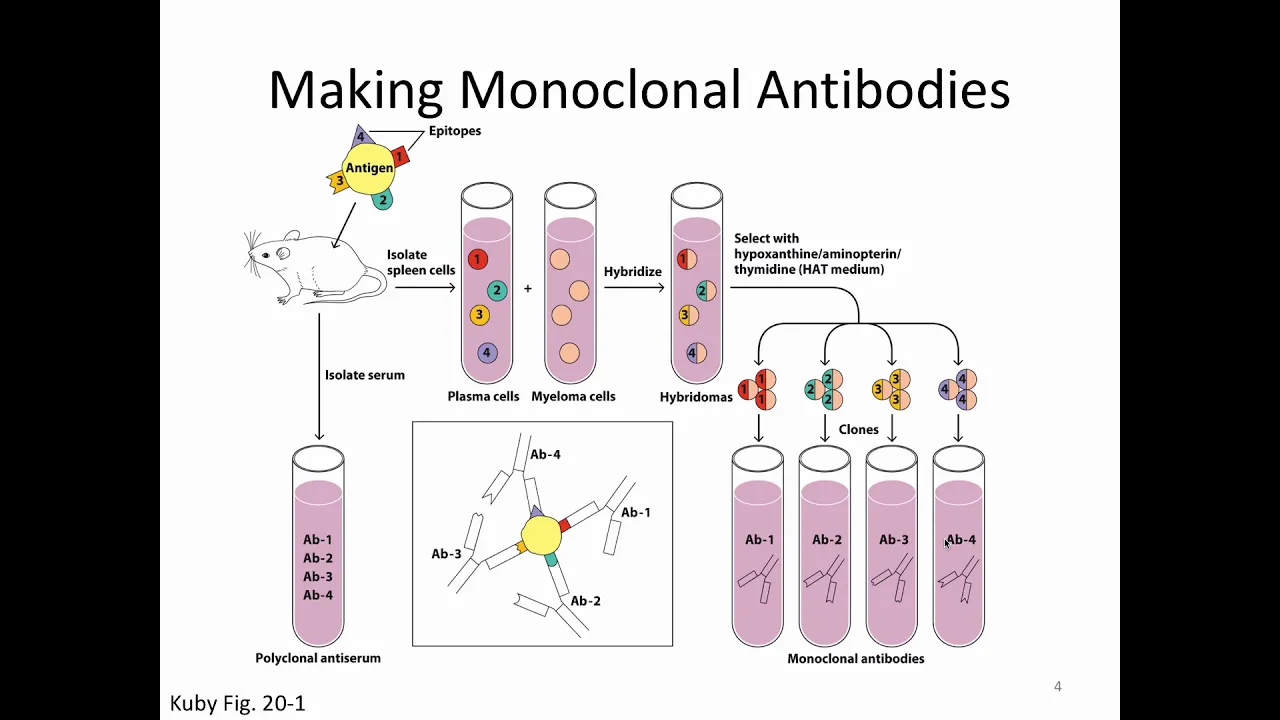
Monoclonal antibodies specifically recognize epitopes on CD markers. Since single antibodies may cross-react with related markers, multiple antibodies are used together for confirmation. A Cluster of Differentiation (CD) is defined as a group of monoclonal antibodies recognizing the same molecular target. Each CD marker receives a numerical designation (CD1, CD2, CD3...) based on discovery order. This systematic approach enables precise identification of cell surface molecules across diverse cell types and developmental stages.
Click to watch →

CELLS, TISSUES AND ORGANS OF THE LYMPHATIC SYSTEM - recorded lecture
6.4K
Cluster of Differentiation (CD) antigens are specific surface markers expressed by different white blood cell subtypes. CD45 is expressed by most white blood cells, while CD4 is unique to monocytes and T helper cells. Flow cytometry uses CD markers to identify cell types in cancer diagnosis and immune disorders. These markers help distinguish between different lymphocyte populations and identify abnormal cell populations.
Click to watch →

ANTÍGENOS INMUNOLOGÍA
2.5K
Cell surface antigens are molecules (proteins, carbohydrates, lipids, glycoproteins) in the lipid bilayer, with red blood cell antigens (blood groups) being characteristic examples. These are classified using the CD system (Cluster of Differentiation), where each number identifies a specific function. CD4 denotes helper T cells, CD8 denotes cytotoxic T cells. Autoantigens are host molecules that can cause autoimmune reactions, including hormone precursors, structural lipids, and mitochondrial proteins. Non-microbial antigens include food antigens, inhaled particles, vaccines, and transplanted tissues.
Click to watch →
Deep Dive
Principles of antigen-antibody specificity and binding affinity.

Anticuerpos (Generalidades).
495
Antigen-antibody binding is rapid, spontaneous, specific, and reversible through non-covalent interactions (hydrogen bonds, van der Waals forces, hydrophobic interactions). Affinity refers to binding strength at a single site, while avidity refers to overall binding strength from multiple interactions. High avidity can result from multiple weak interactions even with low individual affinity. These principles explain how antibodies achieve both high specificity and strong overall binding to eliminate antigens effectively.
Click to watch →

كل تمارين الإستجابة المناعية مع الحل المفصل | من دورة 2007 حتى 2019
53.1K
Antigens (foreign substances) and antibodies have specific binding sites that allow them to recognize and attach to each other. This binding occurs through structural compatibility (structural complementarity) between the antigen and the antibody's binding site. Each antibody is specific to a particular antigen, and this specificity ensures that the immune system can target specific threats without affecting the body's own cells.
Click to watch →
IMUNOLOGIA- Imunoensaios (parte 1)
11.1K
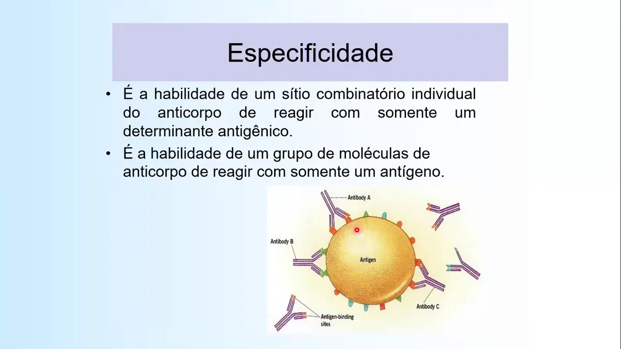
Antigen-antibody binding occurs through non-covalent interactions including hydrogen bonds, electrostatic bonds, Van der Waals forces, and hydrophobic bonds. Molecular complementarity between antibody combining sites and antigen epitopes determines binding efficiency. Greater complementarity increases attractive forces and decreases repulsive forces during molecular collisions. Affinity measures single-site binding strength, representing the net balance of attractive and repulsive forces. Avidity represents total multivalent binding strength across all antibody combining sites. Specificity refers to the ability of individual combining sites to react only with specific antigens. Cross-reactivity occurs when shared epitopes allow antibodies to bind to multiple antigens.
Click to watch →

#Imunologi#Antigen#
670
Affinity refers to the strength of binding between a single antigenic determinant and an antibody. High affinity means strong binding at each site. Avidity refers to the overall binding strength of multivalent antigens with multivalent antibodies, which is the sum of all individual binding interactions. Specificity is the ability of an antibody to recognize and bind to a specific antigenic determinant. Antibodies can distinguish between primary, secondary, and tertiary structures of antigens.
Click to watch →

Radioimmunoassay technique (RIA)
415.8K
Antigens are fragments of bacterial cells or viral particles that circulate throughout the body when infection occurs. Each antigen has a specific complementary antibody that binds to it with high affinity, forming an antigen-antibody complex. This specificity is fundamental to RIA because it ensures that only the target antigen will compete with the labeled antigen for binding to its corresponding antibody, enabling precise detection.
Click to watch →
Deep Dive
Basic cell biology concepts, including cell heterogeneity and the necessity of isolating specific cell populations.
![Bio-Active LVIE Webinar | 10x Genomics: Key Principles of Single Cell Technology [Thai Webinar]](https://i.ytimg.com/vi/vJ6EyLxQ1x8/maxresdefault.jpg)
Bio-Active LVIE Webinar | 10x Genomics: Key Principles of Single Cell Technology [Thai Webinar]
195
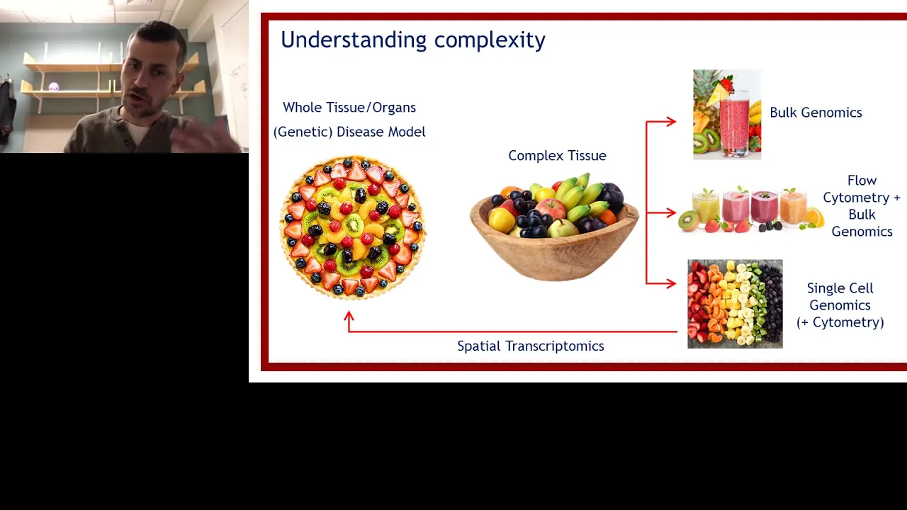
Tissues contain multiple cell types, not uniform populations. Blood contains red blood cells, white blood cells, and platelets; tumors contain cancer cells and stromal cells. Bulk tissue analysis (extracting DNA from all cells together) provides only an average, losing information about individual cell characteristics. Single-cell analysis is necessary to discover cell-specific gene expression patterns, identify rare cell types, and understand how different cell populations contribute to tissue function and disease. This approach enables researchers to study cell functions within complex tissues, identify differences between cell types in different organs, determine cell states and developmental stages, and gain deeper understanding of biological processes at the cellular level.
Click to watch →
Using omics technology to identify important subpopulations of cells
171
The cellular fraction isolated from adipose tissue and the resulting cells grown in culture are heterogeneous, meaning they are not a single uniform population. This heterogeneity is a well-established characteristic of these cell populations. Furthermore, the expansion of this heterogeneous cell population leads to the selection of specific clones and a loss of diversity over time. However, it remains unclear whether this initial heterogeneity of the original isolate (stromal vascular fraction or SVF) or the loss thereof during expansion contributes to or impedes the potential therapeutic use of these cells.
Click to watch →

Igniting Technology_Kumar Wickramasinghe.mp4
423
This section explains why traditional bulk cell analysis fails to capture true biological complexity. While population averages appear uniform with single-mode distributions, individual cells actually exhibit dramatically different expression levels ranging from low to high. This hidden heterogeneity is fundamental to understanding biological processes—most cells may express below a threshold while some occasionally exceed it, creating cells with distinct characteristics. These fluctuations drive tissue organization and various biological processes, explaining why genetically identical organisms like twins or identical cats still show differences. The section emphasizes that detecting these variations at the single-cell level is essential for understanding disease mechanisms and developing targeted therapies.
Click to watch →

Cellular Heterogenity And Tumor Heterogeneity | BioCode: Learn Bioinformatics
1.9K
Cellular heterogeneity refers to cell-to-cell differences based on gene expression patterns. In normal conditions, each cell has a unique expression pattern suited to its function—for example, B cells differ from T cells. In disease states, particularly tumors, this diversity becomes amplified, with cells exhibiting drastically different expression patterns from adjacent cells. This phenomenon originates from a single cell gaining gatekeeping mutations, which then diversifies into multiple populations expressing different genes based on their requirements and the tumor microenvironment. The causes include genetic variation, mixture of cell types, and trans-differentiation, leading to complex subpopulation structures within tumors.
Click to watch →
Tips for successful single cell and single nucleus RNA-Seq - Part 1
2.9K
Cellular heterogeneity arises from multiple sources: (1) Tissue composition varies between organs and even within the same organ across different individuals; (2) Cells exist at different stages of the cell cycle (dividing vs arrested vs differentiated); (3) Some cells are alive while others are already dead; (4) Regulatory networks cause cells to interact and influence each other's states; (5) Gene expression relationships create epistatic interactions where mutations in one gene only manifest if other genes are also mutated; (6) Differentiation paths create additional complexity as cells progress through developmental trajectories.
Click to watch →
Deep Dive
Fundamental physics of magnetism, specifically how magnetic fields exert force on magnetic particles.

สรุป แม่เหล็กไฟฟ้า by P'BEN - BE'nTRUST
41.6K
Magnets are natural (from magnetite ore) or artificial (human-made). Every magnet has two poles: north and south, which cannot exist in isolation. Like poles repel, opposite poles attract. Magnetic field lines emerge from the north pole and enter the south pole. Earth acts as a giant magnet with its magnetic field generated by molten iron rotation in the outer core. The field lines emerge from the geographic south pole (magnetic north) and enter at the geographic north pole (magnetic south). Magnetic flux (Φ) is the total number of field lines through an area, measured in Weber (Wb). Magnetic flux density (B) is flux per unit area, measured in Tesla (T). Electric current produces magnetic fields around conductors. For straight wires, field lines form concentric circles determined by the right-hand rule. For circular loops and solenoids, the field resembles a bar magnet. Charged particles moving through magnetic fields experience Lorentz force: F = qvB sin(θ). Current-carrying wires in magnetic fields experience force: F = ILB sin(θ). Parallel current-carrying wires attract if currents flow in same direction, repel if opposite.
Click to watch →

CSIR NET 2025 & GATE | Electromagnetic Theory - Charge Particle In Magnetic Field | Physical Science
225
Magnetic fields exert force only on moving charged particles, unlike electric fields which act on stationary charges. The magnetic force is given by F = q(v × B), with magnitude F = qvB sin(θ), where θ is the angle between velocity and magnetic field. The direction is determined by the right-hand rule: thumb along velocity, fingers along magnetic field, palm faces force direction. When velocity is parallel or anti-parallel to the magnetic field (θ = 0° or 180°), sin(θ) = 0, so no force acts and the particle moves in a straight line.
Click to watch →
![Halliday 10판(한글) 28~32장. [맥스웰 방정식] 자기장](https://i.ytimg.com/vi/5BvNWqMnJbg/maxresdefault.jpg)
Halliday 10판(한글) 28~32장. [맥스웰 방정식] 자기장
100
A field is a property of space that describes how physical quantities vary at each point. Mass creates gravitational fields, and charged particles create electric fields. Through experiments with small charged particles, scientists discovered that magnets create magnetic fields that exert forces on charged particles. Magnetic forces have unique characteristics: they depend on charge magnitude and velocity, are zero when particles are stationary, and act perpendicular to both velocity and magnetic field directions (Fleming's left-hand rule). The Lorentz force formula F = Q(v × B) describes this interaction, where the cross product indicates the force is perpendicular to both velocity and magnetic field. The magnetic field at a point is defined as B = F/(Qv), measured in Tesla.
Click to watch →

Pré Enem #intensivão2021 - Física - Eletricidade 04
209
Magnetism originated in ancient Greece in Magnesia, where magnetite minerals were found. Every magnet has two poles (North and South) and is always a dipole. The principle of inseparability states that cutting a magnet creates new magnets with both poles. Like poles repel, opposite poles attract. Earth behaves as a giant magnet with geographic and magnetic poles offset by approximately 1,300 km. The Earth's magnetic field is generated by the rotation of electrically charged particles in its liquid iron core. This field acts as a protective shield against solar wind particles, deflecting charged particles and creating the auroras. Ferromagnetic materials (iron, nickel, cobalt) contain atoms that act like tiny magnets and align to form magnetic domains. Magnetic field lines represent the direction and strength of a magnetic field, running from North to South outside the magnet. A charged particle moving through a magnetic field experiences a force only if its velocity is not parallel to the field. The magnitude is given by Fm = qvBsin(θ), where θ is the angle between v and B. The force is maximum when v and B are perpendicular and zero when parallel. The Tesla (T) is the SI unit for magnetic field strength, named after Nikola Tesla.
Click to watch →

física - electromagnetismo
100
Magnetism is a physical phenomenon where bodies exert forces of attraction or repulsion on other bodies. Magnets are bodies that attract iron or nickel objects. There are natural magnets (produced by nature) and artificial magnets (manufactured from iron, nickel, or cobalt compounds). Every magnet has two poles: North and South, where magnetic force is most intense. Opposite poles attract while like poles repel. A magnetic field is the region where magnetism manifests, represented by field lines that emerge from the North Pole and travel to the South Pole. The strength of the magnet determines the size of its magnetic field.
Click to watch →
Prerequisite Knowledge
- Concept 01Understanding of cell surface markers, antigens, and the Cluster of Differentiation (CD) nomenclature system.
- Concept 02Principles of antigen-antibody specificity and binding affinity.
- Concept 03Basic cell biology concepts, including cell heterogeneity and the necessity of isolating specific cell populations.
- Concept 04Fundamental physics of magnetism, specifically how magnetic fields exert force on magnetic particles.
Subsequent Learning
- Step 01Comparing MACS (Magnetic-Activated Cell Sorting) with FACS (Fluorescence-Activated Cell Sorting) regarding purity, yield, and cell viability.
- Step 02Exploring downstream applications of isolated cells, such as cell culture, flow cytometry, and single-cell genomic analysis.
- Step 03Designing experimental workflows using Positive Selection versus Depletion (Negative Selection) strategies.
- Step 04Investigating clinical-scale applications of magnetic cell separation, such as CAR-T cell therapy manufacturing and stem cell transplantation.
MACS core
Depletion
MACS core
0:00Playing Section
- 1
MACS uses magnet, column, and microbeads for gentle cell separation.
- 2
Matrix amplifies gradient 10,000x, reducing stress and aggregate formation.
- 3
Minimal labeling prevents activation, preserving cells in natural state.
Limitations of Column-Based Sorting and the Advantages of Column-Free or Multi-Parametric Alternatives
While MACS is a standard method for cell isolation, it faces significant criticism and competition from alternative technologies. A primary drawback of MACS is its reliance on a column matrix packed with ferromagnetic spheres. This column can subject cells to high mechanical shear stress, risk clogging with cellular aggregates, and cause non-specific entrapment, which can compromise cell viability and yield. Furthermore, despite claims of minimal interference, residual magnetic nanoparticles can remain bound to or internalized by cells, potentially affecting downstream assays, in vivo tracking, or clinical applications. In contrast, column-free magnetic separation techniques (such as EasySep) eliminate the column altogether to reduce physical stress on cells. For complex research requiring high-purity isolation of specific subpopulations, Fluorescence-Activated Cell Sorting (FACS) is often preferred. Unlike the largely binary selection of MACS, FACS allows for precise, multi-parametric sorting based on multiple intracellular and extracellular markers simultaneously, offering a level of specificity and characterization that magnetic isolation cannot achieve.
Deep Dive
Comparing MACS (Magnetic-Activated Cell Sorting) with FACS (Fluorescence-Activated Cell Sorting) regarding purity, yield, and cell viability.

Immunological Methods: Understanding and Enhancement of Pre-clinical Animals Models of Disease
636
Cell sorting extends flow cytometry to physically isolate specific cell populations. Fluorescent Activated Cell Sorting (FACS) uses electrical charges to deflect cells into collection tubes based on fluorescent labeling, achieving high purity but involving shearing forces that reduce yield and viability. Magnetic Activated Cell Sorting (MACS) uses antibody-coated magnetic beads instead of fluorescent labels, passing cells through magnetic columns where labeled cells are retained while unlabeled cells flow through. MACS is faster, gentler (higher yield/viability), and can be performed in biosafety hoods for sterile applications. Both methods achieve comparable purity (~97%) for naive T cell isolation. The choice depends on experimental needs: FACS offers greater flexibility in gate setting for extreme purity, while MACS provides workflow efficiency and cell viability advantages.
Click to watch →
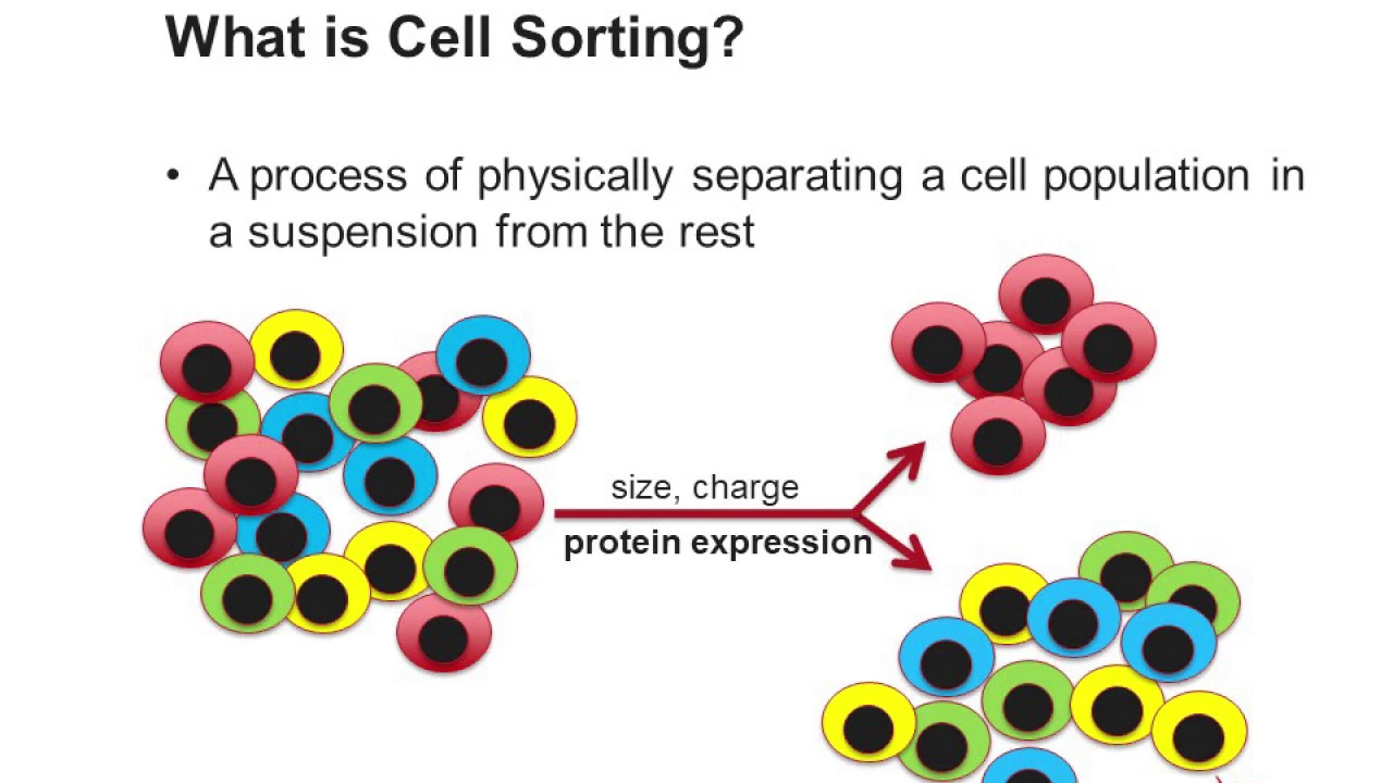
Cell Sorting Using Flow Cytometry
41K
Cell sorting using flow cytometry is a technique that physically separates target cell populations from heterogeneous mixtures by exploiting unique cellular characteristics; while Magnetic Activated Cell Sorting (MACS) uses magnetic nanoparticles bound to monoclonal antibodies for fast isolation of large cell quantities with approximately 90% purity, Fluorescent Activated Cell Sorting (FACS) utilizes multiple fluorescent characteristics through staining with fluorescently labeled antibodies to analyze and sort every single cell separately, achieving high specificity and purity approaching 100%. FACS works by charging a flow stream containing single cells, forming droplets at a constant frequency, and applying an electrostatic field to deflect droplets containing target cells into collection tubes while others are discarded into waste.
Click to watch →
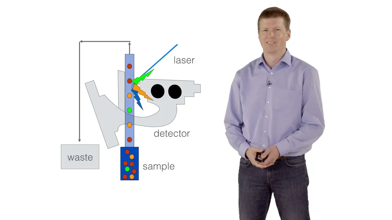
FACS - Fluorescence Activated Cell Sorting - Steffen Schmitt (DKFZ)
34.4K
FACS is a droplet-based cell sorting technology that physically separates and collects single cells or cell populations by charging cells with an electric field and deflecting them into collection tubes using secondary electric fields; the system balances key parameters including sorting speed (up to 9000 cells/second), cell viability, purity, and recovery rate, with optimal settings depending on the downstream application such as establishing cell lines, enriching for specific cell cycle phases, or studying single-cell transcriptomes.
Click to watch →

Fluorescence activated cell sorting (FACS)
158.1K
Fluorescence Activated Cell Sorting (FACS) is a cytometric technique that uses fluorescently-labeled antibodies to identify and separate cells based on their protein expression levels; the technique measures two key parameters: fluorescence intensity (indicating the amount of protein expressed by individual cells) and fluorescence frequency (indicating the number of cells expressing that protein), enabling researchers to isolate specific cell populations for further study.
Click to watch →

Zpracování a využití biologického materiálu pro výzkumné účely od nemocných s monoklonální gamapatií
460
Comparative analysis reveals median purity of approximately 88% for magnetic separation versus approximately 99% for FACS. While FACS achieves superior purity, it is more time-consuming and costly. Financial costs for simple single-marker sorting are comparable, but increase significantly for subpopulation sorting or combined approaches. Research applications extend beyond positive tumor cell fractions to include negative non-tumor fractions such as serum, peripheral blood plasma, bone marrow plasma, and DNA from whole peripheral blood. Flow cytometry analyzes B lymphocyte and plasmacytoma phenotypes. Molecular biology laboratories study gene expression changes at RNA and microRNA levels, hereditary predisposition through SNP analysis, and drug resistance markers. Molecular cytogenetics performs routine cytogenetic abnormality analysis and assesses prognostic significance of chromosomal abnormalities in relation to disease transformation.
Click to watch →
Deep Dive
Exploring downstream applications of isolated cells, such as cell culture, flow cytometry, and single-cell genomic analysis.

Coffee Chat - Failures in Paucicellular Pathology
147
Post-sorting analysis uses flow cytometry-like visualization displaying fluorescent parameters. Keratin identifies tumor cells, vimentin identifies stromal cells, double-positive cells indicate EMT states, and double-negative cells may represent lymphocytes or stem cells. DAPI comparison against stromal cells reveals tumor-specific amplifications and deletions. Downstream applications include: targeted NGS using pools of 100-300 cells, single-cell analysis via WGA and low-pass sequencing, and copy number analysis. Isolated tumor cells provide clean genetic profiles free from stromal dilution, enabling accurate CNV reporting and detection of genetic aberrations previously obscured by healthy cell background.
Click to watch →

Single-Cell Analysis - Powered by REPLI-g: Single Cell Analysis Series Part 1
4.6K
The single cell workflow follows bulk experiment principles but requires specialized adaptations. Primary samples (tissue, blood, embryos) undergo single cell isolation—the critical differentiator. For suspended cells: serial dilution distributes cells to wells following Poisson statistics; micromanipulation visually picks cells; FACS sorts fluorescently labeled cells at ~10,000 events/second. For solid tissues: mechanical dissociation grinds tissue into suspensions, or laser capture microdissection isolates cells from sections. Critical considerations include maintaining cold chains, minimizing storage, preventing contamination (even one contaminant equals 50% signal), and using decontaminated reagents. After isolation, whole genome or transcriptome amplification raises DNA/RNA to workable levels. Downstream applications include targeted resequencing, microarrays, qPCR, and NGS. The amplified DNA serves as a permanent bio-bank for future analysis since the original cell is consumed.
Click to watch →

How to Use the STEMprep™ Tissue Dissociator
1K
The STEMprep™ Tissue Dissociator automates the process of converting solid tissues into high-viability single-cell suspensions by precisely controlling temperature, mechanical forces, and timing parameters, enabling efficient and reproducible sample preparation for downstream applications like cell separation, cell culture, flow cytometry, and immunology research.
Click to watch →
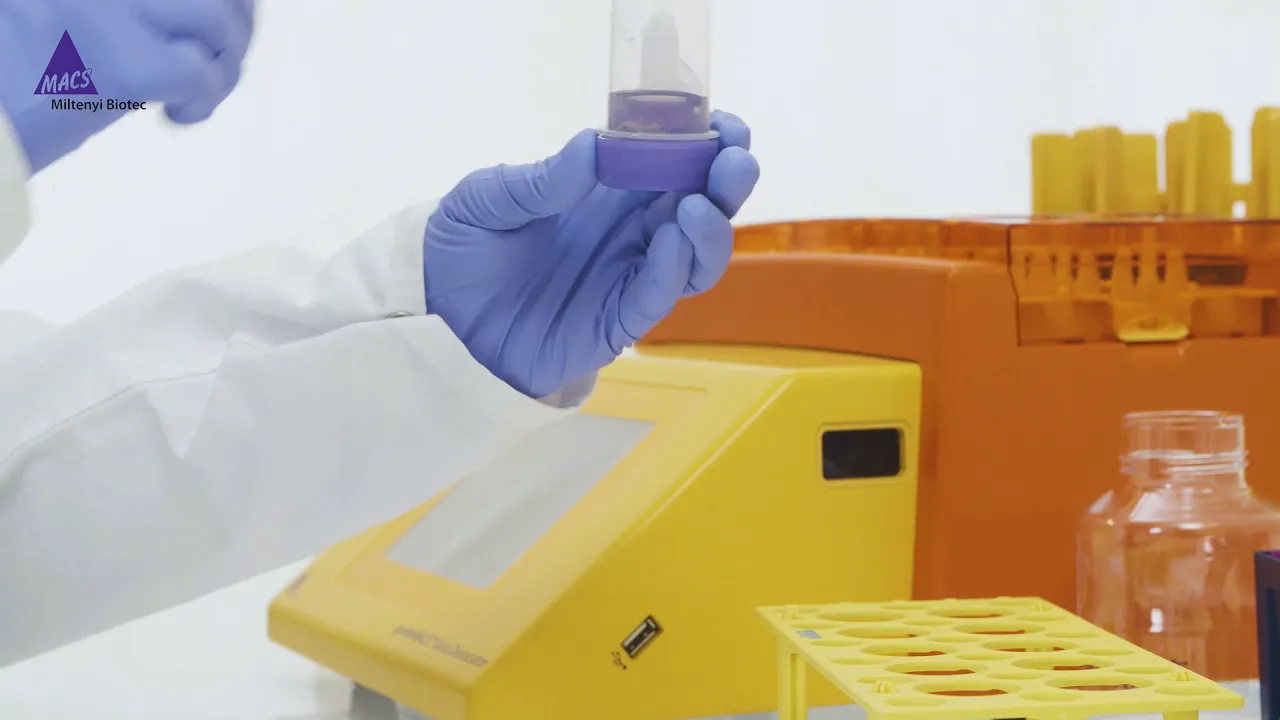
How to dissociate FFPE tissue
4K
This video demonstrates how to dissociate formalin-fixed paraffin-embedded (FFPE) tissue using the gentleMACS™ Octo Dissociator with Heaters and the FFPE Tissue Dissociation Kit to generate single-cell suspensions from human carcinoma sections while preserving important cell epitopes like cytokeratin for carcinoma cells and vimentin for non-carcinoma cells, enabling downstream applications such as mutation analysis by next-generation sequencing or ploidy analysis by flow cytometry.
Click to watch →

Ginkgo Ferment 2024
8.4K
Factorial Bio developed a platform enabling single-cell resolution in genomics, where traditional bulk NGS provides average data across cell populations but understanding biology requires single-cell resolution to characterize differences between cells. This enables high-throughput, high-content single-cell genomics applications including understanding tumor heterogeneity and characterizing CRISPR edits at genome scale. While the science of gut microbiome as a sensor, modulator, and translator of biological signals is established, the ability to identify sources of signals and harness them as precision therapeutics has lagged. Deep neural network analysis is required to identify biological 'dark matter' - patterns and signals that other methods cannot detect. Plant cell culture technology enables production of bioactive compounds (like carotenoids for eye health, flavonoids for heart health) that plants don't naturally produce for human consumption. The process involves taking plant parts, forming callus, creating suspension cultures, and testing against various conditions using transcriptomics and metabolomics.
Click to watch →
Deep Dive
Designing experimental workflows using Positive Selection versus Depletion (Negative Selection) strategies.
RNAi vs CRISPR Screens, May 2015 - CHI (Cambridge Healthtech)
655
Successful pooled screening requires transducing at least 200-fold library complexity (e.g., 10 million cells for 50,000-construct libraries) to ensure adequate statistical power. Two selection strategies are employed: positive selection identifies effectors preventing cell death after treatment, revealing genes important for survival under specific conditions; negative selection (viability dropout assay) identifies effectors underrepresented after growth, revealing essential genes for cellular proliferation. Both approaches compare post-screen effector frequencies to original library baselines.
Click to watch →

ELRIG DD14 Day 2 1000 Ulrich Elling, IMBA
257
In AML mouse models, hairpin constructs cause tumor cell differentiation within days. Competition assays show cells depleted of these genes drop out of co-culture with wild-type controls. Haploid screens are fast (weeks), saturating, and highly specific. Positive selection works well. Negative selection is challenging in diploid cells because shRNA/CRISPR may not achieve efficient knockdown, limiting depletion to 10-fold. Haploid cells enable effective negative selection because each cell has independent mutations, allowing complete depletion of true loss-of-function mutations.
Click to watch →

knockout Mice
707
Two selection strategies ensure only correctly modified cells survive: (1) Positive selection uses antibiotic resistance genes - cells with the knockout construct survive antibiotic treatment while normal cells die, (2) Negative selection addresses random integration using thymidine kinase gene - cells with random integration are killed by converting ganciclovir into a toxic compound, while cells with proper homologous recombination at the target site survive. This combined strategy ensures only cells with correct homologous recombination at the target site are selected for further study.
Click to watch →
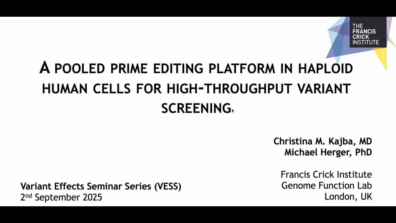
A pooled prime editing platform in haploid human cells for high-throughput variant screening
101
Two complementary screening frameworks were developed: negative selection depletes loss-of-function variants causing cell death, while positive selection enriches variants conferring drug resistance. Negative selection was applied to MARCHB1 variants (chromatin remodeler, 45% UOCS), testing over 1,300 point mutations. Positive selection targeted MLH1 (Lynch syndrome gene), enabling testing of diverse non-coding variants across large genomic regions including deep introns. Function scores were calculated by comparing pegRNA frequencies before and after selection, with control group analysis revealing that widespread editing efficiency variation initially obscured variant differences until filtering retained only active guides.
Click to watch →

A Novel Magnetic Platform for Large-Volume Cell Isolation from Whole Blood and Leukopaks
1.3K
For negative selection using the Easy 250 magnet, the Easy Step isolation cocktail is added to a single-cell suspension in a T75 flask, mixed, and incubated for 5 minutes. Then EasySep rapid spheres are added and mixed. Unlike positive selection, no additional incubation is required for the rapid spheres. The T75 flask is topped up with Easy Set buffer and placed into the Easy 250 magnet for two rounds of separation. The desired fraction is then pipetted off into a new centrifuge bottle for downstream applications.
Click to watch →
Deep Dive
Investigating clinical-scale applications of magnetic cell separation, such as CAR-T cell therapy manufacturing and stem cell transplantation.

Inside Bioprocessing Evolving the future of cell therapy manufacturing
603
Automated magnetic separation systems like the Gibco CTS Dyna select streamline cell therapy manufacturing by providing closed, scalable, and consistent cell isolation and bead removal processes, addressing industry challenges of manual processing inconsistencies, contamination risks, and long throughput times while supporting both autologous and allogeneic workflows.
Click to watch →

Enhance Your Cell and Gene Therapy Workflow with GenScript Tools
241
Cell therapy has emerged as a transformative innovation, with CAR-T therapy achieving remarkable success in treating leukemia. The process involves collecting patient PBMCs, isolating T cells using CD3/CD28 antibodies, activating cells, gene editing via lentivirus, 10-14 day expansion, and reinfusion. Despite over 2,000 clinical trials worldwide, manufacturing faces critical challenges: material costs approach $80,000 per dose, total treatment costs reach nearly $500,000, delivery times average 2-3 months, and only one GMP-compliant supplier exists for cell isolation reagents. GenScript addresses these challenges through magnetic cell isolation beads (100nm, non-toxic, biodegradable) with antibody coating for efficient cell binding. Products include isolation beads for T cells, NK cells, and stem cells, along with Cytosync 1000 instruments. Validation by Legend Biotech ensures optimal isolation performance with comparable purity and yield to competitors while offering better expansion rates and higher memory T cell differentiation.
Click to watch →

Enrichment & analysis of rare antigen & virus-specific T cells
380
The MaxQuant cell sorter employs microchip sorting technology with microwolf for the gentlest cell sorting and highest viability, even for rare and fragile cells. Its closed sterile cartridge system enables GMP-compliant operations essential for clinical applications including CAR T cell therapy and tumor-specific T cell isolation. Clinical trials demonstrate successful isolation of CMV-specific CD8 positive T cells achieving ~94% purity. This technology bridges research and clinical translation by providing reproducible, automated cell sorting suitable for therapeutic cell product manufacturing. The integration of sample preparation, enrichment, and sorting technologies creates complete workflows for isolating defined T cell populations at clinical scale.
Click to watch →

Applying a closed, modular, semi automated system to CAR T cell therapy manufacturing
1.8K
Effective cell therapy manufacturing requires precise cell isolation and selection technologies. The Rotia counterflow centrifuge isolates PBMCs by creating fluidized cell beds where different cell sizes and buoyancies separate naturally. This closed system removes DMSO, red blood cells, and platelets while achieving high t-cell recovery. Dynabeads CD3/CD28 combined with dynamic magnets achieve dual T-cell selection and activation in one step, with 93% average recovery and preserved immunophenotype. An integrated workflow further combines these steps, achieving 75% initial recovery with purity improving to 90-95% by day 3.
Click to watch →

Enabling healthcare innovation – our technology in CAR-T cell therapy | ZEISS
392K
CAR-T cell therapy is a revolutionary personalized cancer treatment where T-cells are extracted from patients, genetically modified to target specific cancer antigens, expanded in number through stimulation, and then reinfused to fight leukemia and lymphoma; automated manufacturing systems like ProCell integrate precision microscopy for real-time cell imaging, enabling decentralized production that improves quality control, reduces labor intensity, and increases accessibility of this life-saving therapy.
Click to watch →
MACS core
Depletion
MACS core
0:00- 1
MACS uses magnet, column, and microbeads for gentle cell separation.
- 2
Matrix amplifies gradient 10,000x, reducing stress and aggregate formation.
- 3
Minimal labeling prevents activation, preserving cells in natural state.
Limitations of Column-Based Sorting and the Advantages of Column-Free or Multi-Parametric Alternatives
While MACS is a standard method for cell isolation, it faces significant criticism and competition from alternative technologies. A primary drawback of MACS is its reliance on a column matrix packed with ferromagnetic spheres. This column can subject cells to high mechanical shear stress, risk clogging with cellular aggregates, and cause non-specific entrapment, which can compromise cell viability and yield. Furthermore, despite claims of minimal interference, residual magnetic nanoparticles can remain bound to or internalized by cells, potentially affecting downstream assays, in vivo tracking, or clinical applications. In contrast, column-free magnetic separation techniques (such as EasySep) eliminate the column altogether to reduce physical stress on cells. For complex research requiring high-purity isolation of specific subpopulations, Fluorescence-Activated Cell Sorting (FACS) is often preferred. Unlike the largely binary selection of MACS, FACS allows for precise, multi-parametric sorting based on multiple intracellular and extracellular markers simultaneously, offering a level of specificity and characterization that magnetic isolation cannot achieve.
Understanding of cell surface markers, antigens, and the Cluster of Differentiation (CD) nomenclature system.
Principles of antigen-antibody specificity and binding affinity.
Basic cell biology concepts, including cell heterogeneity and the necessity of isolating specific cell populations.
Fundamental physics of magnetism, specifically how magnetic fields exert force on magnetic particles.
Comparing MACS (Magnetic-Activated Cell Sorting) with FACS (Fluorescence-Activated Cell Sorting) regarding purity, yield, and cell viability.
Exploring downstream applications of isolated cells, such as cell culture, flow cytometry, and single-cell genomic analysis.
Designing experimental workflows using Positive Selection versus Depletion (Negative Selection) strategies.
Investigating clinical-scale applications of magnetic cell separation, such as CAR-T cell therapy manufacturing and stem cell transplantation.
61K views337likes3:00@Miltenyi_BiotecOriginal Release: 2017-09-08
[Music] whoa hello there I'm captain t-cell and today I will tell you why Mac's technology is the best choice for isolating your cells with Mac's technology you simply need three things a magnet a Mac's column and Mac's microbeads let me show you how it works at the heart of our Mac's technology is the Mac's column which consists of a ferromagnetic matrix the spheres that make up the matrix amplify the magnetic gradient 10,000 fold this allows for minimal labeling with small nano sized magnetic beads got my magnifying glass here mmm that's interesting Wow cells that pass through the matrix within the Mac's column have a lot of space causing no stress to the cells labeled cells hover between the iron spheres while unlabeled cells pass through you may know that there are different ways of isolating cells but let me explain to you what makes Mac's technology so unique due to the minimal labeling with just a few nano sized Macs microbeads there are no aggregates on the cell surface column-free technologies require large amounts of bigger beads to maintain labelled cells in a weak magnetic field this can result in aggregate formation epitope blocking and cross-linking another advantage of Mac's technology is that there is no cell activation during cell isolation as you can see here that is really cool when you want to look at your cells in their natural state in contrast column-free technologies can cause cell activation as shown here do you want to go for a depletion strategy in which you label all the unwanted cells and leave your cells of interest untouched be aware that there is no guarantee for unlabeled targets when using a column-free technology this immunofluorescent staining of isolated target cells clearly shows a green signal of bead remnants on the cell surface if you really want untouched cells check out our micro bead based max cell isolation kits they are the only way to get truly unlabeled cells as you can see here you see there are different ways of isolating cells but why settle for less select the best with max technology [Music]
Up Next

35:44
Lentiviral Gene Delivery Tools: A Comprehensive Webinar Guide
@origenesocial
5.8K views•2022-12-08

12:54
Triumph of Orthodoxy Icon: Byzantine Art & History Explained
@BenCallan
2.1K views•2024-08-06

9:49
FastAPI vs Flask vs Django: Choosing the Right Python Web Framework
@TechWithTim
302.5K views•2024-05-26

1:42
Game of Thrones Opening Credits: A Cinematic Analysis
@gameofthrones
46.3M views•2011-04-18
Related Study Plans & Knowledge Roadmaps
Structured learning paths in General & Interdisciplinary Studies
general-studies
Elevator Engineering: History, Physics & Cities
01From Hoists to Safety Brakes: The Dawn of Vertical Lift
02Hydraulic vs. Traction: The Engineering Physics of Elevators
03Modern Speed, Safety, and Algorithms
+ 2 more learning modules
12h5 modules
Explore Roadmap
general-studies
Rubber: Material Science, History & Geopolitics
01Origins of Natural Rubber & Mesoamerican History
02The Chemistry of Natural Rubber & Vulcanization
03The Geopolitics of the Rubber Boom & Colonization
+ 2 more learning modules
12h5 modules
Explore Roadmap
general-studies
Containerization: Evolution, Logistics & Trade
01Ancient Trade & Bulk Cargo: Amphorae to Barrels
02The Industrial Revolution & Cardboard Packaging
03The Containerization Revolution: Malcom McLean
+ 2 more learning modules
12h5 modules
Explore Roadmap
general-studies
Textile Tech: Evolution, Labor & Global Impact
01Pre-Industrial Textiles: Handloom & Cottage Industry
02Mechanization: The Spinning Jenny to the Power Loom
03The Factory System & Global Labor Transformation
+ 2 more learning modules
10h5 modules
Explore Roadmap