




















Ultrasound imaging relies on piezoelectric crystals that generate sound waves above 20,000 Hz; these waves reflect off tissue interfaces based on density differences, with higher density contrasts producing brighter (hyper-echoic) images. The technology uses three main display modes—B-mode for 2D anatomy, M-mode for motion analysis, and Doppler for blood flow visualization—with different probe types (linear for vascular/musculoskeletal imaging and convex for abdominal/obstetric/cardiac imaging) and frequencies (low for depth, high for resolution) tailored to specific clinical applications. Common artifacts include shadowing, comet tail, reverberations, enhancement, and mirror images, while safety follows ALARA principles with minimal documented bioeffects.
Ultrasound Physics and Instrumentation for Residents
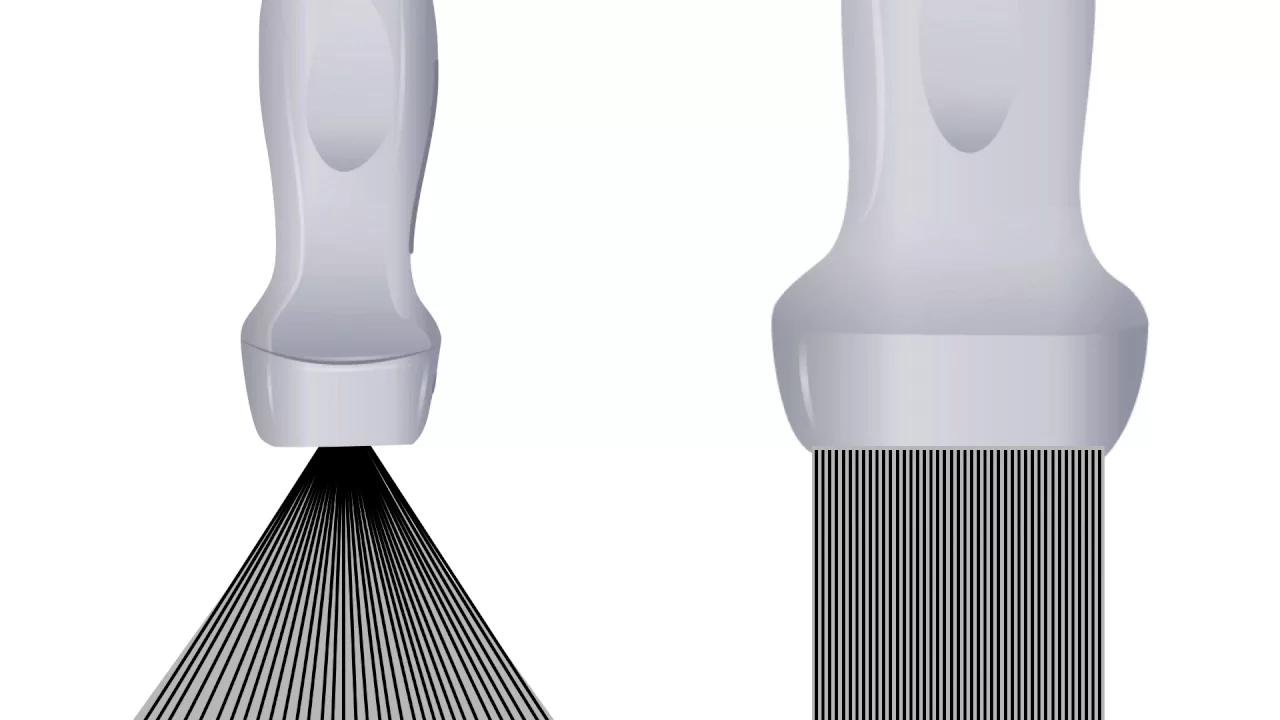
Added:hello I'm Nicholas Cohen a third-year family medicine resident at University Hospital's Case Medical Center I'm also a registered diagnostic medical sonographer and a registered diagnostic cardiac sonographer this module is the first in a series of modules for limited obstetrical ultrasound as part of the Family Medicine Residency curriculum resource presented by the society of teachers of family medicine and the association of Family Medicine Residency directors I had helped putting this module together from Dr Justin Lapin Dr quinland and Dr Stevens this module is focused on ultrasound physics and instrumentation ultrasound is defined as sound that is of a frequency too high to be heard by normal human ear and that frequency has determined to be 20,000 Hertz or 20 khz ultrasound that's used for Imaging is created by a piso electric Crystal a piso electric crystal is basically a substance that when electricity is supplied to it creates a soundwave the Soundwave is emitted or sent out from the transducer it encounters tissue and is reflected back to the transducer the reflected returning Echoes are what creates the image on the screen when sound waves encounter tissue one of two things can happen the sound waves can be transmitted through the tissue or can be reflected back from the tissue what happens depends on whether there's a change in density of the substance that the sound wave is passing through if there's no change in density the sound wave is transmitted if there is a change in density the sound wave is reflected the percentage that the TR that the sound wave is reflected is determined by the difference in the density of the two tissues so if you have two tissues that are extremely different densities you'll have a high reflection so if you look at this image right here which is of the fetus there's a high reflection at the surface where the probe interacts with with the skin there's also a high reflection between the Amic fluid and the skull the difference between the fluid and the skull the difference in density is great so there's a significant amount of brightness or reflection that occurs at that interface the reason we use gel when we're doing ultrasound is to decrease the reflection that occurs when the sound waves leave the transducer and pass through the air into the skin air and skin that interface is extremely differing densities obviously the air is a lot lower density so there's a lot of reflection that occurs unless we can eliminate the air that is would otherwise be between the probe and the skin attenuation is a property of tissues and it causes a decrease in the power or the amplitude of the sound waves as it travels through tissue the more that the the further the sound waves travels the more the sound wave attenuates and the more the sound wave attenu attenuates the less bright the image becomes so you can see in this image right here of the human body the tissue is bright it's brighter towards the probe the probe is here as we go further away from the probe the tissue gets darker in appearance that's because sound waves are attenuating as they travel through the tissue differing tissues have differ differing amounts of attenuation air will attenuate the most again a reason why we use gel to eliminate air from from part of the path that the sound wave has to traveling and water attenuates sound waves the least clinical ultrasound uses a variety of fre frequencies to create images the range of frequencies is typically between 1 and 20 megaherz the most common range is between 2 and 10 mahz the higher the frequency of the probe used the greater the resolution of the image created however the tradeoff is that the higher the frequency of the probe used used the less depth the sound waves can penetrate into the tissue you can see in this image here this is a parasal short access view of the heart's left ventricle taken with two different frequenc probes this is a 3 mahz probe and this is a 5 mahz probe the 3 mahz probe has not as good image resolution as the 5 megahertz probe there are typically three categories of frequencies for ultrasound imaging low frequency intermediate and high frequency probes low frequency probes typically in the range of two or 3 to or 3.5 to 5 5 mahz are typically used when you need to image deeper such as for Trans thoracic abdominal and pelvic Imaging intermediate frequency probes typically in the range of 5 to 7.5 are most commonly used with transesophageal probes for cardiac imaging higher frequency probes specifically 7.5 to 10 mahz are used when you don't need to image deep but but but but uh high resolution is important such as for muscular skeletal applications and for vascular applications when we describe the image created by ultrasound we refer to a structure's echogenicity echogenicity is the brightness of the structure that's produced the more that the sound waves are reflected back from the tissue the brighter the image created we describe images in relation to their surroundings this image which is brighter than its surroundings is hyper eoic this image which is darker than its surroundings is hypo eoic this image which is the same color as the surrounding tissue is called ISO eoic or the same echogenicity in this image is lacks any Echoes at all it's black and so we call it an eoic or without Echoes there are two types of transducers when using ultrasound for clinical Imaging they are the linear probe and the convex probe the linear probe is here and the convex probe is here the convex probe is also called a sector probe the linear probe you can see here is flat on its footprint and its footprint is larger than the convex probe which means that it takes up more of surface area on the skin when it contacts the skin typically linear probes are higher frequency the image that they produce is a rectangular shape and they are better for vascular Imaging and muscular skeletal Imaging demonstrated in this picture is ultrasound guided canulation of the internal jugular vein the other type of probe sector or convex probe has a smaller footprint typically it uses a lower frequency and the image that's created instead of being rectangular like the linear probe is a pi shaped or sector shaped image the trans the convex probe is better for abdominal obstetric and cardiac imaging where for cardiac imaging you need a smaller footprint as well as for abdominal and obstetric where you need to be able to image deeper there are different display modes the three that are most relevant to our applications are B mode M mode and Doppler mode B mode or brightness mode is typically what you think of when you think of ultrasound imaging it's two-dimensional Imaging that shows a structure's Anatomy M mode or motion mode provides a one-dimensional image here you can see a parisal long AIS view of the heart this this is the 2D image here is the m mode image the M mode image is taking only one line it's taking just one line straight down through the heart on the y axis this would be the depth of that line and the xaxis is time so particularly here at the aortic valve it's showing you the opening and closing of valves in in the in relationship sh to time as time progresses M mode is particularly good for obstetric Imaging when you're calculating the fetal heart rate it's also good whenever you're measuring something that needs to be done precisely in relation to time particularly heart valves in cardiac imy Doppler mode is the third display mode we'll discuss Doppler mode measures motion through vessels and toppler gives you both information about the direction of flow as well as the velocity of flow it's important to note that red does not mean artery and blue vein instead you look to your color map when you're using Doppler and it will tell you the direction of flow as well as the velocity using this color map anything above the middle line is flow towards the probe anything below the middle line is the color representing Flow Away from the probe so here's where the this is the top of the um the top of the image is closest to the probe and the bottom of the image is furthest away from the probe as we look at this image here we see red we look to our color map and we can tell that this is flow going towards the probe when we look at this flow we look at our color map and we see that this blue is the color that's below the middle line so we know that this is Flow Away from the probe furthermore we have a velocity map which is the further we go from that middle long the faster the color the corresponding color is is the an increasing velocity so in this color map as we go from red to Yellow we go from slower to faster [Music] velocity when we go from Blue to White we go from slower to faster velocity so in this particular image where we have flow going towards the transducer the yellow in the middle represents faster flow than the flow towards the outer aspect of the vessel which would be [Music] slower artifacts are anything that appear in the image that do not have corresponding anatomy in the tissue being examined the most common types of artifacts and the ones we'll discuss are shadowing Comet Tail or ring down reverberations enhancement and Mirror Image shadowing can be seen here this is a gallbladder and this is a gall stone shadowing occurs when sound hits a very reflective surface there's little sound transmitted through the stone So Below or deep to the stone there's a very little sound that propagates and so very it's it this structure the area below the hyperic area is hypo coic to surrounding tissue [Music] in Comet Tail or ringdown artifact again we have a highly reflective surface and because of the highly reflective surface we actually have a acceleration of the velocity of sound at that interface and that acceleration causes a hyper aoic streak or what's called a comet tail or ring down artifact in our next artifact we have What's called the reverberation reverberations occur when we have two highly reflective structures the sound waves basically pingpong back and forth between the two highly reflective structures and you get a series of lines that are equally spaced apart they are referred to or commonly described as ladderlike or V blind in appearance next we have enhancement enhancement occurs when we have a structure that has weak attenuation so if we have a fluid filed structure or cyst we have sound that's able to speed through this area so there's very little reflection going on here that means that sound that reaches the distal area behind this cystic or Hollow structure has an increased intensity and so appears Hy aoic compared to surrounding tissue a mirror image artifact is created when we have a strong reflector here's our strong reflector the strong reflector creates a second copy of the anatomy the second copy is always in line with the ultrasound beam and it's always deep to or further away from the probe than the actual Anatomy it's also always equidistant from the reflector as the actual [Music] Anatomy next we're going to discuss bio effects ultrasound is safe there's never been documented any human harm caused by Ultras sound for Diagnostic purposes it's important to keep in mind that there is a theoretical risk to ultrasound and the theoretical risk is due to the heat that's produced by the probe the heat that's produced by the probe is described using units called the thermal index the thermal index describes in a unit of measure how much the probe raises the tissue that's being EX examined by De C so a thermal index of one means that the exam raises the tissue by 1° C there's a theoretical risk to using ultrasound during gestation on a fetus at when the thermal index is greater than 1.5 to2 the only way that a thermal index can reach that level the only way that that much heat can be produced by an ultrasound probe is using Doppler and using Doppler for an extended period of time and the only vulnerable time during dation is the first trimester so there's a particularly concerning risk for ultra sound that's used in the first trimester and when Doppler is the Imaging modality that's a reason why Doppler is avoided in first Trester scanning lastly we have a concept called aera as low as reasonably achievable which means that we limit the use of ultrasound to only what is necessary to produce useful images for Diagnostic and therapeutic purposes
Up Next
Ultrasound Physics for Emergency Medicine: Modes, Probes, Artifacts, Orientation
@TakeoKun
359K views•2016-07-05

21cm Hyperfine Transition in Neutral Hydrogen: Radio Astronomy Basics
@AaronRobertParsons
12.4K views•2011-10-13

NMR Spin Physics I: Zeeman Effect, Resonance Condition & Larmor Frequency
@nptel-indianinstituteofsci8064
2.3K views•2024-01-17

Entropy and the Second Law of Thermodynamics Explained
@veritasium
27.5M views•2023-07-01
Related Study Plans & Knowledge Roadmaps
Structured learning paths in Physics