Surgical debridement is a medical procedure where doctors remove dead, damaged, or contaminated tissue from wounds caused by injuries, burns, bedsores, or infected surgical incisions; this removal is essential because dead tissue compromises circulation, invites bacterial infections that compete with healthy cells for nutrients, and can form hardened eschars that prevent healing; during the procedure, the doctor numbs the area, irrigates the wound, evaluates tissue depth, removes unhealthy tissue to create a clean margin, and applies bandages or moist dressings to promote healing.
Surgical Debridement: Removing Dead Tissue to Heal Wounds
Added:Deep Dive
Anatomy of the skin and the four primary phases of wound healing (hemostasis, inflammation, proliferation, and remodeling).
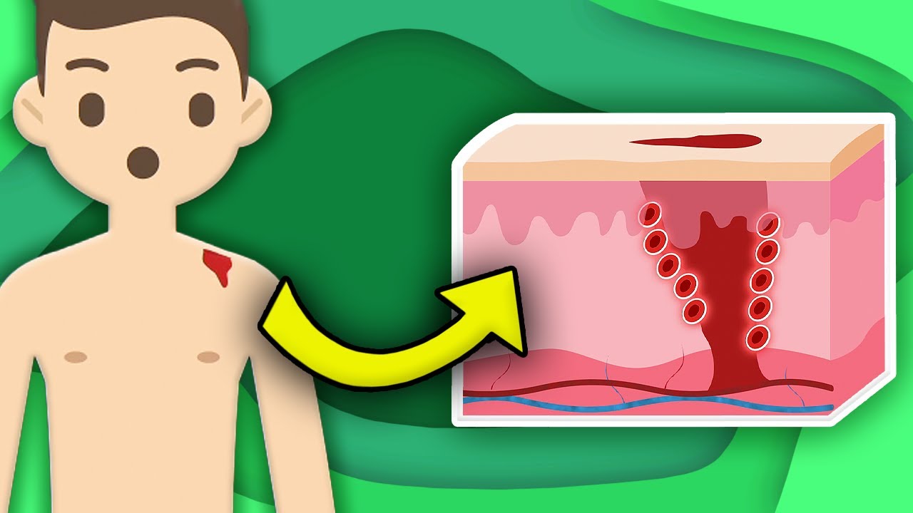
How Does YOUR Skin Heal? | KLT Anatomy
92K
Skin healing occurs through four sequential phases: hemostasis (blood vessel constriction and platelet clot formation to minimize bleeding), inflammation (immune cells and chemical mediators remove debris and bacteria), proliferation (new tissue generation with granulation tissue containing blood vessels, collagen, and fibroblasts), and remodeling (new tissue strengthens and becomes more flexible, with scar tissue gradually fading as collagen fibers realign).
Click to watch →

Conquer General Pathology with GNP Sir I Vet Pathology Online Class
387
Wound healing is a complex biological process repairing damaged tissue through four phases: Hemostasis (initial phase stopping bleeding: blood vessel constriction, platelet aggregation forming clot, fibrin stabilizing clot); Inflammation (recruitment of neutrophils and macrophages to clear debris and fight infection); Proliferation (angiogenesis forming new blood vessels, granulation tissue formation rich in collagen and blood vessels, epithelial cell migration to cover wound); Remodeling (final phase where collagen in granulation tissue remodels, forming scar tissue).
Click to watch →

Learn How Your Body Recovers From Being Hurt Or Sick! | Human Body Songs For Kids | KLT Anatomy
130K
When skin is cut, the healing process occurs in four phases. First, hemostasis happens as blood vessels constrict to minimize bleeding and platelets form clots to seal the wound temporarily. Second, inflammation occurs when immune cells appear at the site, removing debris, bacteria, and foreign substances while bringing nutrients and oxygen via inflammatory mediators. Third, proliferation happens when new tissue is generated to replace damaged tissue, filling the wound with granulation tissue consisting of new blood vessels, collagen, and connective tissue produced by fibroblasts. Fourth, remodeling occurs where newly formed tissue increases strength and flexibility; over time, scar tissue gradually fades as collagen fibers realign and become more organized.
Click to watch →

Fisiologia da Cicatrização
202
The skin is the body's largest organ (15% of body weight) with functions including protection, thermal regulation, sensory perception, and vitamin D synthesis. Structurally, it comprises epidermis (stratified squamous epithelium with four layers), dermis (connective tissue with fibroblasts, vessels, and glands), and hypodermis (adipose tissue). Wound healing occurs in four overlapping phases: hemostasis (immediate clot formation), inflammation (days 0-7 with neutrophil and macrophage migration), proliferation (days 3-24 with angiogenesis and granulation tissue formation), and remodeling (days 21-2 years with collagen maturation and wound contraction).
Click to watch →
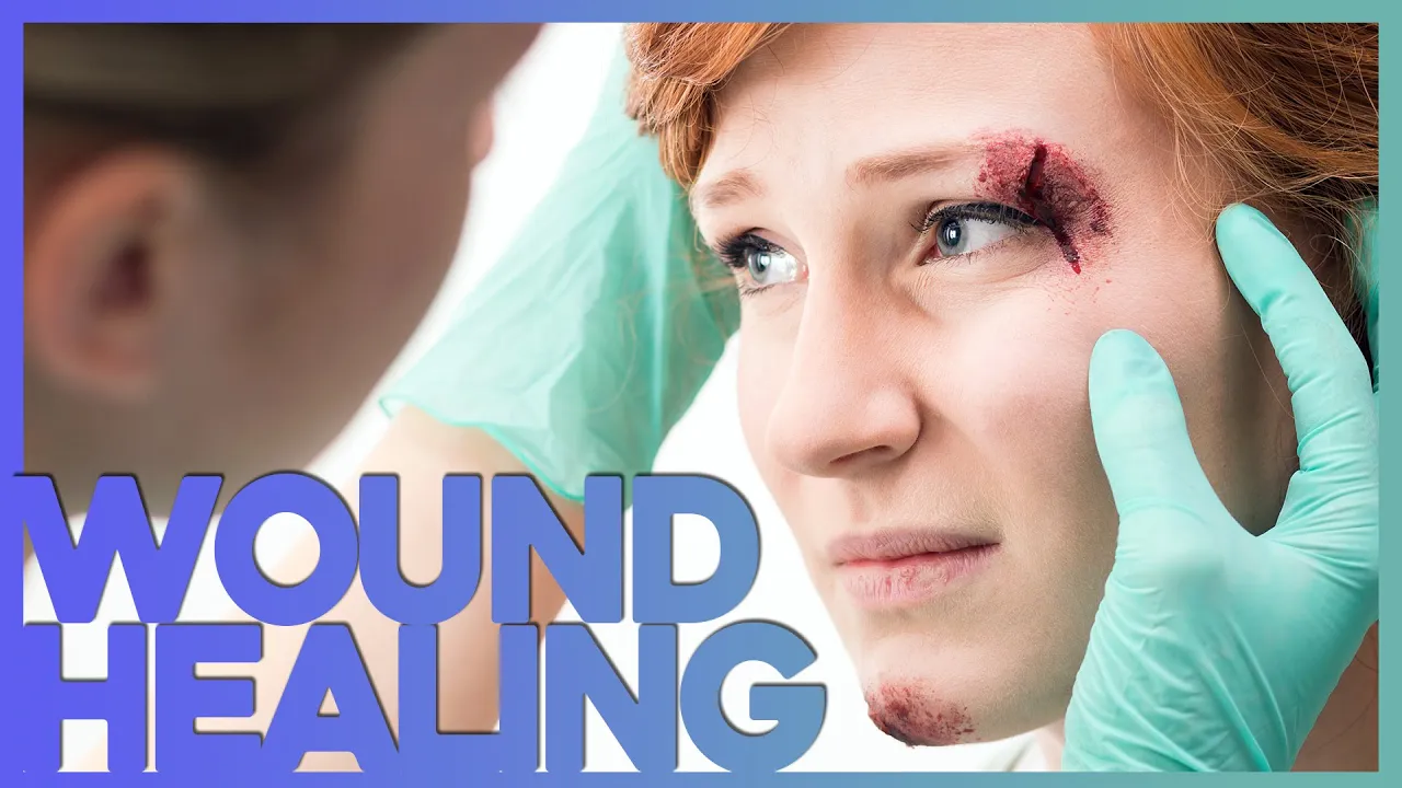
Phases of Wound Healing Explained - How a Cut Becomes a Scar | Corporis
134K
The wound healing process consists of four sequential phases: hemostasis (immediate blood clotting to stop bleeding), inflammation (immune response to fight infection and clear debris), proliferation (tissue repair and regeneration), and maturation (collagen remodeling for scar strength); each phase serves a specific purpose in transforming damaged tissue back into functional skin, with the entire process taking anywhere from weeks to years depending on injury severity and individual factors.
Click to watch →
Deep Dive
The physiological distinction between viable (healthy, vascularized) tissue and non-viable (necrotic, ischemic, slough, or eschar) tissue.

Projeto R1 Enfermagem para Residências - Prof. Bárbara Melo #06
371
Wound assessment distinguishes viable from non-viable tissue. Viable tissue includes granulation tissue (red, moist, vascularized) and epithelial tissue (new protective layer). Non-viable tissue includes necrosis (black eschar), slough (yellow fibrinous material), and devitalized tissue. The RYB assessment method categorizes: Red (viable granulation/epithelium, leave intact), Yellow (slough/fibrin, remove carefully), Black (necrosis/eschar, remove completely).
Click to watch →

WoundRounds Webinar - Wound Assessment & Documentation: A Practical Guide
108.5K
Wound tissue assessment distinguishes viable from non-viable tissue. Viable tissue includes granulation tissue (beefy red, moist, bumpy appearance resembling 'raw hamburger'), clean non-granulating tissue (flat and smooth without bumpy texture), and epithelial tissue (pale pink, flat, representing migrating epidermal cells). Non-viable/necrotic tissue includes eschar (black or brown, can be loose or adherent, moist or dry, hard or soft) and slough (yellow, tan, gray, or white, firmly adherent, stringy). Eschar represents the body's protective mechanism when circulation is inadequate and should not be removed prematurely; it should only be removed when it becomes soft and mushy. Slough requires enzymatic debridement or hydrogel application for removal. A scab is not necrotic tissue—it is dried blood and serum that should be dissolved away by providing moisture. Describing percentages of each tissue type provides objective documentation of wound status.
Click to watch →

Enfermagem Mapeada: Banca FGV! com Victor Roberto
5.9K
Wound tissues are classified as viable or non-viable. Viable tissues include granulation tissue (red, shiny, moist, vascularized) and epithelialization (pink, fragile tissue). Non-viable tissues include necrosis of coagulation (black eschar), necrosis of liquefaction (yellow-green, soft tissue), and fibrin tissue (yellow/white, adherent, stringy). The RYB classification system (Red, Yellow, Black) is commonly used to classify wounds by tissue type.
Click to watch →

PERIPHERAL VASCULAR DISEASE - Case Presentation | | SCALPEL '24 - TWIN CME for UG and PG |
360
The line of demarcation is the boundary between viable (living) tissue and necrotic (dead) tissue in gangrene. The viable tissue appears pale or pinkish, while the necrotic tissue appears dark black. This line is important for determining the extent of tissue loss and planning amputation levels. The presence of a clear line of demarcation indicates that the tissue has been viable long enough to establish this boundary.
Click to watch →

Como identificar os tecidos viáveis e inviáveis numa ferida? #enfermagem #feridasecurativos
5K
In wound healing, viable tissues (granulation tissue, epithelialization, muscle regeneration, and subcutaneous tissue regeneration) actively promote healing through new blood vessel formation, cell migration, and tissue reconstruction, while non-viable tissues (coagulation necrosis, liquefaction necrosis, and fibrinous tissue) delay healing by creating favorable environments for biofilm formation, prolonging inflammation, and preventing new tissue development; debridement is essential to remove non-viable tissue and restore the wound bed for effective healing.
Click to watch →
Deep Dive
Fundamental principles of surgical asepsis, sterile technique, and infection prevention in clinical settings.
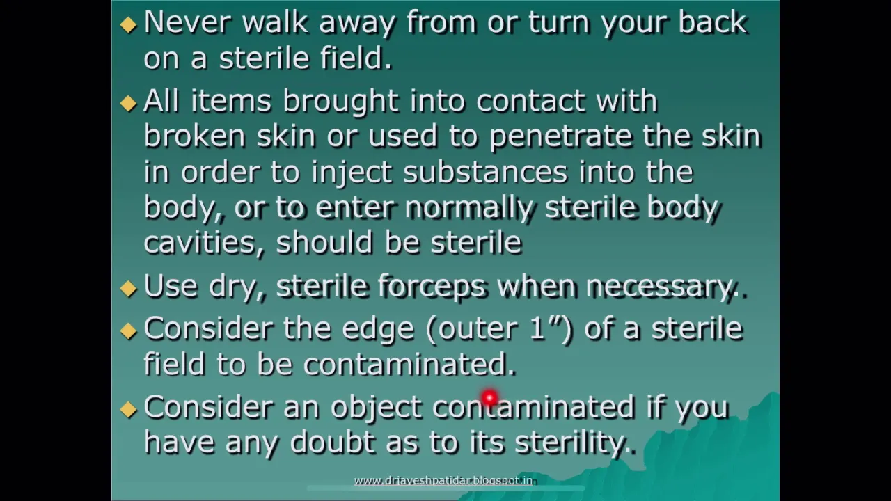
Surgical asepsis
4.2K
Surgical asepsis (sterile technique) is a fundamental infection control practice used in operating rooms, delivery rooms, during surgical procedures, catheterization, and dressing changes. The core principles include: (1) Only sterile objects should contact sterile items; (2) When opening sterile packages, avoid touching the inner flaps with non-sterile hands; (3) Maintain a sterile field at the level of the operating table, with all items positioned at or above this level; (4) Never stand with your back to the sterile field; (5) Avoid talking, coughing, sneezing, or reaching over the sterile area; (6) Consider any item in doubt as contaminated; (7) Immediately rectify any contamination by replacing items with sterile alternatives; (8) Healthcare workers should not be a source of contamination by removing jewelry, keeping nails short, and avoiding nail polish.
Click to watch →

2026문제풀이-16)실기-기본간호ㅣ 간호조무사 강의/무균술/섭취량과배설량
19.6K
Surgical asepsis (외과적 무균술) kills all microorganisms including spores, while disinfection (소독) only kills most microorganisms. Sterile items remain sterile only when in direct contact with other sterile items; once exposed to air or non-sterile surfaces, they become contaminated. The edge of sterile drapes is considered contaminated. Skin cannot be sterilized, only disinfected. This strict protocol prevents surgical site infections.
Click to watch →

محاضره Infection Control كامله
33.3K
Medical asepsis principles include: hand washing before and after all procedures, cleaning and disinfecting shared equipment, maintaining clean environments around patients, and ensuring healthcare providers are free from infectious diseases. Patients with infectious diseases require isolation. Surgical asepsis requires: sterile items only used in sterile areas, personnel wearing sterile gowns and gloves, sterile items at eye level, sterile personnel only touching sterile items, and non-sterile personnel excluded from sterile areas. Sterile items become contaminated when exposed to air for extended periods.
Click to watch →

محاضره أساسيات تمريض/Infection Control
12.4K
Surgical asepsis principles include: (1) Sterile items must only be used in sterile fields, (2) Sterile personnel must wear gown and gloves, (3) Sterile field must be at eye level, (4) Sterile personnel can only touch sterile items, and (5) Non-sterile personnel cannot enter sterile areas. These practices ensure complete pathogen elimination during surgical procedures.
Click to watch →

Principles of Sterile Technique
53.5K
Surgical asepsis follows seven key principles established by professional nursing associations: (1) Scrubbed personnel maintain a sterile field using proper attire including gowns, gloves, masks, and eye protection without contacting nonsterile surfaces; (2) Sterile drapes create the sterile field, covering all equipment except the surgical site, with only the top surface above waist height considered sterile; (3) All items within the sterile field must remain sterile, with compromised packages or dropped items requiring specific handling protocols; (4) Items introduced to the field must be opened and transferred using methods preserving sterility; (5) The sterile field requires constant monitoring and preparation near procedure start time to prevent contamination from airborne particles, liquids, or personnel movement; (6) Personnel movement must maintain field integrity by staying centered and avoiding nonsterile areas; (7) Written policies and procedures must be developed, reviewed annually, and readily accessible to ensure consistent sterile technique application across the surgical team.
Click to watch →
Deep Dive
The role of bacteria in wounds, specifically the progression from contamination and colonization to local and systemic infection.

FERIDAS OPERATÓRIAS | CONHECENDO A INFECÇÃO E DEISCÊNCIA | LIVE #02
1.7K
Once a wound is opened, it becomes contaminated, but low bacterial counts in early phases can actually promote healing through enzymes like hyaluronidase that promote autolytic debridement. However, excessive bacterial multiplication leads to colonization, which is a precursor to infection. Infected wounds present with inflammatory signs including edema, erythema, increased warmth, and purulent exudate. Understanding the progression from contamination to colonization to infection is essential for proper wound management and preventing dehiscence.
Click to watch →

WoundRounds Webinar Series - Dressing(s) for Success: Wound Care Dressing Selection
285.2K
When skin barrier integrity is compromised, normal flora bacteria overgrow and cause inflammation that interferes with healing. Bacteria release endotoxins and proteases that degrade wound proteins and building molecules. The infection continuum ranges from contamination (manageable bacterial presence) to colonization (increased bacteria count but still manageable), then to local infection, spreading infection, and systemic infection where bacteria overwhelm natural defenses. At this stage, bacteria compete with wounds for oxygen and nutrients, causing delayed or no healing. Understanding this continuum guides appropriate antimicrobial intervention selection.
Click to watch →

Tanda Luka Infeksi, Seperti Apa ??? II Part 1
10.7K
Wound infection develops through three progressive stages: (1) Contamination - bacteria enter wound tissue in small numbers; (2) Colonization - bacteria multiply and form strong colonies that resist treatment; (3) Local Infection - the infection becomes established and requires medical intervention. Each stage builds upon the previous one, making early intervention critical for preventing serious infection.
Click to watch →
Introducción al Manejo de las Heridas
1.7K
Infection diagnosis is clinical, not based on systematic cultures. The infection continuum progresses from contamination through colonization to local subtle infection, local classic infection, and systemic infection. Biofilm, present in 80-100% of chronic wounds, consists of bacteria protected by a mucopolysaccharide capsule that resists treatment. The infection creates a vicious cycle: necrotic tissue feeds microorganisms, which form biofilm, which increases exudate, which provides more medium for growth. Breaking this cycle requires simultaneous treatment of all four elements. Urgoclean Silver combines TNC technology with silver's antimicrobial properties and electrostatic debridement to address infection comprehensively. Urgostart Plus uses TNC technology to reduce excess metaloproteases in patients with compromised healing, accelerating wound closure. The earlier these technologies are used, the greater the benefit obtained. Treatment timing is critical - early intervention leads to better outcomes and shorter healing times.
Click to watch →
Managing Infected Wounds and Biofilms: What have we learned in the past year?
1.1K
Chronic wounds are defined by slow or stalled healing progression through phases. The infection continuum framework describes progression from contamination (bacteria present without proliferation) to colonization (bacterial proliferation causing host injury) to local infection (bacteria invading deeper compartments) to spreading infection and systemic infection. This replaces outdated terminology like 'critical colonization.' Most chronic wounds fail to heal due to biofilm problems, which trap wounds in the inflammatory phase by stimulating excessive protein production. Biofilms form as bacterial communities producing protective glycocalyx material, making them resistant to antibiotics and immune responses. They cannot be directly visualized and reform within 2-3 days after debridement. Clinicians must understand this continuum to appropriately manage wound infections.
Click to watch →
Prerequisite Knowledge
- Concept 01Anatomy of the skin and the four primary phases of wound healing (hemostasis, inflammation, proliferation, and remodeling).
- Concept 02The physiological distinction between viable (healthy, vascularized) tissue and non-viable (necrotic, ischemic, slough, or eschar) tissue.
- Concept 03Fundamental principles of surgical asepsis, sterile technique, and infection prevention in clinical settings.
- Concept 04The role of bacteria in wounds, specifically the progression from contamination and colonization to local and systemic infection.
Subsequent Learning
- Step 01The 'TIME' framework for wound bed preparation (Tissue debridement, Infection/inflammation control, Moisture balance, and Edge of wound advancement).
- Step 02Comparative analysis of alternative debridement modalities, including autolytic, enzymatic, mechanical, and biological (maggot) therapy.
- Step 03Advanced wound dressing selection and post-procedural therapies, such as Negative Pressure Wound Therapy (NPWT) and hyperbaric oxygen therapy.
- Step 04Management of potential complications of surgical debridement, such as hemorrhage, damage to underlying structures (tendons, nerves), and osteomyelitis.
Debridement Prep
Post-Op Care
Debridement Prep
0:16Playing Section
- 1
Surgical debridement removes dead tissue from wounds.
- 2
Causes include injuries, burns, ulcers, and infections.
- 3
Local or general anesthesia numbs the area before treatment.
Conservative and Selective Debridement Alternatives
While surgical debridement is highly effective for rapid tissue clearance, it is an invasive, painful, and non-selective procedure that carries risks of damaging healthy viable tissue, bleeding, and infection. Critics and alternative clinical approaches advocate for conservative, selective debridement methods—such as autolytic, enzymatic, or biological (maggot) therapy. Autolytic debridement uses the body's own enzymes and moisture under specialized dressings to naturally liquefy necrotic tissue, preserving healthy structures and minimizing pain. Biological debridement utilizes sterile larvae that precisely consume dead tissue while sparing live cells and secreting antimicrobial substances. These alternative modalities argue that a less traumatic, highly selective approach can achieve comparable healing outcomes with lower patient discomfort, reduced need for anesthesia, and decreased overall healthcare costs, particularly in patients with compromised healing capacity or poor surgical candidacy.
Deep Dive
The 'TIME' framework for wound bed preparation (Tissue debridement, Infection/inflammation control, Moisture balance, and Edge of wound advancement).
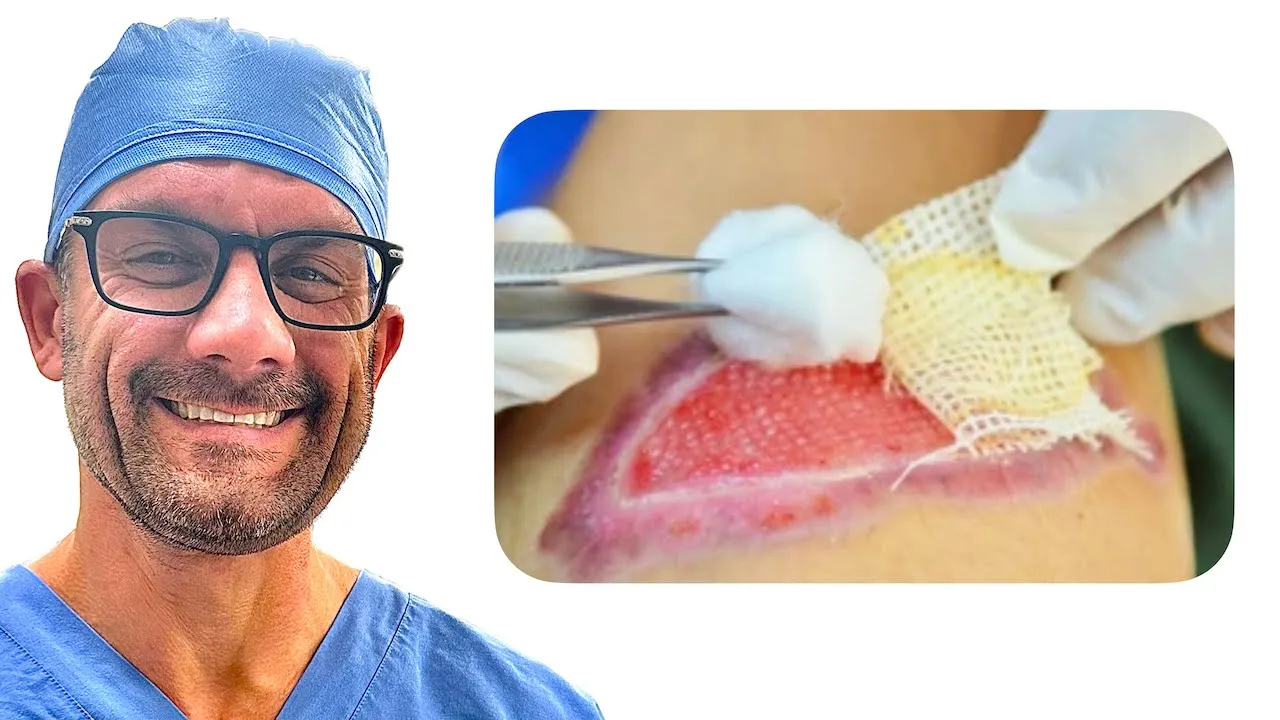
How Surgeons Clean Wounds
83.3K
Wound bed preparation is a holistic approach to removing barriers to wound healing using the TIME acronym: Tissue debridement (removing necrotic tissue through surgical, mechanical, autolytic, chemical, larval, hydro, or ultrasonic methods), Inflammation/biofilm management (addressing biofilms present in 60-90% of chronic wounds), Moisture management (balancing exudate to prevent excessive matrix metalloprotease activity), and Epithelial edge advancement (using modalities like acellular dermal matrix, negative pressure therapy, or hyperbaric oxygen). Success is measured by 20% wound reduction after two weeks and 40% after four weeks.
Click to watch →
Manejo avanzado de heridas, recursos actuales - MVZ Esp Aremi Lujano
10.7K
The TIME acronym provides a comprehensive framework for wound assessment and management: T (Tissue) evaluates non-viable tissue requiring removal; I (Infection/Inflammation) addresses inflammatory processes and infection control; M (Moisture) manages wound moisture balance; E (Edge) evaluates wound edge characteristics. Wound bed preparation involves removing non-viable tissue through surgical, enzymatic, larval, or mechanical methods. Moist wound healing, established by George Winter in 1962, demonstrates that wounds heal faster in moist environments than exposed wounds. Perilesional skin protection prevents maceration from excess exudate. Understanding these principles guides appropriate treatment selection and optimization of healing outcomes.
Click to watch →

WEBINAR NASIONAL METODE PERAWATAN LUKA MODERN DENGAN HIPNOTERAPI
244
Effective wound assessment requires a holistic approach examining the wound bed, wound edges, and periwound skin. The Three Angel Assessment method evaluates: wound bed (color, depth, tissue type, odor), wound edges (epithelialization, thickness, cleanliness), and periwound skin (integrity, maceration, erythema). Wound bed colors indicate healing status: red (healthy granulation), yellow (slough/infection), black (necrosis). Wound measurement includes length, width, depth, and undermining using clock face documentation. The TIME framework guides wound bed preparation: Tissue management (debridement), Inflammation/Infection control, Moisture balance, and Epithelial advancement. Tissue management methods include surgical, autolytic, enzymatic, mechanical, and chemical debridement. Infection control distinguishes normal inflammation from infection. Moisture balance prevents maceration while maintaining optimal healing environment. Epithelial advancement requires healthy, thin wound edges. The framework requires 2-4 weeks for wound bed preparation before epithelialization can occur.
Click to watch →

Ferramenta TIME na avaliação de feridas e coberturas/ Úlcera por pressão ou Lesão por Pressão?
7.4K
The TIME framework is a systematic approach for wound bed preparation developed in 2002 and updated in 2008. It consists of four components: T (Tissue viability), I (Infection/Inflammation), M (Moisture balance), and E (Edema). The framework was developed by the NPUAP, an American non-profit authority on wound care. The first step involves assessing tissue viability and identifying necrotic tissue requiring debridement. Five debridement types exist: autolytic (body's natural healing), enzymatic (using agents like collagenase or papain), mechanical (hydrotherapy or wet-to-dry), surgical (by surgeons), and sharp (by trained nurses). Each method has specific applications based on wound characteristics and tissue type.
Click to watch →
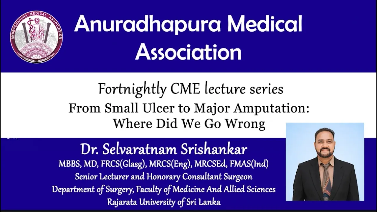
From Small Ulcer to Major Amputation | Dr. Selvaratnam Srishankar - Consultant Surgeon
265
The TIME framework for preventing amputation includes: Tissue debridement (surgical or biological methods like maggots); Inflammation and infection control (distinguishing contamination, colonization, and true infection); Moisture balance (optimal environment for healing); Epithelial edge advancement (cutting crusty edges to allow healing). Biofilm formation prevents antibiotic penetration and requires debridement. True infection is indicated by swelling, pus, pain, and systemic signs. Proper wound culture requires deep swabbing after thorough cleaning.
Click to watch →
Deep Dive
Comparative analysis of alternative debridement modalities, including autolytic, enzymatic, mechanical, and biological (maggot) therapy.
WoundRounds Webinar Series - Dressing(s) for Success: Wound Care Dressing Selection
285.2K
Debridement removes nonviable tissue (slough - moist yellow tissue; necrotic tissue - dried hardened tissue) to restore healthy wound beds and advance healing. Multiple methods exist with different indications: Surgical debridement is rapid but requires appropriate setting and clinician skill. Autolytic debridement uses the body's natural enzymes and is gentle. Enzymatic debridement uses collagenase to break collagen bonds attaching dead tissue. Mechanical debridement with wet-to-dry gauze can traumatize the wound bed. Biological debridement uses maggot therapy effective against MRSA. Selection considers wound characteristics, patient factors, setting, time constraints, infection risk, clinician skill, and cost. Methods can be combined strategically, such as surgical followed by enzymatic debridement.
Click to watch →
VSUR50 Module 8: Management of Open Wounds
2.8K
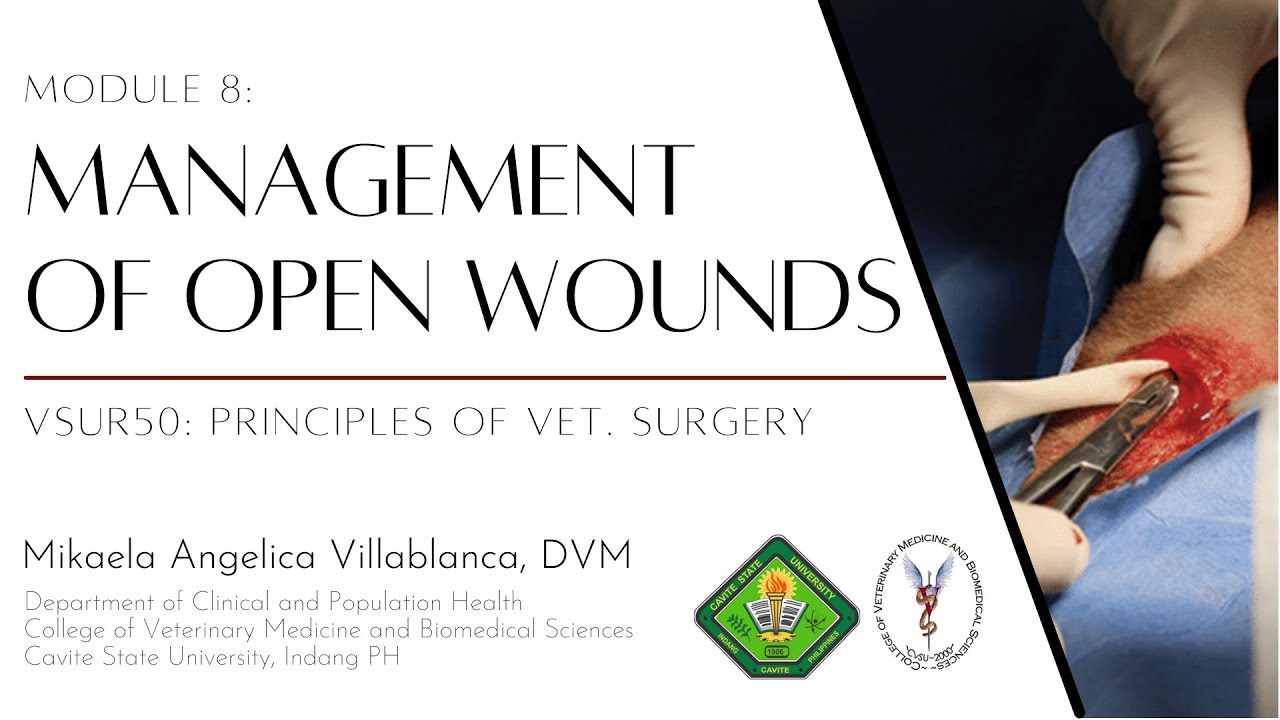
Multiple debridement approaches serve different clinical scenarios. Autolytic debridement uses occlusive bandages (72-96 hours) to activate patient's own enzymes selectively, being less painful and causing less scarring than mechanical methods. Mechanical (wet-to-dry) bandage debridement is cheap but very painful and non-selective. Enzymatic debridement uses commercial agents breaking down necrotic tissue as adjuncts to other methods. Maggot therapy employs fly larvae secreting proteolytic enzymes—each larva consumes up to 75 mg of necrotic tissue daily, applied at five to eight larvae per cm². Tissue preservation prioritizes bone, tendons, nerves, blood vessels, and muscles, while fat tissue can be removed more freely.
Click to watch →

ISSO AQUI NÃO É FIBRINA ENTENDA (Fibrina ou Esfacelo)?
31.6K
Debridement is the removal of necrotic tissue from wounds. There are six types: (1) Autolytic debridement uses the body's own enzymes in a moist environment to selectively remove necrosis; (2) Enzymatic debridement uses exogenous enzymes (papain, collagenase) but is non-selective and can damage healthy tissue; (3) Mechanical debridement includes irrigation with saline and friction techniques during cleaning; (4) Biological debridement uses treated maggot therapy; (5) Instrumental conservative debridement uses instruments performed by trained nurses; (6) Surgical debridement is performed by physicians for extensive necrosis.
Click to watch →

Wound infection, biofilms & antimcrobials Gary Bain
205
This section covers debridement options: autolytic (white blood cells eat dead tissue, slow and inefficient, not for infected wounds), mechanical (ultrasound cleaning systems, effective but expensive), osmotic (honey and hypertonic saline - honey kills bacteria through osmotic action causing bacterial implosion), and negative pressure wound therapy (helps debride and deliver antimicrobials). Maggot therapy is also discussed - laboratory-manufactured maggots eat dead tissue and disrupt biofilms, available in tea bags or free-range. Each method has specific applications and limitations.
Click to watch →

Wet-to-Dry: Substandard of Care Webinar
4.4K
Modern wound care offers multiple selective debridement options beyond wet-to-dry dressings. Autolytic debridement uses the body's own enzymes (neutrophils, macrophages) to dissolve necrotic tissue, requiring moist environments and hydrogels. Collagenase (Santyl) is the only FDA-approved enzymatic debrider, working within pH 6-8 and requiring once-daily application. Biological debridement uses live maggots that mechanically debride while secreting enzymes and promoting fibroblast growth. Mechanical alternatives include whirlpool/hydrotherapy (4-15 psi), ultrasonic debridement, monofilament brushes, and negative pressure wound therapy with instillation/aspiration. Sharp debridement requires sterile technique and should be performed at least twice weekly to prevent biofilm formation within 2-3 days.
Click to watch →
Deep Dive
Advanced wound dressing selection and post-procedural therapies, such as Negative Pressure Wound Therapy (NPWT) and hyperbaric oxygen therapy.

CURSO "LESIONES POR PRESIÓN. ACTUALIZACIÓN, PREVENCIÓN, MANEJO Y TRATAMIENTO"
933
Advanced wound dressings include hydrocolloids (form gel with exudate, maintain moisture, promote autolytic debridement), alginates (highly absorbent, form gel, good for heavy exudate), foams (absorb exudate, reduce friction, provide cushioning), and hydrogels (provide moisture for dry wounds, promote autolytic debridement). Negative pressure wound therapy (NPWT) uses controlled suction to remove fluid, reduce edema, and promote granulation tissue. Hyperbaric oxygen therapy delivers high oxygen concentrations for refractory cases. Selection depends on wound characteristics, exudate level, and patient factors.
Click to watch →

MSN, PEDIA, PHARMA | NORCET Series #1198 | NORCET 9 Mains & All Nursing Exams Special Class
6.4K
Advanced wound healing therapies include: Negative pressure wound therapy (NPWT/vacuum-assisted closure) which promotes early wound healing, reduces edema, prevents necrotic tissue, and increases blood flow using suction devices. Wet-to-dry dressings are used for manual removal of necrotic tissue without suction. Hyperbaric oxygen therapy uses high-pressure oxygen chambers without suction devices. Each therapy has specific mechanisms and clinical applications.
Click to watch →

Negative Pressure Wound VAC Therapy & Wound Dressing: Which Dressing Should You Use?
14.5K
Wound dressing selection depends on wound characteristics: basic gauze for absorbency and debridement, alginates/hydrofiber for moderate drainage wounds, hydrocolloids for fragile skin protection, hydrogels for dry wounds, foams for edematous wounds, and combination dressings for multifunctional needs; negative pressure wound therapy (NPWT) includes basic therapy for wound bed preparation, installation plus therapy for debridement, and incisional therapy for surgical incisions, with troubleshooting alarms requiring checking connections, ensuring proper drape sealing, and replacing discs when necessary.
Click to watch →

ACOME & LAUGHH: Academy Updates - Day 1
2.1K
Advanced wound therapies include Negative Pressure Wound Therapy (NPWT/VAC) applying -521 to -175 mmHg pressure to control exudate, increase blood flow, and reduce infection risk—serving as bridge to surgical closure. Extracellular matrix allografts provide scaffolding from collagen and elastin for slow-healing wounds, requiring weekly application. Hyperbaric oxygen delivers 100% oxygen at 2-3 atmospheres for 90-minute sessions over 5-7 days to correct hypoxia in chronic wounds, though complications include ear trauma and seizures. Emerging technologies include bioelectric dressings with microcurrents. Antibiotics are reserved for infected wounds, not prophylactically, since all wounds are colonized but not all are infected.
Click to watch →
INTERNATIONAL WEBINAR SURGICAL WOUND DEHISCENCE JUNE 21, 2026
110
Advanced wound management includes NPWT (VAC) for promoting granulation and angiogenesis, antimicrobial dressings (silver, iodine-based), and appropriate dressing selection based on exudate level. NPWT is contraindicated for cancer wounds. Dressing selection depends on wound characteristics, exudate level, and patient factors.
Click to watch →
Deep Dive
Management of potential complications of surgical debridement, such as hemorrhage, damage to underlying structures (tendons, nerves), and osteomyelitis.
OSTEOMIELITIS
3K
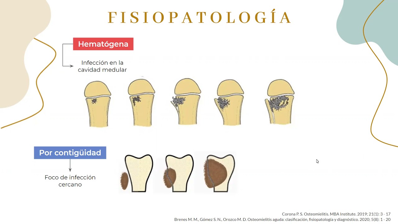
Chronic osteomyelitis is a surgical pathology requiring bone and necrotic tissue removal to eliminate biofilms. The six pillars of surgical treatment are: 1) Surgical debridement (key factor for success, eliminating necrotic bone and ischemic tissues), 2) Microbiological diagnosis (ideally before antibiotic administration, with 3-6 samples from necrotic bone fragments), 3) Dead space obliteration (reducing space excluded from circulation and antibiotic activity), 4) Targeted antibiotic therapy (IV initially, then oral if appropriate), 5) Soft tissue reconstruction (creating biological chamber for revascularization), and 6) Bone reconstruction (for unstable bone defects). Complications include persistent pain, neurological deficits, epidural abscesses, spread to adjacent joints, progression to chronic osteomyelitis, sinus tract-associated neoplasms, and pathological fractures. Early diagnosis is decisive for initiating treatment. Acute osteomyelitis has a good prognosis with adequate treatment (90% cure rate), but 5-10% progress to chronic forms with more unfavorable prognosis, potentially requiring amputation.
Click to watch →

اسس الجراحة/ المحاضرة الخامسة/ مع حل جميع الاسئلة الوزارية دور اول وثاني ( حسب المنهج الجديد 2025)
12.1K
When antibiotic treatment fails, surgical intervention becomes necessary. Surgical goals include: removal of necrotic bone tissue, elimination of bacteria, and reduction of tissue damage. After debridement, the resulting cavity may be filled with bone grafts, bone substitutes, or bone cement to provide structural support and resist bacterial re-infection. Complications include: development of antibiotic-resistant bacteria like MRSA; spread of infection to other organs (lungs, liver, kidneys); damage to growth plates in children; chronic osteomyelitis causing joint destruction and bone deformities; and increased fracture risk in elderly patients. Life-threatening complications include sepsis and organ damage from bacterial spread.
Click to watch →

Musculoskeletal Disorders S23 Copy
168
Surgical intervention with debridement is required for treatment failure, abscess, joint infection, neurological deficits, or spinal instability. Infected orthopedic hardware requires removal. In extreme cases, amputation promotes healing. Complications include sepsis, amputation, and secondary malignancies. Nursing assessment includes vital signs, pain evaluation, wound assessment, and neurovascular assessment. Nursing actions include IV antibiotics, analgesia, thermal therapy, range of motion exercises, fall precautions, and nutritional support. Teaching emphasizes completing antibiotic courses and pain management. Evaluating outcomes focuses on pain control, infection resolution, and limb preservation.
Click to watch →

Osteomyelitis #orthopedic Nursing unit#1 #part-5 #basic concept types of osteomyelitis
889
Complications include bone deformity, pathological fracture, chronic purulent drainage, amyloidosis, sepsis, and sarcoma. Surgical management includes debridement of necrotic tissue, irrigation with saline/antibiotics, and bone grafting to stimulate healing. The treatment protocol involves initial IV antibiotics for 2-3 weeks followed by oral antibiotics for 3 months. Immobilization with casts reduces pain and prevents fracture.
Click to watch →

Infecciones óseas en el esqueleto inmaduro: ¿sabemos identificarlas?
858
Surgical management of osteomyelitis involves an approach proportional to the lesion size. The procedure includes periosteal incision, culture samples, drainage of purulent material, and debridement of necrotic tissue. Some surgeons perform window osteotomies to remove necrotic bone. The cavity may be decompressed with a drain, and a splint is applied for protection. Complications include sepsis and death, persistent infection, deep vein thrombosis, thromboembolism, encephalitis, meningitis, pathological fractures, infection recurrence, chronic osteomyelitis, and septic arthritis in children under 18 years.
Click to watch →
Debridement Prep
Post-Op Care
Debridement Prep
0:16- 1
Surgical debridement removes dead tissue from wounds.
- 2
Causes include injuries, burns, ulcers, and infections.
- 3
Local or general anesthesia numbs the area before treatment.
Conservative and Selective Debridement Alternatives
While surgical debridement is highly effective for rapid tissue clearance, it is an invasive, painful, and non-selective procedure that carries risks of damaging healthy viable tissue, bleeding, and infection. Critics and alternative clinical approaches advocate for conservative, selective debridement methods—such as autolytic, enzymatic, or biological (maggot) therapy. Autolytic debridement uses the body's own enzymes and moisture under specialized dressings to naturally liquefy necrotic tissue, preserving healthy structures and minimizing pain. Biological debridement utilizes sterile larvae that precisely consume dead tissue while sparing live cells and secreting antimicrobial substances. These alternative modalities argue that a less traumatic, highly selective approach can achieve comparable healing outcomes with lower patient discomfort, reduced need for anesthesia, and decreased overall healthcare costs, particularly in patients with compromised healing capacity or poor surgical candidacy.
Anatomy of the skin and the four primary phases of wound healing (hemostasis, inflammation, proliferation, and remodeling).
The physiological distinction between viable (healthy, vascularized) tissue and non-viable (necrotic, ischemic, slough, or eschar) tissue.
Fundamental principles of surgical asepsis, sterile technique, and infection prevention in clinical settings.
The role of bacteria in wounds, specifically the progression from contamination and colonization to local and systemic infection.
The 'TIME' framework for wound bed preparation (Tissue debridement, Infection/inflammation control, Moisture balance, and Edge of wound advancement).
Comparative analysis of alternative debridement modalities, including autolytic, enzymatic, mechanical, and biological (maggot) therapy.
Advanced wound dressing selection and post-procedural therapies, such as Negative Pressure Wound Therapy (NPWT) and hyperbaric oxygen therapy.
Management of potential complications of surgical debridement, such as hemorrhage, damage to underlying structures (tendons, nerves), and osteomyelitis.
1.6M views6.4Klikes2:40@nucleusmedicalmediaOriginal Release: 2011-03-23
your doctor will perform surgical debent to remove dead tissue from in and around your wound wounds can result from a variety of causes including injuries Burns bed sores also called pressure ulcers or decubitus ulcers and infected surgical incisions dead damaged or contaminated tissue can compromise circulation to your wound in addition the unhealthy tissues are prone to infection dead tissues invite bacteria to grow which compete with growing cells for nutrients and interfere with the he healing process the wound's healthy tissues may become infected the infection May develop into a pocket of pus called an abscess that impedes wound healing a wound resulting from a burn may form a hardened crust of dead tissue called an escar that can prevent healthy tissue growth and healing before your procedure your doctor will numb the area around your wound with local anesthetic for more extensive debent you will receive general anesthesia to begin your doctor may use saline or disinfectant solutions to wash or irrigate your wound next your doctor will evaluate the depth of your wound checking the extent of the dead tissue your doctor May remove a rim of tissues from around your wound to create a clean margin of healthy tissue and improve healing at the end of your procedure your doctor will place a dry bandage in your wound to manage any bleeding he or she may also cover your wound with moist dressings to encourage healing after your procedure you should follow wound care directions very carefully be sure that you protect your wound from injury and keep the wound and its dressings clean and dry
Up Next

1:30
Local Anesthetic Infiltration Techniques: Wound Margin Injection
@thelacerationcourse2009
498.4K views•2022-04-25

48:47
Graphic Medicine: Comics for Collaborative Healthcare Communication
@nationalpatientadvocate
189 views•2023-12-04

3:05
Stages of Labor and Vaginal Birth | Childbirth Animation
@nucleusmedicalmedia
52.1M views•2017-08-18

19:45
The Science of Fentanyl Addiction and Overdose Risks
@theanatomylab
9.1M views•2022-09-30
Related Study Plans & Knowledge Roadmaps
Structured learning paths in Medicine
medicine
Abnormal LFTs: Diagnostic Workup & Pathology
01Liver Anatomy and Basic Physiology
02Introduction to Liver Function Tests (LFTs)
03Hepatocellular vs. Cholestatic Patterns
+ 3 more learning modules
12h6 modules
Explore Roadmap
medicine
Analyzing Blood Smears: RBC, WBC & Platelets
01Introduction to Blood Components & Smear Preparation
02Normal Morphology of Blood Cells
03Erythrocyte Pathologies & Morphological Variants
+ 2 more learning modules
12h5 modules
Explore Roadmap
medicine
Mechanical Ventilation: Waveforms & Alarms
01Anatomy and Physiology of Breathing
02Introduction to Mechanical Ventilation & Core Modes
03Ventilator Settings and Waveform Interpretation
+ 2 more learning modules
12h5 modules
Explore Roadmap
medicine
Neuro Exam: Localization & Stroke Syndromes
01Foundations of Neuroanatomy and Vascular Supply
02The Neurological Examination: Step-by-Step
03Principles of Neurological Localization
+ 3 more learning modules
18h6 modules
Explore Roadmap