To properly infiltrate local anesthetic into a wound, insert the needle at the wound margin rather than intact skin, aspirate to confirm absence of blood vessel entry, inject while slowly withdrawing the needle, and reposition the needle within the tissue to avoid multiple punctures; repeat this process on both sides of the wound for adequate anesthesia.
Local Anesthetic Infiltration Techniques: Wound Margin Injection
Added:Deep Dive
Basic anatomy of the skin layers (epidermis, dermis, and subcutaneous tissue) and the pathway of superficial sensory nerves.
Anatomy & Physiology 1: ENTIRE Course Explained in One Video!
66.1K
The skin has three layers: epidermis (outer layer of tough, flattened cells protecting against environment), dermis (middle layer containing blood vessels, sensory neurons, hair follicles, and connective tissue), and subcutaneous tissue (below dermis, composed mainly of adipose/fat cells for insulation and energy storage).
Click to watch →

(2020) L1 | Generalidades | Profesor Eduardo A. Pró | Anatomía 2 | FMed UBA
51.9K
The skin (cutis or piel) consists of two main layers: the epidermis (superficial epithelial layer) and the dermis (deeper connective tissue layer with collagen fibers). The dermis provides structural support and resistance. The subcutaneous tissue (tela subcutánea or hipodermis) lies deep to the skin and contains adipose tissue, connective tissue, blood vessels, and nerves. This tissue provides insulation, energy storage, and cushioning. The subcutaneous tissue contains superficial nerves (nervios superficiales or nervios cutáneos) that provide sensory innervation to the skin. It also contains superficial lymphatics (linfáticos superficiales) for lymph drainage. The cutaneous retinaculum (retináculo cutáneo) is a fibrous structure that anchors the skin to deeper tissues. In some regions like the face, muscles are located in the subcutaneous tissue and insert into the dermis, allowing for facial expressions.
Click to watch →

Anatomy of the skin Cosmetology Stateboard 2021
14.4K
The skin has three main layers: epidermis (outermost, dead layer), dermis (middle, living tissue), and subcutaneous layer (bottom, fat). The epidermis contains five layers from deepest to outermost: stratum germinativum/basal cell layer (new cell birth and melanin production via melanocytes), stratum spinosum/spiny layer (shedding begins and keratinization starts), stratum granulosum/granular layer (fully dead cells with visible keratin), stratum lucidum/clear layer (only on palms and soles), and stratum corneum/horny layer (outermost, water-resistant). The dermis (cutis/chorium) is 25 times thicker than the epidermis and contains two layers: papillary (top, near epidermis) and reticular (bottom). It houses sebaceous glands, sweat glands, nerves, blood vessels, lymph vessels, hair follicles, and erector pili muscles (causing goosebumps). The subcutaneous layer (adipose tissue/fat) provides cushioning, shape, and insulation beneath the dermis.
Click to watch →
GENERALIDADES - Repaso integrador de Anatomía
67K
The skin consists of two layers: the skin itself and subcutaneous tissue. The skin provides protection as physical, immunological, and chemical barrier; mechanical function by enveloping deep structures; and temperature regulation through arteriovenous anastomoses. Anatomically, the skin has three layers: the epidermis (superficial epithelial layer, avascular, nourished by diffusion with strata corneum, lucidum, granulosum, spinosum, and basale), the dermis (connective tissue with blood vessels, lymphatic vessels, nerves, and appendages), and the subcutaneous tissue (adipose layer, membranous layer, and loose connective tissue fascia). The dermis has papillary and reticular layers. Cutaneous appendages include sweat glands and hair follicles. Sensory receptors include mechanoreceptors (pressure, textures), thermoreceptors (Ruffini corpuscles for heat, Krause's corpuscles for cold), and pain receptors.
Click to watch →

احياء الثالث متوسط 2024/ الفصل العاشر / المحاضرة 1 / اعضاء الحس
142.7K
The skin (الجلد) is composed of three layers: the epidermis (البشرة) as the outermost layer, the dermis (الادمة) as the middle layer, and the subcutaneous tissue (الطبقة تحت الجلد) as the deepest layer. Sensory nerves and sensory receptors are primarily located in the dermis layer, where they detect various stimuli from the environment.
Click to watch →
Deep Dive
Pharmacology of common local anesthetics (such as lidocaine and bupivacaine), including their onset of action, duration, and weight-based maximum safe dosages.

chapter : L4 5 / Local anesthesia
177
Local anesthetics are classified by duration (short <1h: procaine, chloroprocaine; intermediate 1-2h: etidocaine; long 2-6h: bupivacaine, ropivacaine, lidocaine) and chemical structure (amide: lidocaine, bupivacaine; ester: procaine, chloroprocaine). Mechanism: reversible sodium channel blockers that bind during resting state, preventing depolarization. pKa (7.3-8.4) determines ionization at physiological pH (7.4); closer pKa to pH means faster onset. Tissue pH affects efficacy, with acidic tissues reducing drug penetration. Potency relates to lipid solubility and is measured relative to lidocaine. Protein binding prolongs duration by keeping drug at the injection site. Clearance rate affects duration, with faster clearance shortening duration. Maximum dose limits: lidocaine 300mg (4.5mg/kg), mepivacaine 300mg, bupivacaine 175mg (2.5mg/kg), ropivacaine 200mg (3mg/kg). Vasoconstrictors (epinephrine) increase maximum safe doses by reducing systemic absorption and widening the therapeutic index.
Click to watch →

Regional Anesthesia basics before you prick the needle
223
Local anesthetic onset depends on pKa (lidocaine 7.7-7.8 faster than bupivacaine 8.1). Duration depends on protein binding (bupivacaine longer). Vasoconstrictors prolong action and reduce toxicity. Bicarbonate accelerates onset by raising pH. Safe doses: bupivacaine 2 mg/kg, ropivacaine 2.5 mg/kg, lidocaine 3-5 mg/kg. Intercostal space has highest absorption risk. Opioids enhance quality and duration.
Click to watch →
07 - Autonomic Pharmacology 1
6.9K
Local anesthetics are classified as esters (procaine, chloroprocaine, tetracaine) metabolized by hydrolysis with shorter duration, or amides (lidocaine, bupivacaine, ropivacaine) metabolized by hepatic enzymes with longer duration. Allergic reactions are rare but more common with esters. Onset relates to pKa (lower = faster), while duration relates to protein binding (higher = longer). Maximum safe doses: cocaine 140 mg, lidocaine 7 mg/kg, mepivacaine 18.5 mg/kg, bupivacaine 3 mg/kg, ropivacaine 3 mg/kg. Ion trapping affects toxicity: acidosis/hypoxia increases brain accumulation, while alkalinization enhances tissue penetration. Overdose presents with CNS toxicity followed by cardiovascular collapse, treated with intralipid emulsion.
Click to watch →

Anestésicos locales y principales bloqueos anestésicos
1.6K
Local anesthetics temporarily and reversibly block nerve impulse conduction by blocking sodium channels in neuronal membranes. They block nerves in sequence: sympathetic nerves first, then pain/temperature, protective sensation, touch/pressure, and finally motor function. Esters (procaine, cocaine) are metabolized in plasma and produce sensitizing aminobenzoic acid, while amides (lidocaine, mepivacaine, bupivacaine) are more stable and commonly used. Onset and duration vary: lidocaine/mepivacaine have immediate onset with intermediate duration, while bupivacaine has delayed onset but longer half-life. Maximum doses are weight-based, and epinephrine can extend duration and reduce bleeding but is contraindicated in acral areas, severe hypertension, coronary disease, and pregnancy. Side effects include local reactions (pain, ecchymosis, infection) and systemic effects from overdose (CNS: tinnitus, metallic taste, convulsions; CV: hypotension, arrhythmia). Allergic reactions range from urticaria to anaphylaxis. Precautions include patient monitoring, allergy assessment, aspiration before injection, and emergency preparedness.
Click to watch →

Medical School | Plastics Rotation
79.7K
Maximum safe doses of local anesthetics must be calculated based on patient weight and concentration. For lidocaine without epinephrine, the maximum dose is 4.5 mg per kg; with epinephrine, it increases to 7 mg per kg. For bupivacaine without epinephrine, the maximum is 2.5 mg per kg; with epinephrine, it is 3 mg per kg. These values must be multiplied by the patient's weight in kilograms and the solution concentration to determine the absolute maximum allowable dose.
Click to watch →
Deep Dive
The principles of aseptic technique and wound preparation to prevent introducing pathogens during injection.

🩺 Mastering Aseptic Technique in Wound Care: Protecting Patients, Preventing Infections! 🛡️
4.4K
The five fundamental principles of aseptic technique in wound care are: (1) Hand hygiene - proper cleaning of hands before and after procedures; (2) Sterile field - maintaining a sterile environment for wound care; (3) Personal protective equipment - using appropriate PPE to prevent contamination; (4) Minimal touch - reducing contact with the wound to prevent introducing pathogens; (5) Cleaning the wound inside to outside - starting from the cleanest area and moving outward; (6) Safe disposal of materials - properly disposing of contaminated materials to prevent infection spread.
Click to watch →

HESI Foundations Boot Camp - 6/11
239
Aseptic technique is used when performing invasive procedures to prevent introducing bacteria or pathogens. Key principles include: (1) One inch around the border of the sterile field is not sterile, only the area inside is sterile; (2) Never reach across the sterile field; (3) Never place hands below waist level; (4) Never turn your back on the sterile field; (5) Never cough, laugh, or sneeze over the sterile field; (6) If contamination occurs, the entire procedure must be restarted. Sterile technique is required for procedures like Foley catheter insertion, while clean technique may be used for NG tube insertion.
Click to watch →

Administración de medicamentos vía parenteral || UMG
107
Perform a single asepsis using the principle of moving from center to periphery. Avoid performing double asepsis, as this may leave microorganisms in the same site. This principle prevents introducing pathogens into the injection site.
Click to watch →

CURSO DE INYECTOLOGIA VIRTUAL
6.6K
Asepsis is the technique used to eliminate pathogens from a specific area. In inyectología, asepsis is performed from the center outward using cotton balls or gauze soaked in alcohol (70%). This prevents introducing pathogens into the injection site. Sterile supplies must be verified before use: check that they are in their original sealed packaging, verify they are sterile, and ensure they meet sanitary regulations. A sterile supply is considered contaminated if it touches any non-sterile object.
Click to watch →
1 Sesión - PRINCIPIOS DE ASEPSIA Y ANTISEPSIA
14.8K
The five principles of aseptic technique for wound care are: (1) perform procedures from center to periphery, (2) work from above to below, (3) move from distal to proximal, (4) proceed from clean to dirty areas, and (5) work from inside to outside. These principles ensure proper infection control during wound dressing changes.
Click to watch →
Deep Dive
Understanding the concept of intravascular injection risks and the physiological rationale behind aspirating before injecting.
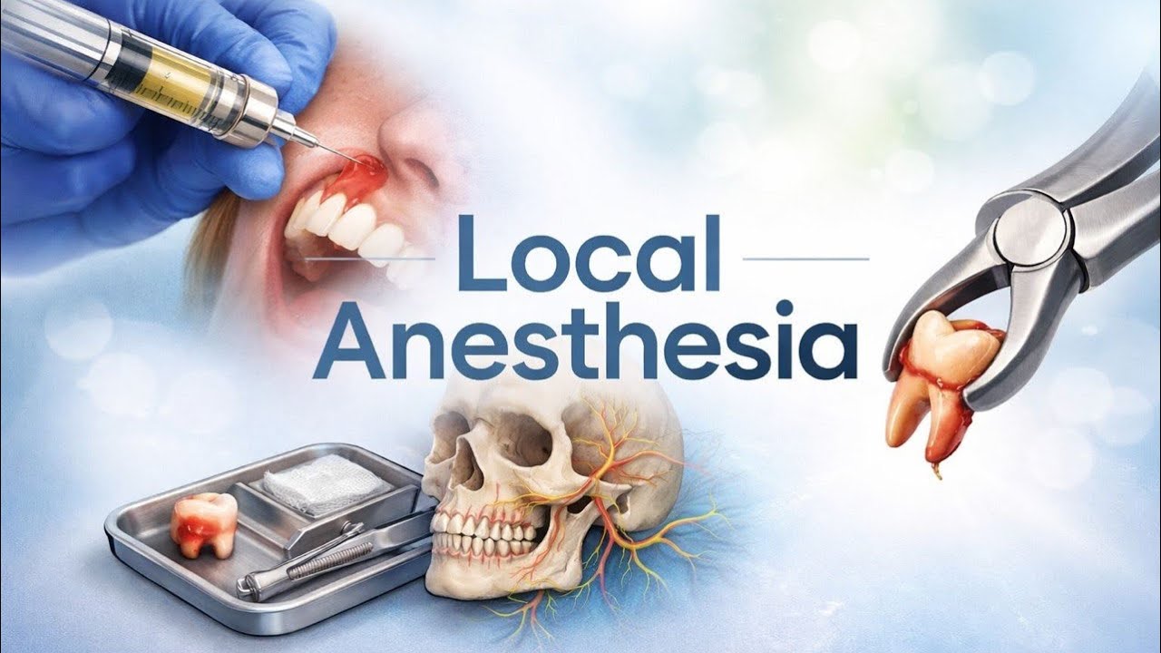
The Armamentarium
160
Aspiration is a critical safety step before injection involving negative pressure to check for blood in the cartridge. If blood appears, the needle is in a blood vessel and the injection must be aborted with complete replacement of needle, cartridge, and injection site. If no blood appears, the injection can proceed. Intravascular injection leads to reduced anesthetic efficacy, decreased duration, and increased systemic toxicity. These risks are particularly dangerous for medically compromised patients with cardiac conditions or compromised liver function. The aspiration should be performed gently by pulling back on the plunger for 2-3 seconds, waiting for blood to appear before proceeding.
Click to watch →

Kyle's vaccine complication
2.6M
Aspiration (pulling back on the syringe plunger after needle insertion) is a technique to check if a needle has entered a blood vessel. Kyle suggested this should be used for intramuscular injections, particularly for mRNA vaccines, to prevent inadvertent intravascular administration. He experienced a metallic taste within 30 seconds of injection, which he believes indicates intravascular administration. Intravenous injections cause immediate systemic effects, while intramuscular injections take approximately 30 minutes or longer for systemic absorption. This could explain why his inflammatory reaction occurred in his heart and joints rather than at the injection site. Nurses expressed surprise at this recommendation, suggesting it is not standard practice for these vaccines.
Click to watch →

Aspiration, more information
256.1K
This extended analysis explores the mechanisms and consequences of inadvertent intravascular injection during vaccine administration. Multiple factors must align for this complication: anatomical variations in blood vessel positioning, metabolic changes affecting vessel elasticity, and predisposition to inflammatory heart disease. Younger males may have more turgid blood vessels, explaining higher rates of adverse events in this demographic. US military data reveals 23 male patients with myocarditis out of 2.8 million doses—a rate of 1 in 121,000. Another series reported 57 patients (myocarditis/pericarditis) among 2 million individuals, or 1 in 35,092. Professional nursing resources recommend aspiration to detect blood return, contradicting current guidelines. The fundamental principle of first do no harm supports incorporating aspiration as a safety measure to prevent potentially fatal intravenous vaccine administration.
Click to watch →
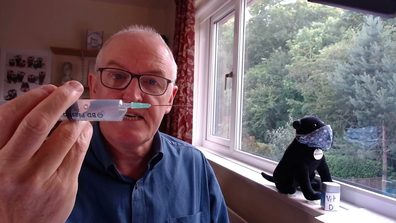
New evidence
906.1K
Safe intramuscular injection technique requires aspirating (pulling back on the plunger) before injecting the vaccine to ensure the needle is not in a blood vessel. If blood appears in the syringe during aspiration, the needle has entered a blood vessel and the injection should not proceed. This simple technique prevents inadvertent intravascular administration, which can lead to serious complications including thrombosis with thrombocytopenia syndrome.
Click to watch →

Que pasa si no aspiro antes de inyectar
196.3K
Before administering an intramuscular injection, healthcare providers must aspirate (pull back on the syringe plunger) to check if blood enters the needle; if blood appears, it indicates the needle has entered a blood vessel, and the injection must be aborted and a new syringe used to prevent accidental intravenous injection, which can cause cardiac arrest when anesthetics like lidocaine enter the bloodstream.
Click to watch →
Prerequisite Knowledge
- Concept 01Basic anatomy of the skin layers (epidermis, dermis, and subcutaneous tissue) and the pathway of superficial sensory nerves.
- Concept 02Pharmacology of common local anesthetics (such as lidocaine and bupivacaine), including their onset of action, duration, and weight-based maximum safe dosages.
- Concept 03The principles of aseptic technique and wound preparation to prevent introducing pathogens during injection.
- Concept 04Understanding the concept of intravascular injection risks and the physiological rationale behind aspirating before injecting.
Subsequent Learning
- Step 01Wound closure and repair techniques, such as simple interrupted, running, or subcuticular suturing.
- Step 02Advanced regional anesthesia techniques, including digital blocks, facial nerve blocks, or field blocks for larger lacerations.
- Step 03Recognition and emergency management of Local Anesthetic Systemic Toxicity (LAST), including the use of lipid rescue therapy.
- Step 04Clinical guidelines for the use of vasoconstrictors (like epinephrine) combined with local anesthetics, including absolute and relative contraindications in distal extremities.
Wound Anesthesia
Wound Anesthesia
0:00Playing Section
- 1
Demonstrates injecting anesthetic into wound margins.
- 2
Covers needle insertion, aspiration, and injection technique.
Perilesional Infiltration and Regional Nerve Blocks
While direct wound margin infiltration is widely practiced, many clinicians advocate for perilesional (intact skin) infiltration or regional nerve blocks as superior alternatives. Critics of wound margin injection argue that inserting a needle through an open, potentially contaminated wound bed can seed superficial bacteria deeper into sterile subcutaneous tissues, increasing the risk of infection. Additionally, direct infiltration causes localized tissue edema (swelling), which can severely distort wound margins and hinder precise cosmetic alignment during suturing. In contrast, perilesional infiltration—injecting through the intact skin surrounding the wound—and regional nerve blocks avoid disrupting the wound bed. Regional nerve blocks target proximal nerves to anesthetize a broader area, completely eliminating local tissue distortion at the injury site. This technique reduces the number of painful needle punctures for the patient and is highly preferred for cosmetically sensitive areas, such as the face, where anatomical distortion must be minimized.
Deep Dive
Wound closure and repair techniques, such as simple interrupted, running, or subcuticular suturing.
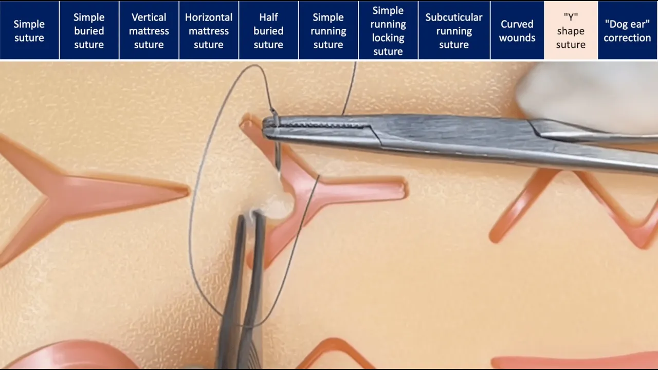
Suturing techniques
437.1K
This video demonstrates multiple suturing techniques for closing cutaneous wounds, including simple interrupted suture (most common technique), simple buried suture (for deep dermal closure), vertical mattress suture (for tension reduction in swollen or high-risk wounds), horizontal mattress suture (faster than two simple sutures), half-buried suture (for plastic surgery to avoid disrupting blood supply), simple running suture (continuous closure), simple running locking suture (for tension control in areas like scalp), subcuticular running suture (cosmetic closure with no external material), curved wound closure (compensating for unequal lengths), Y-shape suture (complex geometric closure), and dog ear correction (marking desired scar location and repositioning tissue).
Click to watch →

SIM SUTURE - 5. Running Simple Sutures
1M
Running simple sutures are a faster alternative to interrupted sutures for wound closure, where a single continuous suture line connects multiple wound edges instead of individual knots; the technique involves starting with a simple suture at one end, then placing subsequent sutures perpendicularly through the skin at each dot while maintaining parallel alignment to create a neat, railroad-track appearance that patients prefer, with the final knot tied using the apex of the previous suture as the grasping point.
Click to watch →
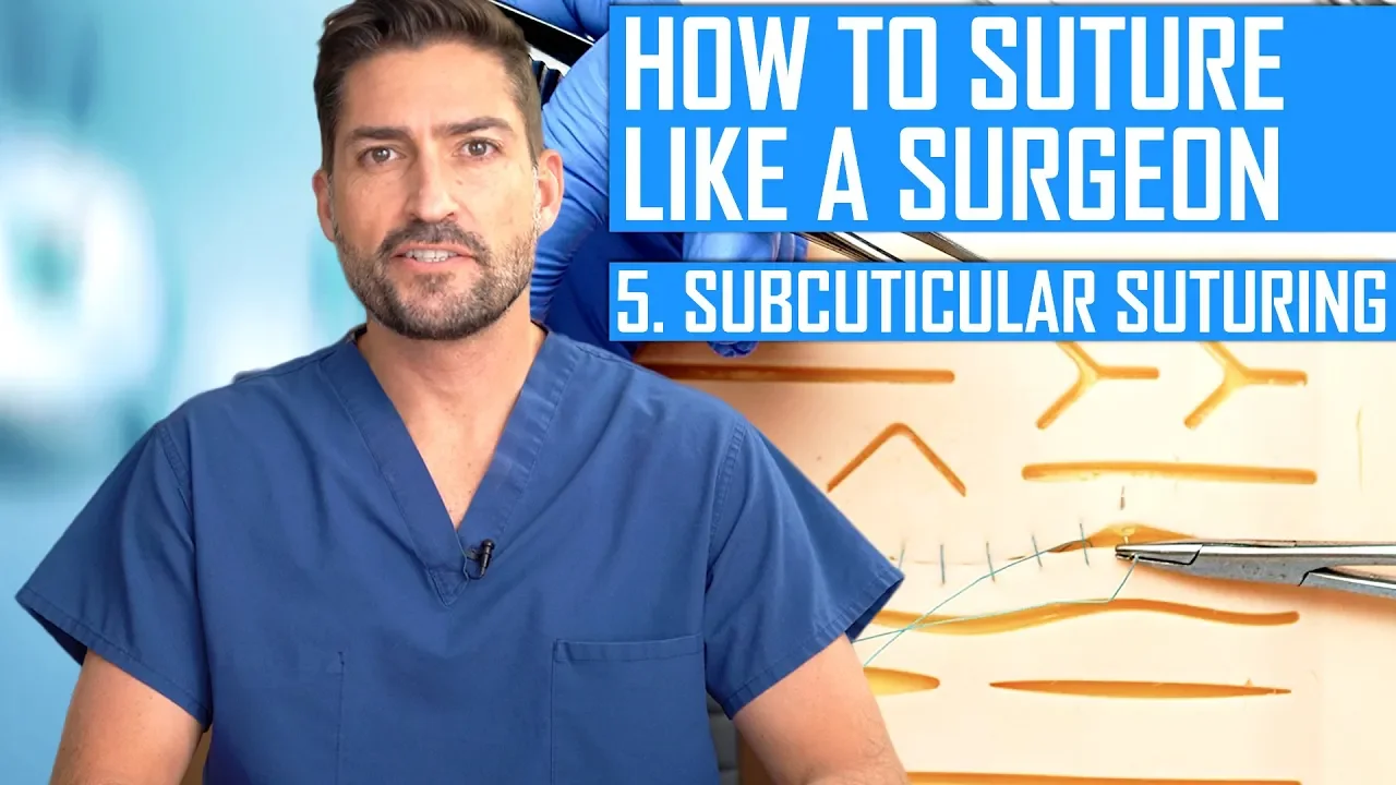
How to Suture Like a Surgeon: Subcuticular Suturing
255.4K
Subcuticular suturing is a surgical technique where the needle passes through the dermis layer (the white layer beneath the epidermis) rather than the epidermis itself, with stitches placed parallel to the skin surface and knots tied deep within the dermis to create an invisible closure; the technique requires consistent depth, proper suture exit on the same side for interrupted sutures, pulling lengthwise along the incision rather than perpendicular, and can be performed as either interrupted or running sutures depending on incision length.
Click to watch →

Running Subcuticular Suture | The Cadaver-Based Suturing Self‑Study Course
337.7K
The running subcuticular suture is a plastic surgery technique using absorbable Vicryl 5-0 sutures that leaves no visible skin marks. The procedure begins by burying the initial knot deep in subcuticular tissue at the distal wound edge, exiting at the dermal epidermal junction. The suture runs in a serpentine pattern through the subcuticular tissue, taking equal bites on alternating sides of the wound while avoiding epidermal buttonholing. Three knots are used initially to minimize tissue reaction. The final knot is buried as the suture approaches the proximal wound edge. The last piece of suture is pulled through and cut on the skin surface to bring the wound edges together. Tissue glue or steri-strips can be applied over the closure. Knots are buried to prevent spitting and reduce scarring.
Click to watch →
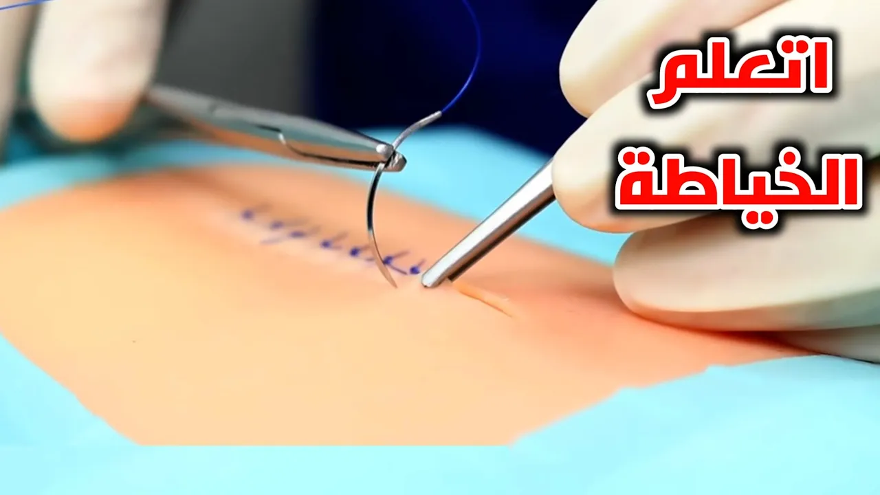
تعلم خياطة الجروح في 5 دقائق_Simple Interrupted Suturing - Perfect Wound Closure
480.4K
This video demonstrates the complete process of simple interrupted suturing for wound closure, including local anesthesia administration with lidocaine, proper needle insertion at 20-45 degrees, creating the first stitch by pulling the thread through the wound edges, tying a secure knot by wrapping the thread twice around the needle holder and pulling in opposite directions, leaving approximately 3cm of thread, and placing subsequent stitches 1cm apart until the wound is fully closed, followed by cleaning, covering with gauze and bandage, and removing sutures after 10 days.
Click to watch →
Deep Dive
Advanced regional anesthesia techniques, including digital blocks, facial nerve blocks, or field blocks for larger lacerations.
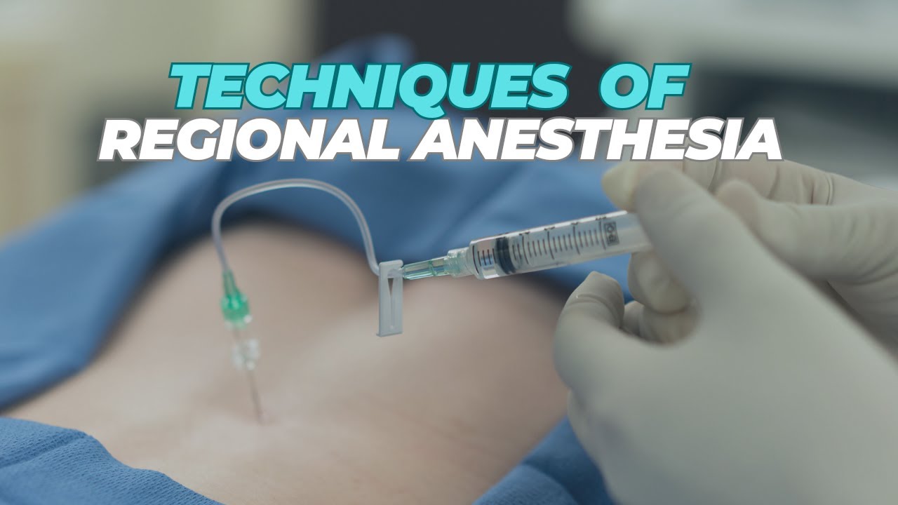
Regional Anesthesia | Techniques
1.2K
Peripheral nerve block techniques include digital nerve blocks targeting finger/toe nerves for digit procedures; plexus blocks injecting near nerve networks (brachial, lumbar, sacral, celiac, stellate ganglion) for larger limb or regional anesthesia; field blocks infiltrating around surgical sites (inguinal hernia example); and infiltration blocks directly injecting into subcutaneous tissue to block cutaneous nerves supplying skin sensation. These techniques provide targeted anesthesia by blocking pain signal transmission at peripheral nerve sites.
Click to watch →
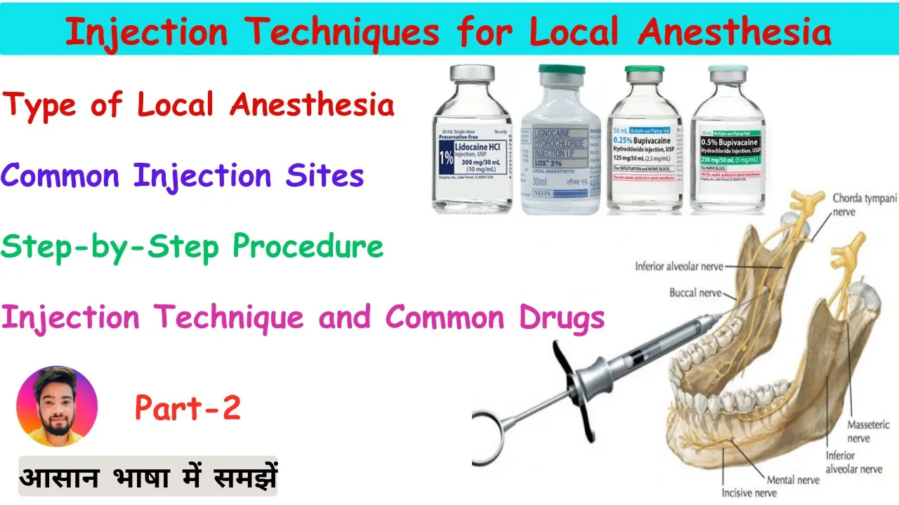
Local Anesthesia Injection Techniques in Hindi : Nerve blocks, Sites, Safety & Procedure
698
This extensive section covers advanced regional anesthesia techniques. Neuraxial anesthesia includes spinal anesthesia (injecting 1.5-3ml directly into CSF at L3-L4/L4-L5 interspace using 25-27 gauge spinal needles for C-sections and lower body surgeries, with post-procedure flat positioning to prevent CSF leakage) and epidural anesthesia (injecting into epidural space using 18-gauge Tuohy needles at T7-L4 levels with loss of resistance technique). Specialized regional anesthesia covers dental blocks (inferior alveolar, mental, infraorbital nerves for tooth procedures) and facial blocks (infraorbital, mental, supraorbital, auriculotemporal nerves for cosmetic procedures). All techniques require sterile technique, aspiration before injection, and monitoring for complications.
Click to watch →
Ophthalmic Anaesthesia: Dr V V Jaichandran, Chennai
7.3K
Subtenons block technique: topical anesthesia, eyelid speculum, inferior nasal quadrant entry 3-4mm from limbus. Create buttonhole in conjunctiva/Tenon's layer with closed scissors to form thin channel through equator. Insert 19G 25mm blunt cannula along scleral curvature and inject 3-4ml local anesthetic. Advantages: less pain, semi-blind procedure, lesser volume (3-5ml), no IOP rise, achievable eyelid akinesia. Disadvantages: unsuitable for repeat surgeries due to scarring, variable akinesia, requires surgical skill. Four facial nerve block techniques: O'Brien's (anterior to tragus above mandibular condyle), modified O'Brien (inferior to earlobe), Atkinson (along inferior edge of zygomatic bone, needle passed upward toward ear), and infiltration (above eyebrow/below inferior margin).
Click to watch →
The Master Practitioner: Nerve Blocks You Need to Know | The Advanced EM Boot Camp
39K
This video teaches essential regional nerve blocks that emergency physicians can perform without ultrasound, including the three-in-one femoral nerve block (for hip fractures), lateral femoral cutaneous nerve block (for meralgia paresthetica and road rash), ulnar nerve block (for hand lacerations and foreign bodies), infraorbital and superior orbital blocks (for facial repairs), scissor blocks (for ear and penile procedures), and ankle blocks (typically requiring ultrasound guidance). The key advantages of nerve blocks over local anesthesia include reduced lidocaine usage (avoiding toxicity at 4.5 mg/kg without epinephrine or 7 mg/kg with epinephrine), better pain control for extended procedures, and decreased administrative burden compared to procedural sedation. These blocks are particularly valuable for facial repairs (cosmesis priority), hand procedures, and orthopedic trauma management.
Click to watch →
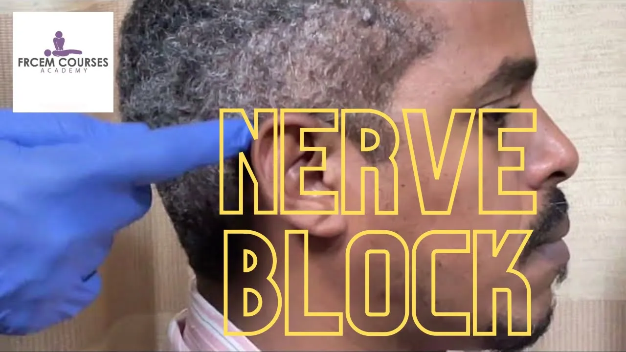
MRCEM OSCE: Nerve block (2)
6.1K
This comprehensive section covers multiple nerve block techniques essential for regional anesthesia. Facial blocks include supraorbital nerve (2.5 cm lateral from midline at eyebrow level), infraorbital nerve (45-degree injection below foramen until bone contact), and mental nerve (lateral approach at 45 degrees). Ear blocks target three nerves: greater auricular (1 cm below earlobe), lesser occipital (1 cm posterior to ear), and auriculotemporal (1 cm anterior to tragus). Wrist blocks include median nerve (between flexor carpi radialis and palmaris longus tendons) and radial nerve (subcutaneous infiltration to distal wrist dorsum). Digital blocks require 4 ml total (1 ml per side), injected from dorsum toward palmar aspect, with critical safety consideration of lidocaine dosing (3 mg/g without adrenaline) to prevent ischemia.
Click to watch →
Deep Dive
Recognition and emergency management of Local Anesthetic Systemic Toxicity (LAST), including the use of lipid rescue therapy.

Local Anesthetic Systemic Toxicity (LAST)
653
Local Anesthetic Systemic Toxicity (LAST) is a life-threatening emergency caused by excessive plasma concentrations of local anesthetic, leading to neurologic and cardiovascular collapse; early recognition of subtle neurologic symptoms (tinnitus, circumoral numbness, metallic taste, muscle twitching) is critical before cardiac involvement, followed by immediate cessation of injection, airway management with 100% oxygen (avoiding hyperventilation), and rapid initiation of Lipid Emulsion Therapy (20% solution) with specific dosing protocols: 100 mL bolus over 2-3 minutes for patients >70 kg or 1.5 mL/kg bolus for patients <70 kg, followed by 0.25 mL/kg/min infusion, with repeat boluses and rate doubling allowed if unstable, while avoiding vasopressin, beta-blockers, calcium channel blockers, lidocaine, and large-dose propofol; continuous monitoring is essential post-rescue due to potential redistribution and recurrence.
Click to watch →
Local Anaesthetic Systemic Toxicity (LAST) | Critical Incident – 3
107
Local Anaesthetic Systemic Toxicity (LAST) is a rare but life-threatening emergency occurring when local anesthetic enters the bloodstream in toxic amounts, causing central nervous system and cardiovascular collapse; immediate management requires stopping the injection, calling for help, managing ABCs, and administering intravenous lipid emulsion (1.5 ml/kg bolus followed by 15 ml/kg/hour infusion), with prevention through careful patient assessment, ultrasound-guided injection, and aspiration to avoid intravascular placement.
Click to watch →
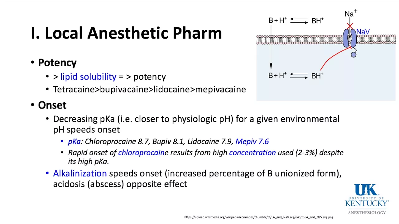
University of Kentucky Anesthesiology Pharmacology Keyword Review 2018 Part 1 of 3 - (Dr. Schell)
61.6K
Local anesthetic toxicity presents with circumaural numbness, tinnitus, twitching, seizures, coma, respiratory arrest, and cardiac toxicity. Treatment follows a systematic approach: establish airway and hyperventilate with 100% oxygen, suppress seizures with benzodiazepines (avoid propofol in hypotensive patients), and address hypoxia/acidosis/hypercarbia. Lipid emulsion therapy (20%, 1.5 mL/kg bolus then continuous infusion) is the definitive treatment. Avoid vasopressin, calcium channel blockers, beta blockers, and additional local anesthetics. For VFib, avoid lidocaine and consider amiodarone. Prolonged resuscitation and cardiopulmonary bypass may be required.
Click to watch →

Local Anesthetic Systemic Toxicity (LAST)
575
Local Anesthetic Systemic Toxicity (LAST) is a rare but potentially fatal complication of regional anesthesia caused by excessive local anesthetic entering systemic circulation, characterized by initial central nervous system symptoms (perioral numbness, metallic taste, tinnitus, dizziness, seizures) followed by cardiovascular collapse (hypotension, dysrhythmias, cardiac arrest), with bupivacaine being particularly dangerous due to its high lipid solubility and strong cardiac binding; effective management requires immediate cessation of the anesthetic, supportive care, and administration of Intravenous Lipid Emulsion (ILE) according to ASRA guidelines (100 mL 20% ILE bolus over 2-3 min plus 200-250 mL infusion over 15-20 min for patients >70 kg, or 1.5 mL/kg bolus plus 0.25 mL/kg/min infusion for those <70 kg), which acts as a 'lipid sink' to sequester the anesthetic and provide fatty acids for myocardial function.
Click to watch →

Local Anaesthetic Pharmacology Refresher & L.A.ST
842
Local anesthetic systemic toxicity (LAST) presents with CNS or cardiovascular symptoms, or both. CNS signs include perioral paresthesia, metallic taste, seizures, and respiratory arrest; cardiovascular signs include conduction disturbances, hypotension, and cardiac arrest. Risk factors include high lipid solubility, patient factors (extremes of age, pregnancy, organ disease), and technique factors (paravertebral, fascial plane, continuous catheters). Treatment requires immediate cessation, airway management, oxygen, and early lipid emulsion therapy (100 mg bolus then 250 mg/15 min infusion). Benzodiazepines control seizures; small epinephrine doses (<1 mcg/kg) manage cardiovascular issues; avoid vasopressin.
Click to watch →
Deep Dive
Clinical guidelines for the use of vasoconstrictors (like epinephrine) combined with local anesthetics, including absolute and relative contraindications in distal extremities.

Odonto em foco - Questões comentadas FGV - Anestésicos Locais com Suellen Rocha
10.4K
Absolute contraindications for vasoconstrictor use include uncontrolled cardiac insufficiency, uncontrolled hypertension, acute myocardial infarction within 6 months, recent coronary artery bypass surgery, unstable angina pectoris, certain arrhythmias (Wolff-Parkinson-White syndrome), untreated congestive heart failure, and uncontrolled hyperthyroidism. Relative contraindications include systolic BP above 160 mmHg, diastolic above 100 mmHg, history of myocardial infarction more than 6 months ago, controlled arrhythmias with pacemaker, controlled diabetes, controlled hypertension, and controlled cardiac insufficiency.
Click to watch →

Anestésicos Locais- Aula 3/4: Critérios de escolha, farmacocinética e efeitos adversos
4.3K
Vasoconstrictors reduce systemic absorption of local anesthetics, decreasing toxicity and prolonging anesthesia duration. Epinephrine (adrenaline) is the most potent and commonly used vasoconstrictor, acting through alpha-1 adrenergic receptors with a half-life of 1-3 minutes. Noradrenaline has slightly less potency and may cause more significant cardiovascular effects. Phenylephrine is 20 times less potent than epinephrine and is a pure alpha-agonist, useful in cardiovascular disease patients. Phentolamine is 6 times less potent than epinephrine and is associated with prilocaine. Vasoconstrictors are contraindicated in uncontrolled hypertension, heart failure, recent MI/stroke, uncontrolled diabetes, hyperthyroidism, pheochromocytoma, and sulfito sensitivity.
Click to watch →

LIVE #019 - Farmacologia dos anestésicos e dos vasos constritores
66.3K
Vasoconstrictors have absolute contraindications where they should never be used: Uncontrolled hypertension (blood pressure not managed medically), Uncontrolled hyperthyroidism (thyroid hormone levels not stabilized), and Uncontrolled diabetes mellitus (blood glucose not regulated). In these cases, vasoconstrictors can worsen underlying pathology. Patients must be stabilized medically before elective dental procedures involving vasoconstrictor-containing anesthetics.
Click to watch →

Vasoconstrictores y Anestésicos
5.7K
Vasoconstrictors are pharmacological agents that delay local anesthetic absorption, increasing duration and creating bloodless surgical fields. They have pH 3.3-4 (vs 5.5-6 without vasoconstrictor). Types include catecholamines (adrenaline/epinephrine) and synthetics (phenylephrine). Contraindications include hyperthyroidism, uncontrolled diabetes, uncontrolled hypertension, recent childbirth, glaucoma, coronary insufficiency, pheochromocytoma, and poorly vascularized areas (ears, nose, genitals) due to necrosis risk.
Click to watch →
Anestesia em Pacientes com Comprometimentos Sistêmicos
25.1K
Epinephrine is the most effective and safest vasoconstrictor for dentistry, but its use requires understanding contraindications. Absolute contraindications include hypertension (>160/100 mmHg), recent MI (<6 months), recent stroke (<6 months), CABG/stent placement, unstable angina, certain arrhythmias, uncontrolled CHF, uncontrolled hyperthyroidism, sulfa allergy, and illicit drug use. Alternative vasoconstrictors include felypressin (prilocaine 3% with 0.03 UI) or no vasoconstrictor. The principle that 'the difference between medicine and poison is dosage' applies universally.
Click to watch →
Wound Anesthesia
Wound Anesthesia
0:00- 1
Demonstrates injecting anesthetic into wound margins.
- 2
Covers needle insertion, aspiration, and injection technique.
Perilesional Infiltration and Regional Nerve Blocks
While direct wound margin infiltration is widely practiced, many clinicians advocate for perilesional (intact skin) infiltration or regional nerve blocks as superior alternatives. Critics of wound margin injection argue that inserting a needle through an open, potentially contaminated wound bed can seed superficial bacteria deeper into sterile subcutaneous tissues, increasing the risk of infection. Additionally, direct infiltration causes localized tissue edema (swelling), which can severely distort wound margins and hinder precise cosmetic alignment during suturing. In contrast, perilesional infiltration—injecting through the intact skin surrounding the wound—and regional nerve blocks avoid disrupting the wound bed. Regional nerve blocks target proximal nerves to anesthetize a broader area, completely eliminating local tissue distortion at the injury site. This technique reduces the number of painful needle punctures for the patient and is highly preferred for cosmetically sensitive areas, such as the face, where anatomical distortion must be minimized.
Basic anatomy of the skin layers (epidermis, dermis, and subcutaneous tissue) and the pathway of superficial sensory nerves.
Pharmacology of common local anesthetics (such as lidocaine and bupivacaine), including their onset of action, duration, and weight-based maximum safe dosages.
The principles of aseptic technique and wound preparation to prevent introducing pathogens during injection.
Understanding the concept of intravascular injection risks and the physiological rationale behind aspirating before injecting.
Wound closure and repair techniques, such as simple interrupted, running, or subcuticular suturing.
Advanced regional anesthesia techniques, including digital blocks, facial nerve blocks, or field blocks for larger lacerations.
Recognition and emergency management of Local Anesthetic Systemic Toxicity (LAST), including the use of lipid rescue therapy.
Clinical guidelines for the use of vasoconstrictors (like epinephrine) combined with local anesthetics, including absolute and relative contraindications in distal extremities.
498.4K views1.8Klikes1:30@thelacerationcourse2009Original Release: 2022-04-25
Okay. So, what we're demonstrating is how to put anesthetic into a wound. So, I've already drawn up my anesthetic into the syringe. I'm using a 25 gauge needle. And there's a lot of different ways to do this, but the principle is the same. Essentially, you're going to insert the needle right into the wind margin, right into the edge of the wind in the tissue. aspirate a little bit to make sure that you're not in the blood vessel. And then you simply put a little bit of the anesthesia underneath the skin. And sometimes you can see the tissue just kind of well up a little bit and then you know that it's where you need to be. So you can advance the needle, aspirate, and then inject while you're withdrawing.
And you can kind of reposition the needle a little bit so that you don't have to do multiple sticks.
Okay.
And then I'm going to go over here on this side and do the same thing. Insert the needle into the wound margin.
Aspirate and aspirate and inject.
And sometimes it'll come out of the opening. Reposition, advance the needle, aspirate, and inject.
And that is it.
Up Next
8:24
Circle of Willis Anatomy and Stroke Types | Blood Supply to the Brain
@armandohasudungan
858.8K views•2015-09-07

48:47
Graphic Medicine: Comics for Collaborative Healthcare Communication
@nationalpatientadvocate
189 views•2023-12-04

10:28
Neuroanatomy: Central and Peripheral Nervous System Divisions Explained
@AKLECTURES
136.2K views•2014-09-20

3:05
Stages of Labor and Vaginal Birth | Childbirth Animation
@nucleusmedicalmedia
52.1M views•2017-08-18
Related Study Plans & Knowledge Roadmaps
Structured learning paths in Medicine
medicine
Abnormal LFTs: Diagnostic Workup & Pathology
01Liver Anatomy and Basic Physiology
02Introduction to Liver Function Tests (LFTs)
03Hepatocellular vs. Cholestatic Patterns
+ 3 more learning modules
12h6 modules
Explore Roadmap
medicine
Analyzing Blood Smears: RBC, WBC & Platelets
01Introduction to Blood Components & Smear Preparation
02Normal Morphology of Blood Cells
03Erythrocyte Pathologies & Morphological Variants
+ 2 more learning modules
12h5 modules
Explore Roadmap
medicine
Mechanical Ventilation: Waveforms & Alarms
01Anatomy and Physiology of Breathing
02Introduction to Mechanical Ventilation & Core Modes
03Ventilator Settings and Waveform Interpretation
+ 2 more learning modules
12h5 modules
Explore Roadmap
medicine
Neuro Exam: Localization & Stroke Syndromes
01Foundations of Neuroanatomy and Vascular Supply
02The Neurological Examination: Step-by-Step
03Principles of Neurological Localization
+ 3 more learning modules
18h6 modules
Explore Roadmap