

















![DƯỢC LÝ [CTUMP] - Thuốc Giảm đau OPIOID | PGS.TS Phạm Thành SuôL](https://i.ytimg.com/vi/Jqxf0JAEPgo/maxresdefault.jpg)
Pain signals travel through neurons via electrical action potentials and chemical synaptic transmission; opioids relieve pain by binding to G-protein-coupled opioid receptors, which then activate G proteins that inhibit neuronal signaling through multiple mechanisms including blocking voltage-gated calcium channels and potassium channels, ultimately dampening the pain signal.
Opioid Pain Signaling and Neuronal Pathways | PDB-101 Molecular Mechanisms
Added:Structure and function of neurons, including synaptic transmission, action potentials, and neurotransmitter release.
The basic biology of G-Protein Coupled Receptors (GPCRs) and intracellular signal transduction cascades (such as cAMP signaling).
Introductory pharmacology concepts, specifically the definitions of receptor agonists, antagonists, and competitive binding.
The physiological pathway of nociception, defining how pain signals are detected and transmitted from the periphery to the central nervous system.
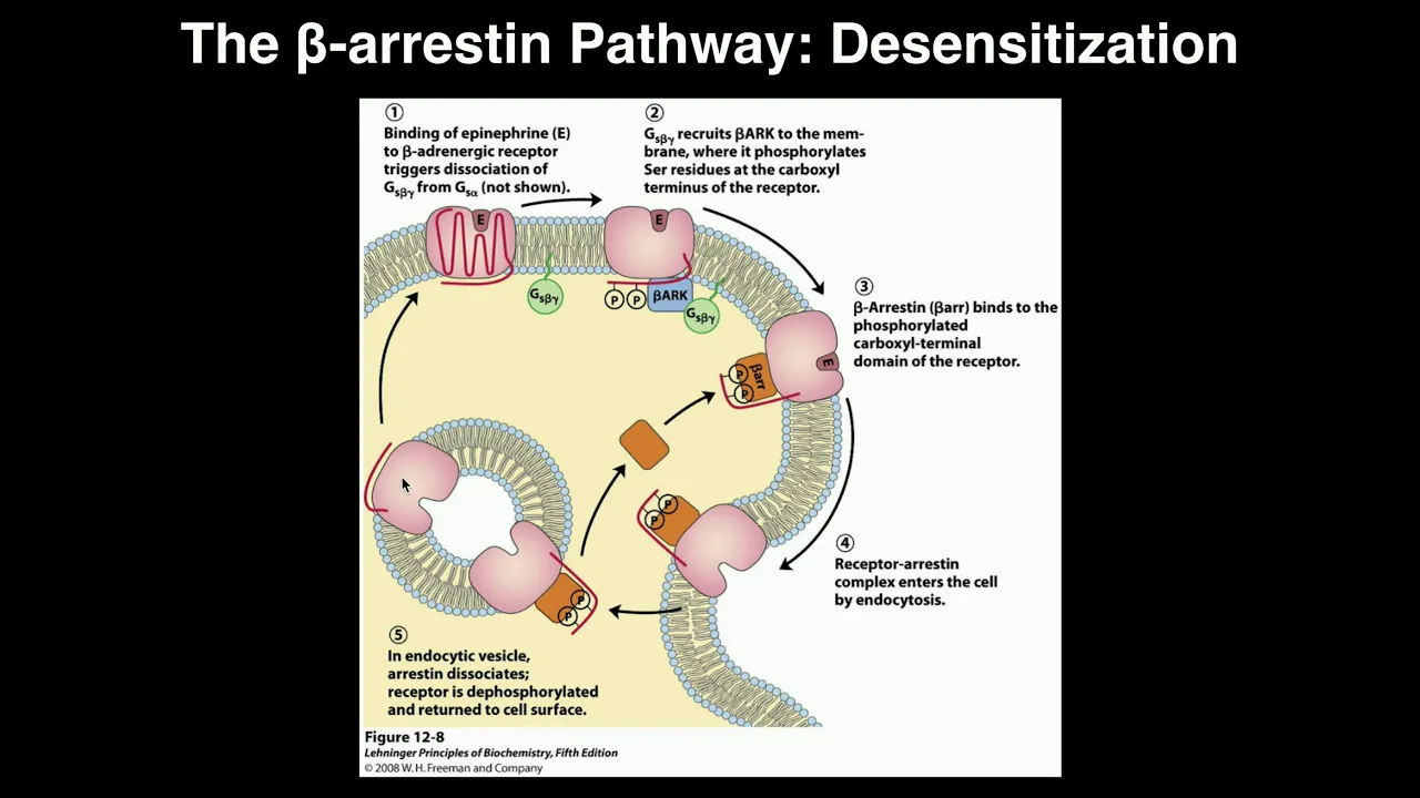
The cellular and molecular mechanisms of opioid tolerance and receptor desensitization, including the role of beta-arrestins and receptor internalization.
The neurobiology of addiction and physical dependence, focusing on how opioids affect the mesolimbic dopamine pathway and reward circuitry.
Advanced clinical pharmacology of analgesics, comparing the efficacy and side-effect profiles of full mu-opioid agonists, partial agonists, and mixed agonist-antagonists.
Public health and therapeutic approaches to treating opioid use disorder (OUD), such as medication-assisted treatment (MAT) using methadone or buprenorphine.
34.1K views1Klikes8:41Original Release: 2021-06-22
[Music] Pain is one of the most trying experiences of life.
On the cellular level it is communicated via special neuronal pathways.
On the molecular level however, pain is communicated like any other sensation- via a set of electrical and chemical signals facilitated by complex molecular machinery.
Along the neuronal membrane, the signal travels much like electric current along a wire.
This process is mediated by membrane proteins called voltage-gated ion channels.
Voltage-gated ion channels open or close in reaction to changes in membrane voltage or membrane potential. These proteins have special voltage sensing domains that are coupled to ion channels. When a neuron is at rest, its ion channels are closed.
A cell membrane potential is generated by concentration gradients of positively charged sodium and potassium ions across the membrane.
When the neuron is at rest, the overall concentrations of ions and proteins create higher potential in the outside and lower on the inside.
This state is referred to as resting potential and the membrane is said to be polarized.
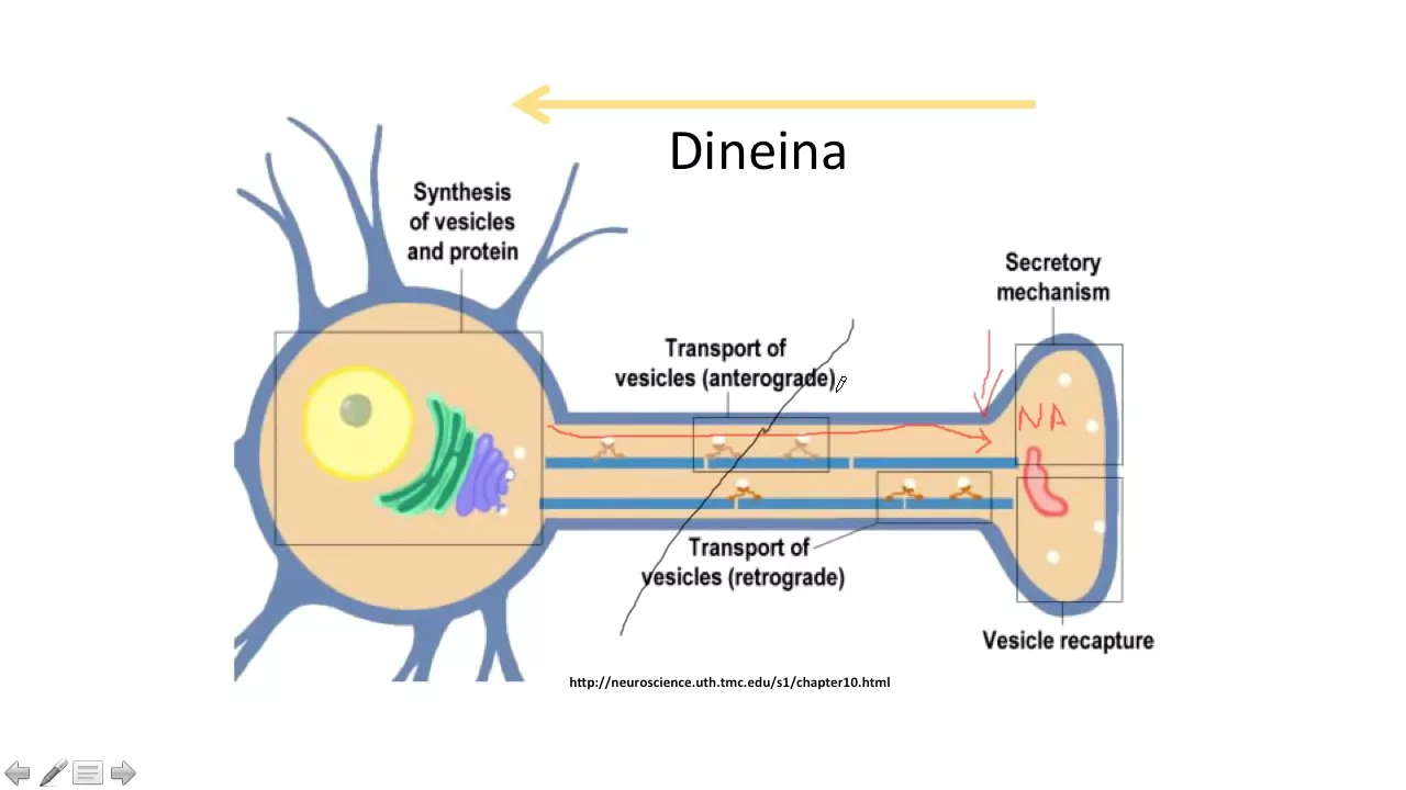
To transmit signal the neuron creates an action potential which travels down the axon.
The action potential is mediated by the opening of the voltage-gated sodium channels, which allows sodium to enter the neuron.
This depolarizes the membrane in the local area.
Neurons then repolarize the membrane in several ways stopping the action potential.
First, voltage-gated sodium channels close and become inactivated.
Then, to counteract the positive charge of the sodium ions, potassium channels start to open allowing potassium ions to exit and lowering the voltage across the membrane.
Finally, after a short refractory period sodium channels open the inactivation gates and ion pumps restore the resting potential in preparation for the next signal.
In this way the signal travels along the neuron - sodium channels open and trigger one another to generate action potentials and potassium channels then stop the action potential in the local area.
Once the action potential arrives at the end of the neuron, it is transferred to the next cell at a place called a synapse.
The synaptic region contains membrane vesicles filled with small molecules called neurotransmitters, which will conduct the signal upon release into the synaptic cleft.
This process is dependent on the presence of calcium ions inside the cell.
Because intracellular calcium is sparse while extracellular calcium is abundant, voltage-gated calcium channels are essential in this process.
The positive charges of the traveling action potential activate these proteins causing the ion channels to open and allowing calcium inside the cell.
Calcium then activates other proteins that help the vesicle to merge with the neuronal membrane, creating an opening for the neurotransmitters to enter the synaptic cleft.
There are many types of neurotransmitters.
One of them, glutamate, is shown here binding to its receptor.
This receptor has an ion channel which opens upon glutamate binding and allows ions to enter the cell initiating a new action potential that will travel along the axon, thus allowing the message to be passed along.
These signals can be modulated on the molecular level causing us to feel less pain or no pain at all.
Our bodies produce natural painkillers known as endorphins and enkephalins and dynorphins.
The peptide showed here is an encephalin.
These natural painkillers are mimicked by opioid drugs.
One of the most widely used opioids is morphine, which is derived from the opium poppy.
Both natural painkillers and opioids activate the same inhibitory receptors called opioid receptors.
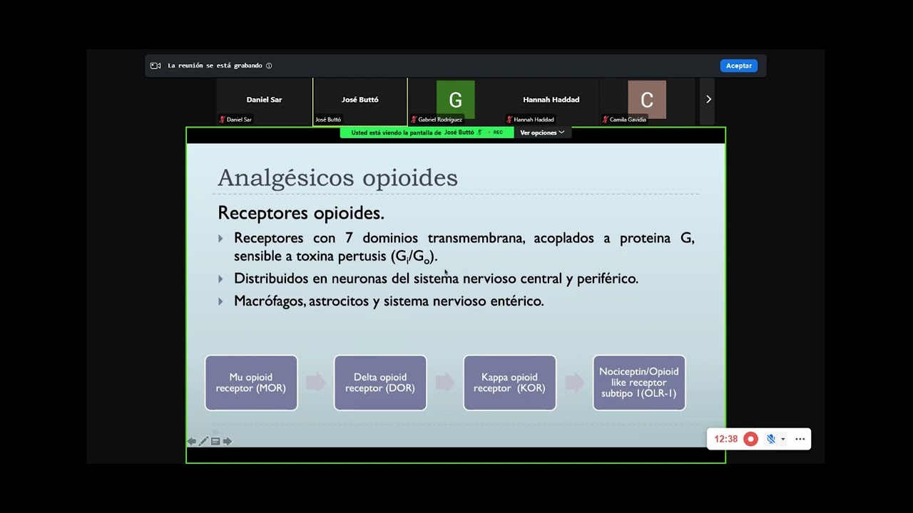
Opioid receptors are part of a large class of g-protein-coupled receptors or GPCRs.
GPCRs are nested in cellular membranes with the opioid binding site on the outside of the cell. Inside the cell, the receptors are coupled to G proteins. G proteins have three distinct subunits alpha, beta and gamma.
An inactive G protein has GDP bound to its alpha subunit.
Upon opioid binding the GDP is released, and GTP binds instead, releasing the g protein from the receptor, causing the alpha subunit to separate from the beta and gamma subunits.
The beta-gamma subunit interacts directly with ion channels, stopping the signal. For example, the beta gamma subunit can bind to special potassium channels, causing them to remain open and allow potassium to exit the cell. This lowers the electric potential inside the cell and eventually leads to the dissipation of the action potential.
The beta gamma subunit can prevent neurotransmitter release by binding to voltage-gated calcium channels. This prevents the activation of the channels.
As a result, calcium is unable to enter the cell, and the release of neurotransmitters is halted.
There are several types of g-alpha subunit that couple to different GPCRs.
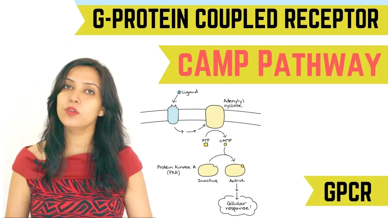
Two types of G-alpha subunits can interact with the enzyme adenylyl cyclase.
These are inhibitory and stimulatory g-alpha.
Opioid receptors are always coupled to inhibitory g-alpha.
In the presence of stimulatory g-alpha, adenylyl cyclase uses ATP to produce cyclic AMP. Cyclic AMP is often called a second messenger because it amplifies the signal generated by the first messengers, the neurotransmitters.
Cyclic AMP can activate some ion channels indirectly via protein kinases, Both activities augment the ion flow regulating the neuronal signal. 0:06:45.440,1193:02:47.295 and activate other ion channels directly.
When opioid receptors are activated, the G-alpha inhibitory subunit inhibits adenylyl cyclase, preventing the production of cyclic AMP.
As a result, the signal is dampened.
Opioid drugs are a great boon in pain management when used for a limited time and as prescribed by a physician. However, the possibility of overdose is a danger as opioids can deactivate neuronal circuits other than those responsible for pain signaling, Causing severe side effects or even death.
During an overdose, drugs like naloxone are used to save lives.
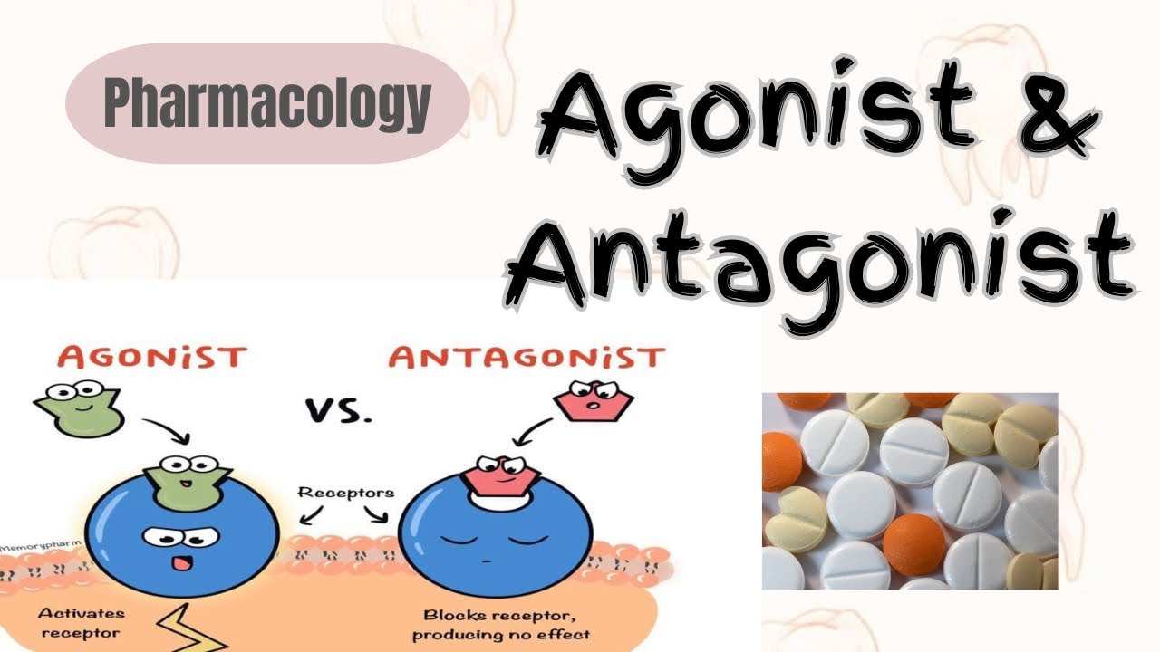
Naloxone is similar in structure to opioids and binds in a similar location on the receptor, but does not activate the G protein.
As a result, it makes the receptors unavailable to opioids.
By studying opioid receptors, scientists are currently trying to discover new, non-addictive compounds that can bind to these receptors in order to create safe painkillers.
[Music]
Up Next

87:42
Understanding Pain and the Brain | Neuroscience Lecture
@uctv
135.3K views•2008-01-03

50:12
Bacterial Communication: Quorum Sensing and Biofilm Formation Explained
@JHUAAP
12.6K views•2011-06-28

5:35
Enteric Nervous System Explained: The Gut's Brain | Neurobiology Lecture
@alumniu6029
438 views•2018-09-12

7:08
Bacteriophages: Earth's Deadliest Killers and Future Antibiotics
@kurzgesagt
34.6M views•2018-05-13
Related Study Plans & Knowledge Roadmaps
Structured learning paths in Biology
biology
RNAi: Biogenesis, RISC & Gene Silencing
01Molecular Biology Foundations: DNA to Protein
02Introduction to RNA Interference (RNAi)
03Molecular Biogenesis of miRNA and siRNA
+ 2 more learning modules
10h5 modules
Explore Roadmap
biology
Mycorrhizae: Signaling, Biophysics & Ecology
01Foundations of Mycorrhizal Symbiosis
02Molecular Signaling and Root Colonization
03Biophysics and Bioenergetics of Nutrient Exchange
+ 1 more learning modules
11h4 modules
Explore Roadmap
biology
Circadian Clocks: TTFL, SCN & Periphery
01Foundations of Molecular Biology & Gene Expression
02Introduction to Chronobiology and Actograms
03The Core TTFL: Molecular Architecture of the Clock
+ 3 more learning modules
14h6 modules
Explore Roadmap
biology
Population Genetics: Math, Drift & Selection
01Genetic & Probability Foundations
02The Hardy-Weinberg Principle and Baseline Models
03Mathematical Modeling of Natural Selection
+ 3 more learning modules
18h6 modules
Explore Roadmap