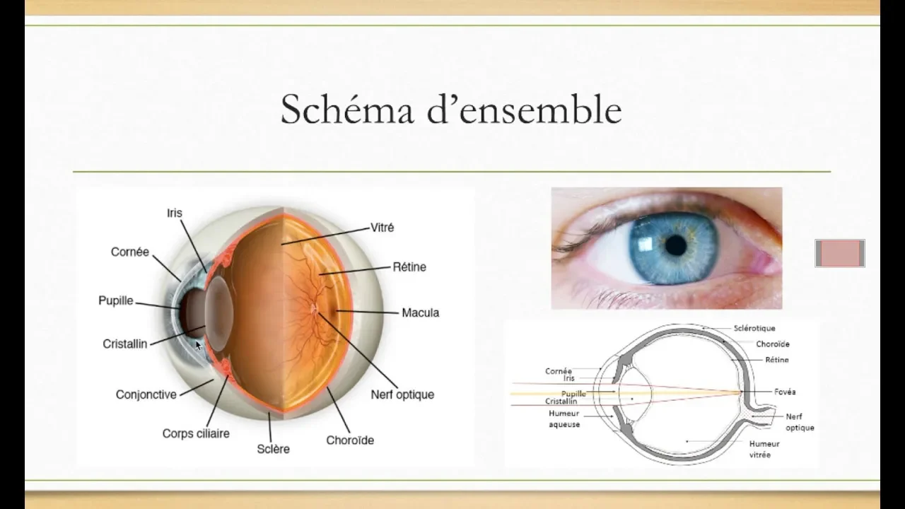




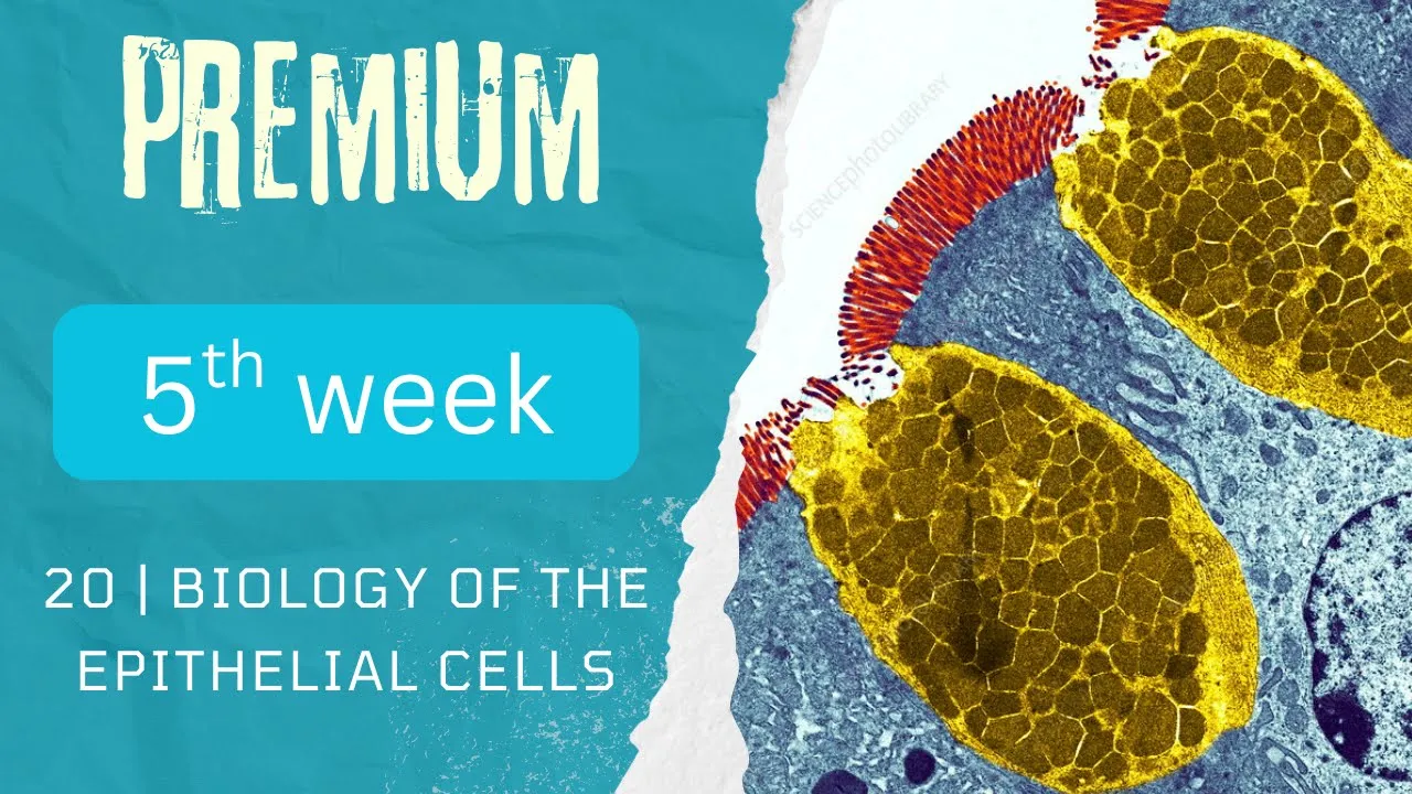







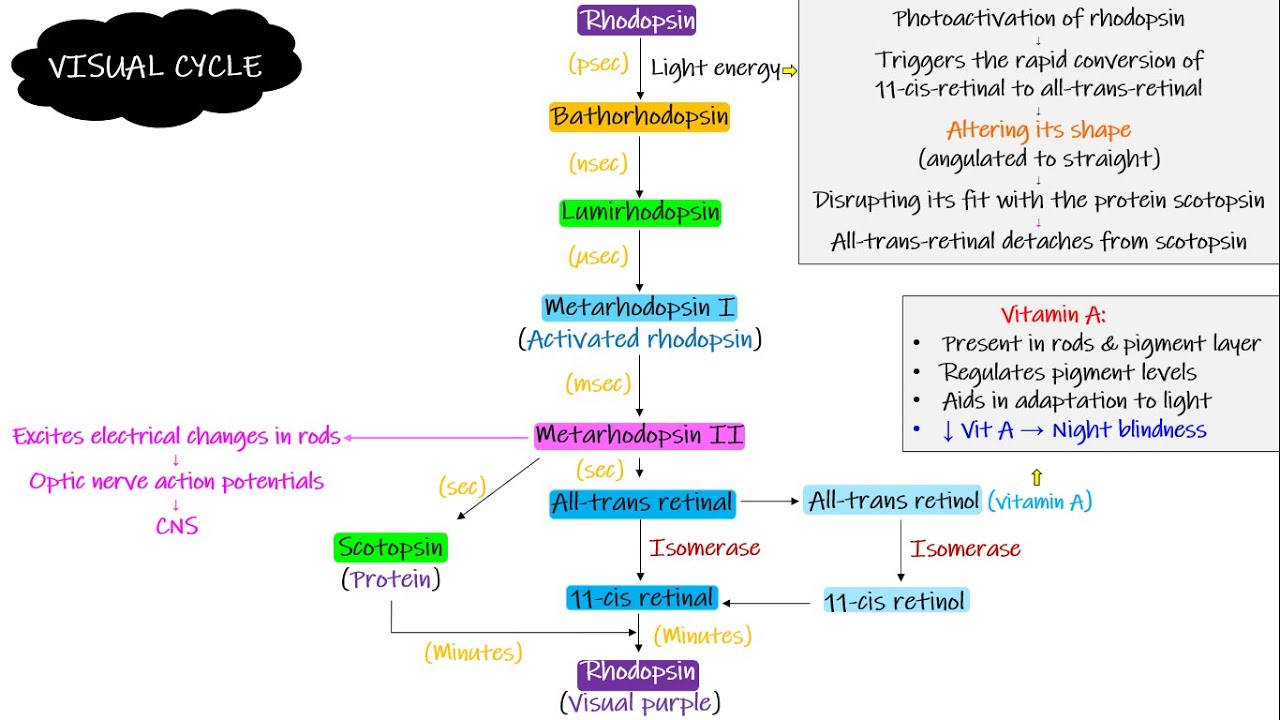

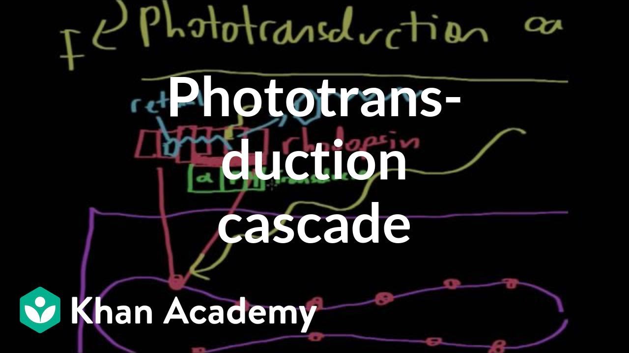







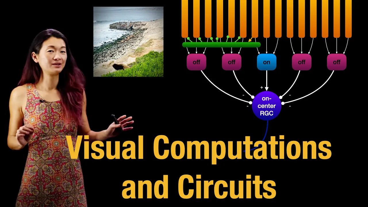



The retinal pigment epithelium performs four critical functions: acting as a blood-retinal barrier, absorbing excess light to prevent internal reflections, regenerating bleached visual pigments (rhodopsin in rods and iodopsin in cones), and maintaining photoreceptor outer segments through phagocytosis; rods (100 million) are more abundant and sensitive to low light with multiple rods connecting to one bipolar cell, while cones (5-7 million) are concentrated in the fovea centralis with one-to-one connections enabling color vision and high visual acuity.
Retina Anatomy: Key Points on Rods, Cones & RPE Functions
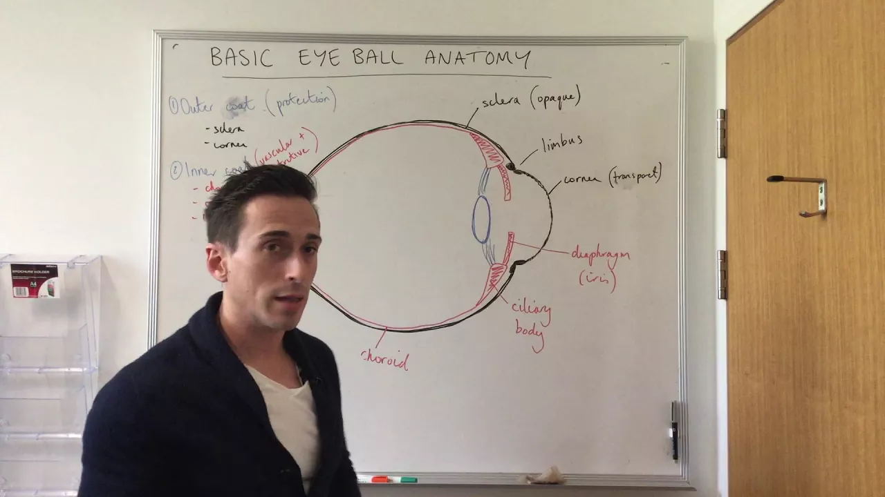
Added:Basic anatomy of the human eye, including the localization of the retina relative to the cornea, lens, choroid, and vitreous body.
The fundamental concept of sensory transduction, specifically how physical stimuli are converted into electrical signals by receptor cells.
Basic cell biology concepts, particularly the structure and function of epithelial cells and active transport mechanisms.
Elementary physics of light, including the visible spectrum, wavelengths, and how the eye's refractive media focus light.
The detailed biochemistry of the visual cycle (phototransduction cascade), focusing on rhodopsin, transducin, phosphodiesterase, and cyclic GMP.
Clinical pathologies of the retina, such as Age-Related Macular Degeneration (AMD), Retinitis Pigmentosa, and Diabetic Retinopathy.
Complex retinal processing circuits, including the roles of horizontal, bipolar, and amacrine cells in establishing receptive fields and lateral inhibition.
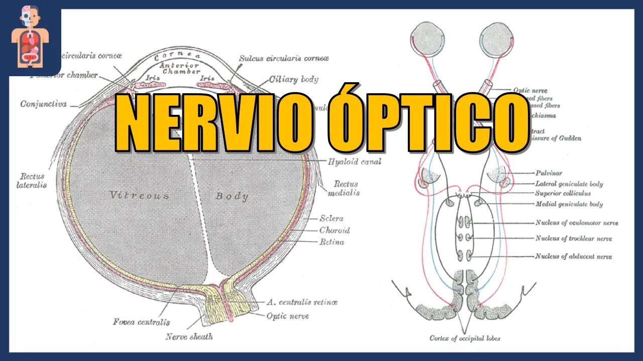
The central visual pathway, tracing neural signals from the optic nerve, through the optic chiasm and lateral geniculate nucleus (LGN), to the primary visual cortex.
275.9K views3Klikes1:16:08Original Release: 2015-12-16
so we were talking about structure of retina right we'll very briefly review it and then continue with four few more important points already you have watched two videos about the structure of retina and I know you must be remembering uh well I told you about the basic structure of retina one way to look at it is that there is a receptor yes of course after endothelium after epithelium pigment epithelium yes receptors then by cells and then gangon cells once you make this chain right then you will add High you remember and then you will add man right I didn't explain these two points let's do it rapidly and that if this is the epithelium which epithelium pigment epithelium right pigment epithelium today we'll go into detail of pigment epithelium it structure and its function right and now in pigment epithelium what are these special what are these okay first I will make the nuclei so that and in these cells of course there are lot of melanoses as well not melanite melanosomes which are the melanin filled vules here which basically absorb the Stray light right and what are these tight junctions and these are melanosomes and here again tight junctions melanosome and again tights right so then what is there RBG what is that yes receptors photo receptors I will just draw a basic structure again and then we'll go to some points in detail right and we'll draw The receptors here uh this is yes this is Rod and let's suppose this is also a rod right and here I can draw a cone right we can make two rods more also now I'm making a little fast so outer segment yes in a segment and nuclei and yes spheral a synaptic terminal in the case of Rod this is spheral and in case of it is pedical right and this is inner segment and here is right so R then B BR R which cells okay I'll make them all with the same color so these are bipolar cells now here it is important rather I must show that rods usually have more convergence rods have usually more convergence to the bipolar cell for example there may be one bipolar cell connecting to multiple rods right okay I will make this Al here so that yes these are bipolar cells and after that what is there RB G gangon cells gangon cells and from here what is this going n fiber is that right yes please what is your question that con have one to one connection with a B yes cones have one to one connection we'll talk about that of of course for sure so you want me to keep it one and we can make it like this now you happy thank you okay he's right that uh in the meula especially in phobia cus con one con one bipolar one gangon cell this is a private line for the cor business rods are usually having a public life multiple rods connected with one bipolar and then to gangan cell is that right and these as of some function importance we'll discuss it later now RBG then we come to high yes horizontal cells amagine cells Interplex form so what are these cells which are connecting here horizontal cells right and what are these cells connecting here Emma crine cells and what are these cells interplexiform cells is that right then we come to M okay I will make it black these were the gal cells M for M cell as we discussed M cells have the cell body here and they have one process going to the outer area right and these processes yes connect with the inner inner segment right in the same way inner segment and in the same way they come over here and connect with the inner segment of rods and cones so these are the ends of outer ends of cells and this line This wavy line which appears basically the are Zona adherence what is this line called Li outer limiting membrane is that right and there peripheral processes they go out as foot plate plates and yes Li this is inner limiting membrane and here is the vitus on this side there is Vitus on this side there is chide and from Vitus as I told some collagen and proteoglycans are also added over here is that right so this was M cell m one then a this is what we did in discussed in last lecture a stands for asites it stands for asites asites are basically cells which are star shift cells and they are mostly in the neuronal layer cells and in gangon layer cells they are also gal cells and they are short processes and these processes are supporting the neuronal structures as well as they're supporting the gangan layer structure and these processes also envelop the capillaries present over here but capilaries which are present in the inner part as I will discuss later they are having some processes receiving on outer side processes from estro site some processes coming from M cells then there is another type of cells these are micro Gia what are these micro Gia and microa are basically macro phases right which are present in central of system and as you know the neuronal this layer of the retina is basically part of Central Central no system so micro glar also there and micro glar actually uh spread in this is asite throughout the retinal layers so microa may be here or maybe here or maybe here at multiple area right these are actually ftic cells when retina is damaged these cells increase in their number and start fying the damage or uh the tissue which they want to remove from there after the injury am I clear so now RPG stand for receptor this these these cells three three and three nine cells there are more cells also but these are the nine most important cells which are contributing to the contributing to the retinal neuronal layer because never forget epithelial layer which was derived from the outer layer of optic optic cup and neuronal layer was derived from the inner layer of the optic Cup right right so these are the nine cells any question up to this so this is complete now there are few more important point I will go to some important points in detail for example the function of pment pigment epithelium let's talk about retinal pigment epithelium what are the important functions of this epithelium it has many functions and many very important functions if those functions are disrupted then even neuronal layer cannot be made maintained well right and blindness can result so what are the most important function of retinal pigment epithelium one function is if you start from this side that this layer of epithelium act as a barrier between the Cho capillaries and the neuronal retina let me make Cho capillaries here listen previously I told you this is short suppose posterior shorty arteries they enter into chide and in the chide they make short cly arteries they make capillary Loops which are basically cides so I will draw cides here that let's suppose this is short uh C poster short artery and it will divide into multiple sinoses is that right they will divide into multiple sinos sides and these cides will of course eventually drain into what vertical veins is that right they will eventually drain into vertical veins now the point which I want to tell you this is actually people a arteries come on the Cal side divide into arterioles then into smaller uh you can say vessels and eventually Coro capillaries which are not really capillaries they are very wide caliber capillaries that is why they are called sinusoids These are the cides and these cids are having endothelial cells which are fenestrated which are fenestrated and there are big interendothelial gaps also right if I draw one let's suppose this is endothelial cell then here is another endothelial cell there's a gap in between those endothal cells of these cocal capillaries plus there are you can say holes present within a cytoplasm so we say that Coro capillaries are highly porous capillaries highly porous capillaries uh which are having fenestrations it means apertures within the endothal cell cytoplasm and also interendothelial gaps so it means these capillar endothelium is not working as blood retina barrier so so many substances can percolate out from here right but the real barrier is here real barrier is here that from the outer aspect of retina from the Coro capillary blood which is moving there the real barrier is put by what is this retinal pigment epithelium and their tight junctions in between and these epithelial lining determines that which substances should be allowed to go into outer retina and we should not be allowed to cross am I clear now these actually Cho capillaris give to the nutrition to the outer part of retina right especially outer five layers outer five layers these are the inner layers it means outer half what are those five layers as you know one one part of is epithelial cell second is photo receptor component of the rods and Cone then there is outer limiting membrane then there is outer nuclear layer and then there is outer why they're becoming more first is yes let's start with the first layer is epithelium right second layer is photo receptors okay I missed it here and third is outer and fourth is this nuclei fourth is the outer nuclear and fifth is outer PL form so actually you can say from here blood supply to the outer retina outer five layers or outer half of the retina is indirectly because itelf blood does not enter here only substances diffuse in and diffuse out so blood supply to the outer five layers of the retina is basically provided by the Coro capill am I clear then here I would love to mention that as I mentioned previously that retina has double blood supply right from Outer side Cho capillar is a supply and from inner side Central linal artery again we'll discuss that later but there's one bloodprint barrier on this side another Blood pint barrier is on inner side on outer side blood pint barrier is provided by retinal pigment epithelium or inner side let me explain for example this is a branch of central retinal artery it has capillaries which are going in okay and coming back to V here is the vein and this is the central retinal artery now these substances which come through or blood which come through the central ltin artery it passes through these capies within the retinal inner layers and drain into Central retinal vein now here is also blood brain blood retinal barrier but here the barrier is made by endothelial cells look here these are endothelial cells right number one these endothelial cells are not fenestrated endothelial lining is not fenestrated secondly these endothelial cells have tight junctions they have tight junctions so the tight junctions which were present here into retinal pigment epithelium here the tight junctions are present in between endothelial cells so in inner circulation or Central retinal artery and vein related micro circulation the blood retinal barrier is maintained mainly by endothelial cells and their Arrangement that here the endothelial cells are not fenestrated and Indo inter endial gaps are not there because those gaps are tightly stitched with they're tightly held with tight junctions right so now what what did we learn up to now that retina has outer five layers it has inner five layers outer five layer has own blood supply that is chois inner five layers of their own blood supply that is Central retinal artery and its system outer retinal blood supply has its own blood retina barrier right which is retinal pigment epithelium inner retinal vascular circulation has its own what is this Blood retinal barrier and that is by the endothelial cells which are not fenestrated and and endothelial cells without interendothelial gaps as they're held together tightly by the tight junctions am I clear so how many blood retinal barriers are there two is that right any question up to this there's no so we come back that this retinal pigment epithelium function number one was it was acting as yes what blood retinal barrier right then second function second function is that uh light is coming from this side look light is coming from outside and it hit the inner retina first then light passes through the in a retina right it passes through multiple layers until Light reaches the Rock and cones and activate them and extra light is absorbed by retinal pigment epithelium this is very important that extra light which is coming from here which is not used by Rod and Cone that should be absorbed by retinal pigment epithelium why the reason being let's suppose here the light is coming right light has to pass through multiple layers it has to pass through external limiting membrane then through gangan layer cell then it has to pass through yesx outer plexiform inner plexiform layer then inner nuclear layer and multiple layers eventually it will reach to the rod and Cone and then light will activate them and produce nerve impulses right and actually rod and cor are the cell which convert the optical image into neuronal activity retinal neuron activity but extra light which is passing through that should be absorbed by retinal pigment epithelium why because if this light is not absorbed then this light will internally reflect here there here and image quality will degrade image quality will degrade so one of the very important function of retinal pigment epithelium is that it absorbs the extra light right so that there is not undue what internal reflections of Light Within the eyeball and image quality should not degrade there should not be too much glare is that right now here the another example is of albinos albinos are the patients or persons who don't make melanin and in albinos retinal pigment epithelium does not have melanosomes in albinos when like when when they enter into bright room light enters through the retinal layer it activate the rod and Cone but when it hit what is this retinal pigment epithelium it is not absorbed and light has internal glare and internal Reflections and uh their Optical image sensing quality degrades and that is why albinos even with the maximum Optical correction they can't have uh 20x 20 VN is that right am I clear so what was the second function that was light absorption light ABS absorption now the third function of it third function of retinal pigment epithelium now it has one more function that is related with the functional aspect of Rod and Cone actually it means maintains the integrity and functionality of photosensitive part of Rod and Cone how you know when light falls on the rods and Cone it bleaches the visual pigment you know visual pigment which is present in Rod is ropin and in the cones it is iodopsin when light Falls uh on the inner part sorry this outer segment and on the outer segment of Rod and Cone when light falls over here the visual pigments which are present over here they undergo bleach reaction and during that process they activate the nerve impulses but those pigment and their derivatives are taken up by epithelial cell and those are regenerated and recycled to be used by the outer segments is that right you know vitamin A is very important for this process right vitamin A is uh retinol is the part of visual pigments is it right and uh this epithelial cells retinal pigment epithelial cell do store vitamin A as well and also they're able to convert all trans retinol into 11 CIS retinol that we will discuss in detail when we talk about functions of rods and cones and how they perceive the light and generate the current but for a while you just believe me that visual pigment which is present over here visual pigment is a photosensitive chemical when light falls on out these light sensitive part of the rod and Cone visual pigment present over here it bleaches and during this process uh impulses are produced which go to the central nervous system is it right but those bleached pigments are recycled by the help of r pigment epithelium and they are recycled and regenerated to be used again so this is one more function of retinal pigment epithelium what is that function regeneration of bleached visual pigment visual pigment it lead to Regeneration of bleached then another important not only here it is managing the function of the rod and Cone it also manages the structural aspects of Rod and Cone actually rod and Cone here are the disc let me make one rod outer segment big this is inner segment of the rod and this is the outer segment of the rod now outer segment of the of course here is thec nucleus of the rod and here it is synaptic terminal now it is having a lot of disc about 1,000 disc right and these disc are Flo into its distal most part but here there are membrane and folding originally when dis are made they made as membrane infolding later on these membrane shelf detach and they are freely floating now this is the outer segment here it is inner segment inner segment here is having mitochondria right in this component and more near to the nuclear side in this area it has endoplasmic reticulum and this is making what the endoplasmic reticulum is making proteins right so visual pigment is being synthesized here opsins and other things so what really happens that let me draw the functionally this is outer segment outer segment and this is inner segment and inner segment this part which is having mitochondria is called ellipsoid part ellipsoid part and this part which is concerned with protein synthesis this is called myoid part so this is a very brief structure of Rod and Cone so what is really happening proteins are being synthesized here and then transported to outermost segment is that right here for these visual pigment and proteins they are added to the membranes membranes they are embeded in the membranes and then these membranes are loaded with visual pigment if I make visual pigment black so this is a part of these membranes visual pigment or here it is ropin is that right now what really happens as time passes by more and more disc are coming and older disc they are eaten up by retin retinal pigment cell and you may find here a fig in which a tip of outermost part of the rod that is foro sensitive part of Rod or cone the tips are eaten by this epithelium so it means they helping in what is this maintenance of Rod and Cones the older components are fyos by retinal pment epithelial cell and fresh components are being synthesized is that right so it is not only playing H you can say reactivating the visual pigment recycling the visual pigment or regenerating the bleached V visual pigment it also is what is this fyto the tips of rods and cones right so that uh new components keep on making and older components are taken by these cells and degenerated am I clear so it means they are also concerned with the turnover of Rod and Cone turn over of outer segments of Rod and Cone the one more function retinal pigment epithelium is really very complex functionally right in the beginning many just back decades back we were doctors were just thinking it's just an epithelium which is absorbing the light then we came to know it absorb the light also and it also act as a barrier then we came to known it also recycles the or regenerates the visual pigment and we also saw under the microscope that these cells are having tips of the rods and cones and they are catabolizing them so that new can be made and now we know that this epithelium also produces growth factors growth factors and these growth factors are yes released to Coro and also these growth factors are released to the rod and Cone layer and these growth factors are very important for maintenance of the whole this structure why because growth factors which are produced by uh retinal pigment epithelium number one they act on the endothelial cells of the Cho capillaries and maintain their integrity and health and functional and structural function right aspects plus these growth factors also work on rod and Cone and also maintain their structure and function am I clear and in the end don't forget that it also produces some amuno supressive substances right so we can say retinal pigment epithelium has many important functions and if these functions are disrupted when retinal pigment epithelium is destroyed right then retina neuronal layer cannot be maintained well and that eventually leads to blindness any question up to here yes why does it produce immunosuppressive effect the reason being that it does not allow sere inflammation here because if there's sere inflammation in reaction to any pathology as you know rod and con outer segment and EP these epithelial cells they very Loosely attached so if inflammation is there what will happen fluid will come and retinal detachment will occur so in this way they don't try to they try to suppress inflammatory reactions here or microbiological and immunological reactions here so that there's not too much drama of inflammation in this very important part and delicate part of our biological system is it clear okay after this now we go to the little detail of Rod and Cone and their differences the differences between rod and Cone the next point now here I will make one rod yes this is its outer segment as we know already right and here it is inner segment and here it is nucleus now here is a structure which is cone now in the cone actually their dis don't detach from the membrane the in the cone dis don't detach from the membrane so they remain like this am I clear now what are the differences between rod and Cone there's so many differences let's talk one by one number one rods are more abundant there are about 100 million rods and there are about five million cones 5 to 7even Millions cones rods are more abundant number two of course rods outer segment is Rod shaped and Cone outer segment is cone shape conical but this is not true because the cones which are cones which are present in mecula phobia Calas they are Rod shaped they are Rod cones cones which are present in the phobia centralis because phobia centralis is the part of the retina with maximum visual Acuity lot of cones are very tightly packed you just imagine rods are like boys men and Cones are like women but the cones which are present in the phobia Cal they're like smart models they're very slim and smart and Broad type apparently you understanding what I'm saying that in the peripheral retina rods are Rod shaped rods are always Rod shaped but cones in the peripheral retina are conical but cones which are present in phobia and tras are very their inner segment is smart this also component smart so that too many cones can be put into phobia centralis to enhance the visual Acuity I will talk about that later why fobia C and meula that is Central V has sharpest Q right but let's come back so generally speaking rods are Rod shaped and generally speaking cones in the peripheral area conical and fat they are like fat ladies right but Con in the phobia andas or meula are very smart smart models why your oh my God these boys are having pupilos spermia wow very bad okay so this is about more difference these are 100 million these are 5 million these are Rod shape these are conical shape yes these are having ropin as its visual pigment what is therein ropin do sin I should write it sin and this is iodopsin now visual pigment in all the rod is similar right but visual pigment in the con is different from the rod that is why it is not called ropin it is called iodopsin but actually visual pigment which is present in different cones there's three types of visual pigment which is present in the cone and these three some visual pigment respond better to the blue light some respond better to green light and some respond better to Red Light actually we can say visual pigments which are present in cone right which are present in red blue and green so-called green cones actually those visual pigment have slightly different spectral sensitivity the sensitivity to the wavelength spectrum light wavelength spectrum is slightly different is that right now go back 100 million 5 million Rod shape cone shape ropin iodopsin all of them have same now another point this is concerned with black and white V rods are concerned with black and white V but cones are concerned with color V let me explain what what is really meant by this coloran and Rod and Cones it will be better explained if I tell you let's suppose here is the retina and what do you think what is it optic dis blood vessels and let's suppose here is mecula lutea and this point is fobia centralis right now fobia centralis and area around it it is called mecula mecula is concerned with the central V functionally and other part of retina is concerned with the peripheral V now important point is if I show cones by the cones by the Red Dot then actually fobia is very very rich in cones highest density of rather it is have only cone so we can say phobia centralis and meula it's extremely rich in cones we can call it Rod free area or cor only area core only area right opposite to that the peripheral most part I will show Rod by the black dot per most part of the what is this retina it is only Rod only area what is this Rod only area and in between in central area and most peripheral area rest of the area has mixture it has some cones and some rods it has some cones and some rods rods is that clear so we can say if we start from the FIA andas we are sitting only on the cones and if you start walking outside on the way we'll see progressively cones concentration drop and Rod concentration increased and when you reach at the periphery you find that is rods only am I clear in due to this reason the cones provide different visual aspect and rods provide different right the visual aspects which are given by the cones we also call it Cent V and visual aspects which are determined by the rod we call it pereral peripheral V now what is the real difference there are so many things let me tell you the difference between MEC V and peripheral VM or we can say Central V and peripheral V or VN by the cones and VN by the rods right let's discuss it now as we discussed previously that with one bipolar cell let's suppose here is one bipolar cell one bipolar cell may be connected with many rods these are I'm showing rods but in the this area maybe two or three cones may be connected with one bipolar bipolar right but in the meula especially in the FIA andas cones become very small smarty they become very SM Smarty And they are connected oneon-one with the bipolar cells is it clear let's remove this component we just remember to compare peripheral retina Central retina is that right or we can say Rod connections and Cone connections of course in the end there is what gangan cells now now we can say every cone in phobia Centras has its private line going to the central nervous system go one cone talking to one talking to one hello bipol cell talking to one gangon cell so these are private lines going very private and personal line of course they models you know they have their demands and they're okay and these rods you know many rods can manage one bipolar cell and be happy with it right so these are multiple rods connected with one bipolar now this thing has some functional differences this anatomical Arrangement produces some differences number one is what do you think cones will be more sensitive to light or rods will be more sensitive to light who will no no no again cones will be more sensitive to light or rods you think cones why one to one no no no no no it's not good let me explain let me explain actually rods are more sensitive now apparently you think it should be the cone but actually rods are more sensitive to light because Rod can activate bipolar cell at very low light at low illumination why let me explain if there is very dim light if there is very dim light suppose now when very dim light let's suppose illumination is 100% illumination is 5% we just think ha illumination is 100 unit hair illumination is 5 unit I will make more simple example we take two situations you go when you are uh walking at night outside the house right that is dim V dim light right for example now in that case suppose five is light is only uh one unit okay one unit right and when you come enter into room from the dark area or you're standing in a room and this is darkened room light is suppose only one unit and when you put the light on switch on light suddenly increases let's suppose it goes to three times more three unit is that right am I clear now imagine in very dim light in very dim light when one unit is there when one unit fall on this it cannot activate because it's very little illumination it cannot activate one cone and even if this cone is very little activated it does not produce enough current to activate the bipolar cell and light is very low it may activate Cora very little because it produces what graded potential but potential is so low that it cannot activate the bipolar cell so do you think when room is darkened such a low illumination fail to activate single cone and single cone does not produce enough current to activate the B yourself am I clear now we come to the rod very little light but it will activate it multiple rods are connected with right here it was one unit falling one unit falling one unit falling one unit is not enough to activate this chain here it is one unit falling one unit one unit one unit one unit one unit many one unit produce very very little CRS which are added together and activate the system is that clear yes or not so what happen rods have low sensitivity rods at low illumination level fire and give the signals why because multiple rods are connected with one bipolar multiple rods are connected with one bip bipolar I will make the rods black so that this diagram more become more clear multiple rods are connected with one bipolar every Rod with very little current added together can activate the bipolar and then current goes in dim light but when there's very dim light one cone cannot produce enough potential to activate the bipolar so at very low illumination cones fail but Rod work is that clear one of the reason for this is that the multiple rods connected to one bipolar there's more convergence and here it is private line personal line just imagine that if telephone line is having public line with many people it will too much rush but if it's private line it is not active all the time except when you're talking to your right am I clear so now I asked a question cones are more sensitive to the light illumination dim light rods are more sensitive rods so rods ODS are more sensitive photo receptors that is why at night or at low light only rods are working and that is why low light when pupil dilate peripheral part of retina become illuminated with dim light and you able to see with dilated pup activate your rods right but rods have one problem they don't respond to different wavelengths in color for example if you are sleeping right with light off in your room and suddenly a very pretty girl with very colorful dress enters in your room again you all of you are having sperm coming out of your eyes right control a very pretty girl with cl dress up properly but colorful and very decent makeup she enters in your room if you open your eye and it is dark of course it's romantic but still control if it's dark do you think at this moment your rods will be working or cones rods will be working I'm talking about rods not Rod then your rods will be working yes serious seriously attend the lecture rods will be working in your eye actually right do you think your cones will work at that very moment if there's no light on no so you may feel that there is a lady or girl or even you just see there's a human being depending on how low light is but can you tell what is the color of her dress what kind of lipstick and uh makeup and other things she has done no can you see the detail of her face can you recognize her that that beautiful girl is your girlfriend or sister or mother or what in very dim light you cannot if she doesn't talk because rods don't see the detail why they don't see the detail because even though they are activated at very low illumination right but the problem with the rod is that multiple rods are connected to one line so either this Rod is activated or that Rod is activated same message is going so details cannot be deifer or inferred you understand it but what you what you will do if that lady come you'll put the the light on or not I hope she will put the light on right and if she puts the light on suddenly everything become colorful you get it you will see what is the color of her dress what is the type of lipstick she has what type of hair ribbon she has you it reminds me few things okay anyway so why because as soon as you put the light on cones are activated because illumination level goes up when cones are activated cones have number one different cones are activated at different wavelength of light so they they can deifer the color and secondly as cones are very much concentrated in the FIA and tras but they are having one-onone private lines so they able to see the detail you understanding but rods cannot see the detail that is why if you're holding the book suppose and studying at night suddenly light goes if power goes you may still feel you can look at the book but you cannot read Because details are missing is that right but as soon as you put the light on fine details can be seen R what happens cones become active is that right so cones are more so rods are more sensitive so they work in dim light we call it scotopic V or night V and remember Rod pigment depends a lot on vitamin A and if someone has chronic vitamin A deficiency Rod ropin level cannot be maintained well and rods don't work well such person has problem in problem to see the objects in dim light we call this night blindness what we call it night blindness is that right so rods are more sensitive this is an advantage actually I feel Rod is a very primitive system it's better for the night when we were in Jungle life there like night animals we cannot see the detail much we cannot see the colors much but we can still see even at low light but as mammals evolved animals evolved I think cone system as a modern system came it was able to differentiate the colors and it was able to see the details but it has one disadvantage it needs more elimination so that is why if at night you're standing along the road you may see car is passing by or truck is passing by but if light is very dim you cannot tell what is the color of the car or can you tell but if like day daytime if cars and trucks are passing by not only you see their detailed structure you can also see their color am I clear let's come back of course con are chical fat rods are smart but cones in the meula OR phobia centralis are very super smart super models is that right cones are only 5 to 7 million it varies from person to person and rods are about 100 million your millionaire as far as rods and cones are concerned is that right rods are more in the periphery and they're absent in the center con are maximum in the center and they are absent in peripheral most part of the retina rods are multiple rods connected with one bipolar and one gangan cell line cones in the fobia central especially cones are on oneon-one arrangement with bipolar and gangon cells is that right now I will especially talk about that in the macular area uh this Central retina not only it has color V it also has more visual acut what is visual Acy ability to see the structures in detail this is a function of cones not Rod because when you put the light off you cannot see the details you may see the shades but cannot see the detail you put the light on cones become active and you are able to see the detail structural detail or color detail both structural detail and color detail of the objects is that right if someone asked you that what is the point of Maximum Acy sharpest Acy that is FIA centralis but why one number one reason is that okay meul meula first I make the statement meula has sharpest visual Acuity or highest highest visual acute meula has especially phobia centralis the center of the meula right this point sharpest visual Acuity what are the reasons of course some reasons are very simple you can say number one that uh a lot of cones lot of cones tightly packed tightly packed and only cones only cones and tightly packed cones as cones are this is one reason second reason one reason was con are SMY as I explained and lot of cones are very tightly packed so lot of uh better detail can be taken up by those cones and sent to the centrer of a system is that right secondly another thing actually in the f Central inner layer of the retina are displaced why let me explain it if uh here is here is your Cera right here is your uh chide and here is your pigment pment EP epithelium right now here where there is phobia right what happens it is only core area right actually bipolar cells and other cell layer they are like that cones have very long processes and and here is their bipolar cells you get it yes and here are their suppose ganglion cells so actually here retina classical 10 layers are not present classical 10 layers are not present inner five layers are displaced pulled displaced outward why so that light can directly hit the cones that is the only point in the retina where inner layers inner layer means inner layer of these layers of what is this they are pulled aside so light can directly fall on the cone so what are the layers here here's the pigment epithelium and first you can say up to the uh outer plexiform layer is there but the gang nerve fiber layer is on the side gangan cell layer is on the side bipolar cell and related all structures are pushed on the side so one reason why visual acut is so much because when light is going directly to the cones it is not passing through the inner layers of retina so light scatter is less so details are better better appreciated here am I clear so one more reason is why this why this is very with sharpest Acy or highest Acy one reason lot of cones two reason cones one on one line with bipolar and gangan personal lines so little area taking more signals third that innermost layer of the retina displaced away from the phobia Calas so light can directly hit the photo receptors cones then there are other reason even retinal inner blood supply is not present here so this mecula is taking its blood supply only from Coro retina main mainly from Coro retina but from Central leral Al the blood supply is not coming to macular maula because if vascular system is added and the top of it that will also scatter the light and reduce the visual Acuity so if someone ask you Central retina mecula having at Center phobia centralis that is the point of sharpest accurity why it is sharpest accurity number one reason the only cones number two lot of cones number three cones there have oneon-one Arrangement line personal lines with bipolar gangon cell and then retina layers are displaced on the side and there's no blood supply on inner respect they only dve this is the part of retina we drive blood supply only from Coro right other part look here's retinal vessels they are also giving blood supply here but here up to here they will come but not in this area am I clear yes any question up to this okay so this was about mecan now we come to another concept that we have learned about this EP function we have also talked about in detail uh rod and Cone structural detail right we didn't talk about how the electrotonic cuns are produced that we'll talk in some other lectures right then we have also talked about that uh meula ltia and phobia Andis and why it is sharpest Acy right and what are the differences in rod and Cone now we come to another point something special about the ganglion cells where are these gangan cells these are here right these are gangan cells as I told you in previous lectures the ganglia cells are the only cells in retina which are having sodium gated voltage gated sodium channels so they're able to produce the action potentials all other cells only produce graded potentials is that right and they can send action potential to the central nervous system previously it was thought gangon cells are just picking the information from here and taking to the central n system but now we know more things gangan cells not only take up the information through the bipolar cell and transfer to the central nervous system they also have two more important function number one some of the ganglion cells some of the gangan cells they can sense the movement of the object for example Optical image when it is passing through previously it was thought only rod and Cone can make the image and sense the image that is true only rod and Cone can make the image but gangan cell when image is slipping on the retina when subject is moving the image will Optical image will slip on the retina some of the gangan cells can sense that so then form the central of system about the movement direction of movement and one of the very latest uh information about the gangan cell is some of the gang not all some of the ganglion cells they're having also visual pigment they also respond to the light but they don't make the image they respond to illumination and those gangan cells send special signal signal those gangan cells which can sense the sense the illumination of the light they send the information to the hypothalamus to the Supra chmatic nucleus to control the Circadian rhythm day and night Rhythm they can talk about they can inform the central system about day and night is that clear so a simple doctor will think gangan cells are just taking the information from the bipolar cell and passing to the central of a system but now we know two more things number one some of the gangan cells are sensitive to the d Optical image when it is moving but more important than that some of the ganglion cells also have photopigment all illumination these gangan cells start sending signals the special gangan cells start sending signal to the hypothalamus there is retino hypothalamic Pathway to the Supra cosmatic nucleus and that nucleus when receiving information from these determines it daytime or night time and control the Circadian rhythm your sleep and wake cycle cortisol levels in the blood with circadian rhythms and many other activities am I clear okay now we move far further this is nerve fiber layer remember these nerve fibers which are present in the retina right they are not melinated thank God they are not melinated because myin is very refractile and if these fibers are melinated right even though they will conduct faster but they will because m is very refractile to the light that will lead to degradation of the image is that right but as soon as these fibers of the gangan cell enter into optic nerve immediately they become heavily melinated by olgod dendr sites right that is why diseases we destroy theod dendrite Central demalation like multiple sclerosis can damage the optic nerve is that right yes am I clear yes right so after this we come to one more area that we have talked about this pigment epithelium we know about rod and Cones we know about bipolar and then gangon and the nerve fiber now I want to specially Focus the nerve fibers from the gangan cells right the exons what are their final destinations in central nervous system right let's come to that from the retina the fibers where they go in the central nervous system let me make the midbrain here pawns Medela midbrain what is the superior culus inferior culus right here is preum nucleus preal nucleus under the superior culus and of course here is third nerve nucleus and there is Dinger westall nucleus you know things like that okay but here at the top there must be what is this structure Thalamus and let's suppose put here is lateral geniculate body lateral okay let me make it again this is lateral geniculate body right and here is of course uh your hypothalamus interior and posterior pitutary is that right and here is I will put your beautiful eye and there is the other eye there and this is optic and what is this gasma okay let me make uh this with different color so it stands out these are the optic this is other eyeball of course you remember there are two eyeball Okay so here is hypothalamus and this is what optic this is optic nerve optic gasma and this is optic track and this optic track mainly end up into lateral geniculate body okay lateral geniculate body now and of course this will also end up on the other side on the lateral genate body okay now and here what is this structure this is hypothalamus this interor P poster P and M body am I clear to this structure any question anyone who's confused about this diagram it's clear okay okay I will remove this too so it becomes clear to you now this is hypothalamus and from lateral nucleate body from here this is going optic radiation this pathway is clear yes and this is the let's suppose retina retina and these are the fibers coming is that right optic n same here any question it's clear to you now what is the destination of fibers from the gangan cells from here first the classical pathway the pathway which makes a visual image the conscious perception of the optical images right that pathway I will make it okay with which color okay I will make it with the green pathway okay this green pathway start from here from Rod and Cone bipolar cell and gangan cells and this will go to the central nous system as you know some fibers will cross over here right first optic nerve then optic chasma then optic track they finish at lateral geniculate body is that right kangan cellon started from here and reach up to lateral geniculate body from here next fiber start and went to the visual cortex where is the Karine Fisher at the occipital lobe is that right I will not go into detail visual pathway right now I'm just trying to tell that these fibers from gangan cells where they end up into central nervous system now this information that gangan cells Central exons through the V optic nerve and optic chasma and optic track they terminate at lateral geniculate body almost all doctor knows that all good doctor know that this pathway and then from here fibers are eventually projected to the visual cortex there you have the conscious perception of image optical image am I clear but there are other Pathways also let me make the other pathway with the green that some of the fibers from here they take information they they cross also they take information and before reaching to the before reaching to the lateral geniculate body They Se and these fibers come to the spiculus and preal nucleus yes it means these fibers did not go to lateral geniculate body these blue fibers did not participate into conscious formation of image they took degree of Illumination of light and gave this information to the midbrain they did not pass through lateral geniculate body before the lateral genate body through the superior brachium these fibers came to the midbrain and they ended up into spicular and preal nucleus and they are connected with the ainger westall nucleus let's suppose this is Dinger westall nucleus and through this parasympathetic fiber go and they will control parasympathetic fibers going to the silary gangon and post ganglionic fibers eventually contr control the celarus muscle and sphin pupil so it means these gangan fibers are giving information at subconscious level to the midbrain to activate the motor pathway for light for light reflex and accomodation reflex is that clear for light reflex and also contribute to the accommodation reflex right then one more pathway I will tell green pathway is a visual image making pathway this was for commutation reflex and then some of the gangan cells from here they take the information and they go directly to hypothalamus to the Supra cosmetic nucleus the hypothalamus this is your biological clock this is able to me day and night by the total stimulation daytime is different and total stimulation to this ganglion at night is different or sleep time is different and Supra cosmetic nucleus along with hypothalamus along with pineal gland which is present over here right which releases melanin it control the Circadian rhythm so what did I tell you I was talking about the central destinations of retinal pathway which was actually the only output fiber from the Ral pathway were the exons from Red gangam cells so most of the fibers go to the lateral geniculate body and eventually they activate the pathway which taking information to the signals to the visual cortex for the conscious perception of image some fibers from here this blue pathway go to the of course they also go bilaterally right uh these fibers because in the gasma crossover these blue fibers they reach to the midbrain right and they control the through the spirio cicular and preal nucleus and eventually uh information going to the Dinger vesal nucleus they control accomodation accommodation reflex and night reflex especially pil reflexes and celarus muscle reflexes then next is that some fibers some of the gangan cells I told you gangan cells can also have photo receptors or photo sensitive pigment or visual pigment and with degree of Illumination they are G firing and these are giving information to hypothalamus to the Supra cosmatic nucleus and that determines the Circadian rhythm is that right one thing which is more important in the end and that is when I talked about the retinal artery that I told you previously Central ltin artery enter through the optic nve and eventually enter here and gives deviance you remember that I told you that uh yes this was optic disc and this was your mecula and retinal artery this is upper Dean lower Dean and it has nasal upper nasal lower nasal and upper temporal and lower temporal divion here again I will stress The Arc in such a way that they spare the meula because meula only get supply from Coro capillaris does not get the blood supply from the retinal artery and its branches secondly about the retinal artery a very important thing that it is end artery and its branches are also end artery end artery mean that let's suppose if this part of retinal artery is blocked by thrombos or due to any reason dependent retina in the respect of the dependent quadrant of retina will not be a bble to get any blood supply from other collateral circulation there's no collaterals from there so we say retinal artery itself is an end artery and its branches are also end arteries why we call them end arteries because when one branch or the artery itself is blocked there sere damage to the VN because there's no collateral circulation uh spurting the blocked vessels territory any question you have up to this yes what is the role of horizontal and amine cell okay very important thing he's saying what is the role of horizontal and ocine cells let's go back they are basically mainly predominantly inhibitory cells is that right now this was let's suppose your epithelium I'm making a very simple diagram these are let's suppose your rods and cones right rods and cones and I'm making a very simple diagram and here are your bipolar cells right now what are the function of horizontal cells as I told you there are inhibitory they are connecting the they are connecting laterally this the hocine network they are in H gabic fibers now what they are doing let's suppose light falls on these three image light is coming to this now as all photo receptors are connected with each other bipolars are connected if light is signals are going from here and here it is possible signal inappropriately spread laterally so these cells inhibit the lateral spread of currants so that uh you should be able to make image properly am I right is it clear so basically they inhibitor secondly they enhance for example if the strong signals going from here they inhibit the surrounding so they become L lateral inhibition they become more suppressed so this image forming mechanism stands out and enhances the contrast in visual analysis am I clear in the same way way amagine cells are they have many functions but they are also inhibitory and they don't allow the inappropriate spread of the electrical current within the retinal neural network any more question interlex form what are the importance of Interplex form okay now you are putting a very special thing actually Interplex form from Outer Plex outer plexes they are giving current in Reverse d direction to the inner plexus now actually we know for average doctor optic nerve has only fibers which are going to the central L system from retina to Central L system recently they have discovered there are some fibers which are coming from the central nervous system to the retina this is called Sentry pital no centri Fugal is going from retina to the central of the system cental flow previously it was believed that optic nerve has only fibers which are going from the retina to the central nervous system but now recently they discover that from Central nous system fibers are coming to the retina also right don't overload your system anyway and these very few fibers they come and connect with the interlex form and so interlex form is part of the cental flow and those signals which are coming from here they are modulating the sensitivity of different part of retina towards light am I clear any more question any more question class dismas
Up Next

15:06
Time-Restricted Eating and Optimal Health | Emily Manoogian, PhD
@TEDx
185K views•2018-10-22

50:12
Bacterial Communication: Quorum Sensing and Biofilm Formation Explained
@JHUAAP
12.6K views•2011-06-28
89:06
Cell Membrane Transport Mechanisms Explained | Cell Biology
@DoctorNajeeb
393.7K views•2021-03-18

7:08
Bacteriophages: Earth's Deadliest Killers and Future Antibiotics
@kurzgesagt
34.6M views•2018-05-13
Related Study Plans & Knowledge Roadmaps
Structured learning paths in Biology
biology
RNAi: Biogenesis, RISC & Gene Silencing
01Molecular Biology Foundations: DNA to Protein
02Introduction to RNA Interference (RNAi)
03Molecular Biogenesis of miRNA and siRNA
+ 2 more learning modules
10h5 modules
Explore Roadmap
biology
Mycorrhizae: Signaling, Biophysics & Ecology
01Foundations of Mycorrhizal Symbiosis
02Molecular Signaling and Root Colonization
03Biophysics and Bioenergetics of Nutrient Exchange
+ 1 more learning modules
11h4 modules
Explore Roadmap
biology
Circadian Clocks: TTFL, SCN & Periphery
01Foundations of Molecular Biology & Gene Expression
02Introduction to Chronobiology and Actograms
03The Core TTFL: Molecular Architecture of the Clock
+ 3 more learning modules
14h6 modules
Explore Roadmap
biology
Population Genetics: Math, Drift & Selection
01Genetic & Probability Foundations
02The Hardy-Weinberg Principle and Baseline Models
03Mathematical Modeling of Natural Selection
+ 3 more learning modules
18h6 modules
Explore Roadmap