













![[REPLAY WEBINAIRE] Accès Vasculaire, webinaire pour les professionnels de santé](https://i.ytimg.com/vi/eKGt_-dqNm8/maxresdefault.jpg)



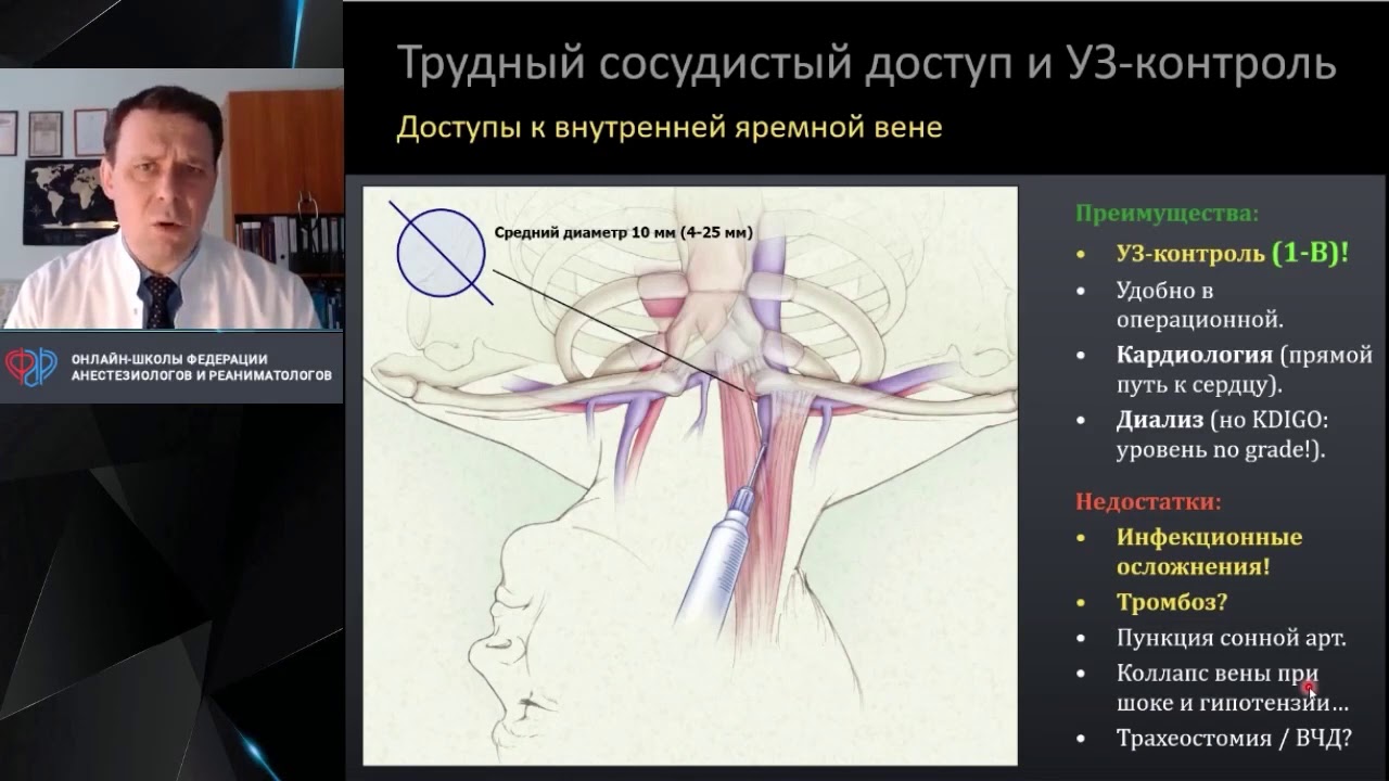
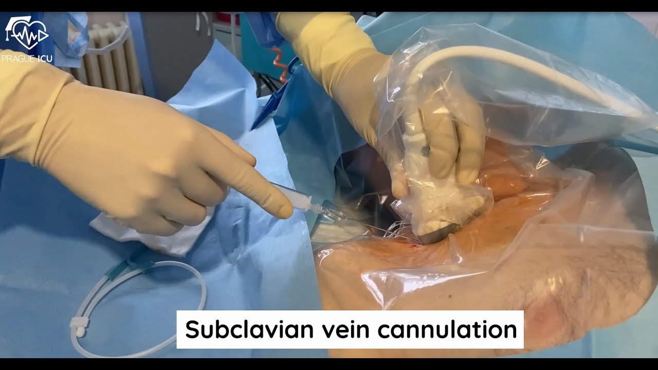

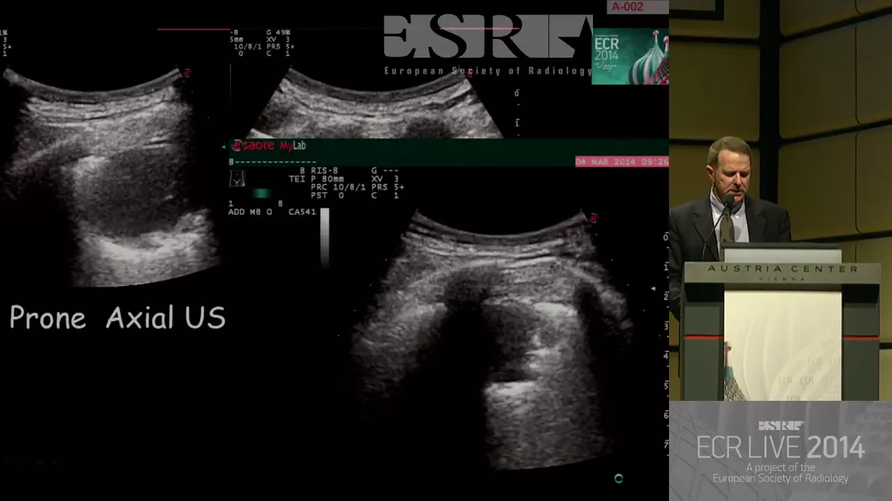



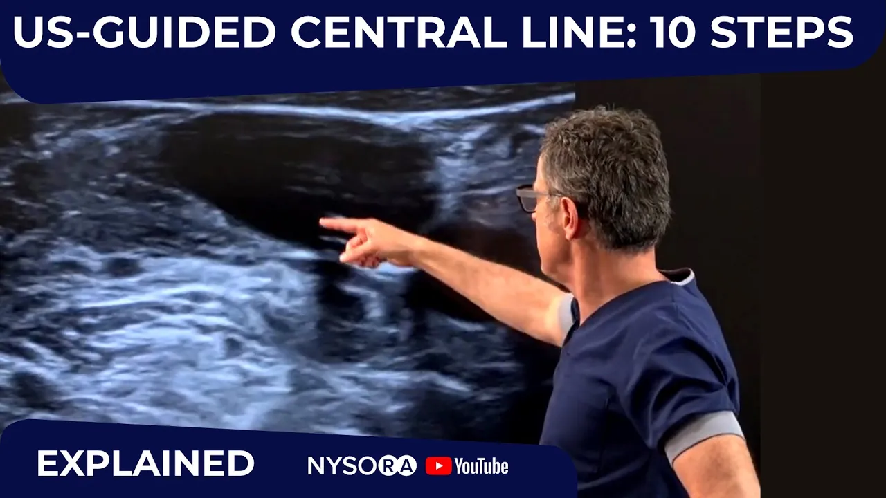
Bedside ultrasound guidance for internal jugular vein cannulation is superior to traditional landmark techniques because it allows precise pre-procedural visualization of vascular anatomy, revealing significant anatomical variations where the internal jugular vein may overlap the carotid artery; using a combination of short-axis (circular vessel appearance) and long-axis (tubular vessel appearance) ultrasound views with Doppler assessment enables accurate differentiation between vein and artery, reduces puncture attempts, lowers complication rates such as pneumothorax and hematoma, and is recommended by the US Agency for Healthcare Research and Quality as one of the top 10 patient safety practices.
Ultrasound-Guided Central Venous Access: Internal Jugular Vein
Added:Gross anatomy of the neck, specifically the spatial relationship between the internal jugular vein, carotid artery, and sternocleidomastoid muscle.
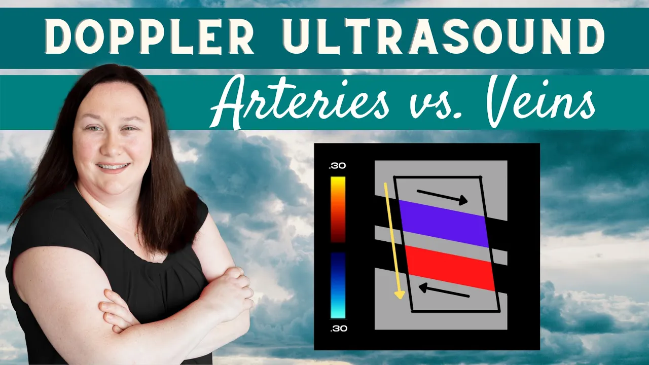
Basic ultrasound principles (knobology), including how to distinguish veins (compressible, non-pulsatile) from arteries (non-compressible, pulsatile) under imaging.
Standard indications, contraindications, and clinical scenarios requiring central venous access versus peripheral access.
Fundamental principles of maintaining a strict sterile field (asepsis) and preventing central line-associated bloodstream infections (CLABSI).
The step-by-step Seldinger technique for catheter insertion, including guidewire advancement, tract dilation, and securing the line.
Post-procedure validation protocols, specifically interpreting chest radiographs to confirm correct catheter tip placement in the lower superior vena cava.
Applying ultrasound-guided cannulation techniques to alternative anatomical sites, such as the subclavian, axillary, and femoral veins.
Identification, prevention, and emergency management of acute procedural complications like pneumothorax, arterial injury, and air embolism.
456.2K views2.5Klikes8:04Original Release: 2010-08-20
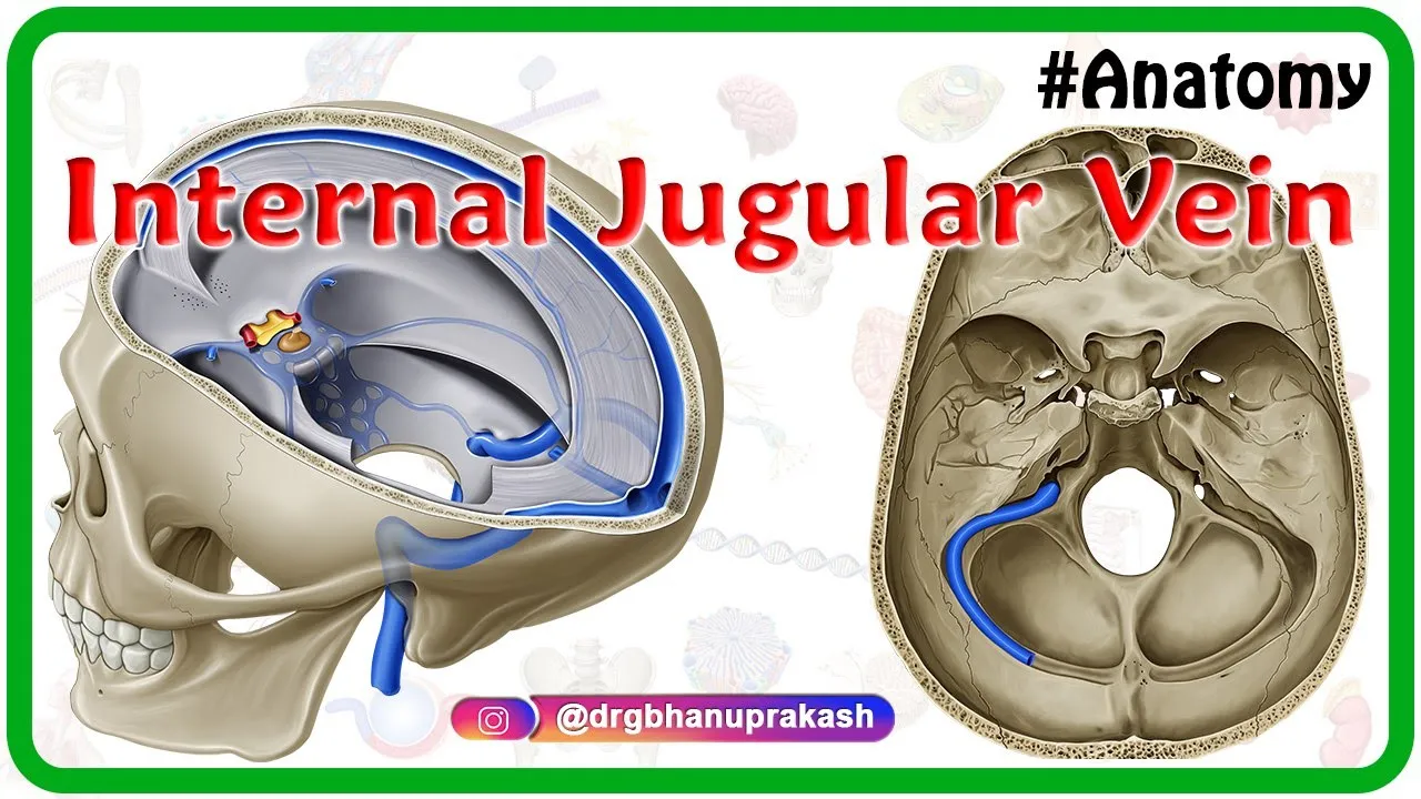
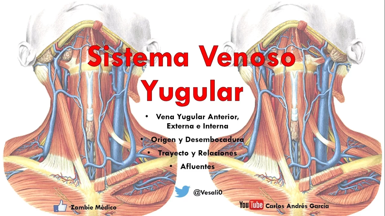
hello my name is Phil Pereira and I'm the emergency ultrasound coordinator at the New York Presbyterian Hospital in New York City and welcome to sound bites cases today's module is going to look at the use of bedside ultrasound for placement of central Venus catheters specifically the internal jugular vein in the neck so the question is why use ultrasound for Central Venus access and why not just use the traditional Landmark technique well interestingly multiple research studies now show a decreased number of puncture attempts are needed using ultrasound guidance and there's also a lower complication rate such as lowering the risk of pneumothorax and hematoma the US Agency for healthc care research the ahrq recommends ultrasound guidance for Central lines right up there in the top 10 patient safety practices ultrasound will allow precise determination of the anatomy of the vascular structures in the neck prior to a puncture attempt here's the middle triangle of the neck that serves as the standard approach for canulation of the internal jugular vein we see here the branches of the sternomastoid muscle the sternal head medially and the clavicular head laterally here we're putting our finger into the triangle of the neck and this indentation between the muscle heads would be the standard approach for placement of the needle we see here that the clavicle forms the in inferior boundary of the middle triangle of the neck within the middle triangle of the neck run two very important vascular structures and as per the textbook orientation of the kateed artery to the internal jugular vein we see in the image here that the kateed artery should run medial to the internal jugular vein which lies lateral to the artery However unfortunately there's great variability in human anatomy and many times the internal jugular vein can overlap the cored artery as shown in the drawing here notice the variation and location of the internal jugular vein to the kateed artery and many times the internal jugular vein is located on top of the kateed artery making it difficult to cannulate thus it's important to look with ultrasound before canulation attempts to avoid puncture to the cored artery here's the highfrequency linear type array probe that we'll be using to best map out the internal jugular vein before puncture attempts notice the probe marker there to the side of the probe here are the orientations that we can place the high frequency probe in relation to the internal jugular vein for vascular line placement here to the left we see the short axis configuration with a probe perpendicular to the vessel and notice that the vessel will appear on the ultrasound screen as a circle as a vessel will be cut and on to the right we see the long axis configuration and note the probe placed along the long AIS course of the vessel The Vessel therefore on the screen will appear as a tubular structure as shown here in the image to the right here's the highfrequency linear type array probe placed over the middle triangle of the neck over the internal jugular vein and kateed artery now I like to have the probe positioned in a side to side orientation with a marker dot oriented towards my left as I stand at the head of the bed the reason for that is then the orientation of the probe marker will line up to the orientation of the screen indicator dot which we see here is oriented towards the left on the ultrasound screen thus the left side of the the probe will Orient directly to the left side of the screen and this will allow us to orient ourselves as we place the needle underneath the patient's neck and culate the vein here's a typical appearance of the internal jugular vein and cored artery in a short AIS configuration taken with a B mode or grayscale image note lateral here towards the left and medial to the right here we notice the internal jugular vein in a location more lateral and superficial to the cored artery which lies deeper and medial to the vein we can see the depth markers to the side and we note the internal jugular vein at about 1.5 CM depth now we can apply Doppler sonography to further differentiate the two structures and here again we notice the internal jugular vein lying lateral and superficial to the cored artery we note the dopplerography steady pulsations of the internal jugular vein that vary with respiratory pattern and we can also see the cored artery with the pulsations with each heartbeat differentiating the two structures we can also press down with a probe to differentiate the two structures the internal jugular vein should compress completely while the more muscular outer walls of the cored artery should keep it open with compression of the probe here's another video clip showing the internal jugular vein and cored artery in a short AIS configuration notice here that this internal jugular vein is much more distended than in the last patient here we see that the internal jugular vein is located more superficially at about 0.5 cm and that it overlaps the kateed artery medially highlighting the fact that the 's great variability in the course of the internal jugular vein in relation to the kateed artery even within the same patient we're running the probe from position high within the neck in which the internal jugular vein is seen more laterally to a position more inferiorly in which the internal jugular vein comes to rest more immediately on top of the kateed artery here's a different patient in which the internal jugular vein is seen smack on top of the kateed artery notice here we'll Place Doppler flow to confirm the cored artery shown here deeper to the more superficial and internal jugular vein in this patient it would be extremely difficult to cannulate the internal jugular vein without puncturing the cored artery best to attempt canulation in another area of the body one pea can be used to further distend the internal jugular vein and make it a better Target for a canulation attempt is to have the patient Val Salva or hum notice here in the image to the left the patient is bearing down and notice that the internal jugular vein becomes much bigger as the patient pushes down in the image to the right note the the relatively small caliber of the internal jugular vein notice that it's almost as big here as the kateed artery but that it becomes much more distended as the patient Bears down using the Val Salva technique can make it a much better Target for placement of the large canulation needle here's the highfrequency probe placed in a longitudinal or long AIS manner on the patient's neck notice here that it's running along the course of the internal jugular vein as it runs up and down the patient's neck by convention here I like to have the probe marker towards the patient's head therefore I know where it lines up on the ultrasound screen notice here as a screen indicator dot is towards the left that Superior on the internal jugular vein will be located towards the left of the screen and inferior will be located towards the right of the screen here's a long AIS view of an internal jugular vein I have the probe marker going more distally or Superior within the neck so to the left is distal and to the right is proximal notice the internal jugular vein that appears like a tubular structure on the ultrasound screen and we see the blood flowing here from left to right here's a video clip again a long axis configuration in a different patient and here we see a much more distended internal jugular vein that's lying on top of the cored artery notice the swirls of blood in the internal jugular vein showing the course of the blood flow from high within the neck to the left low within the neck here to the right in conclusion thanks for tuning in for part one of ultrasound guided Central Venus access I hope I'm been able to score the point that ultrasound is very helpful in determining the relative anatomy of the internal jugular vein and cored artery prior to an invasive procedure as a textbook anatomy of the vein to artery is often Incorrect and it's best to use a combination of short and long exis views prior to a puncture attempt to best Define the anatomy so I hope to see you back in the future as Sono access continues and we return in central Venus access part two
Up Next

4:34
Ultrasound Needle Guidance: Mastering the In-Plane Approach
@gulfcoastultrasound
33K views•2022-06-02

12:54
Triumph of Orthodoxy Icon: Byzantine Art & History Explained
@BenCallan
2.1K views•2024-08-06

9:49
FastAPI vs Flask vs Django: Choosing the Right Python Web Framework
@TechWithTim
302.5K views•2024-05-26

1:42
Game of Thrones Opening Credits: A Cinematic Analysis
@gameofthrones
46.3M views•2011-04-18
Related Study Plans & Knowledge Roadmaps
Structured learning paths in General & Interdisciplinary Studies
general-studies
Elevator Engineering: History, Physics & Cities
01From Hoists to Safety Brakes: The Dawn of Vertical Lift
02Hydraulic vs. Traction: The Engineering Physics of Elevators
03Modern Speed, Safety, and Algorithms
+ 2 more learning modules
12h5 modules
Explore Roadmap
general-studies
Rubber: Material Science, History & Geopolitics
01Origins of Natural Rubber & Mesoamerican History
02The Chemistry of Natural Rubber & Vulcanization
03The Geopolitics of the Rubber Boom & Colonization
+ 2 more learning modules
12h5 modules
Explore Roadmap
general-studies
Containerization: Evolution, Logistics & Trade
01Ancient Trade & Bulk Cargo: Amphorae to Barrels
02The Industrial Revolution & Cardboard Packaging
03The Containerization Revolution: Malcom McLean
+ 2 more learning modules
12h5 modules
Explore Roadmap
general-studies
Textile Tech: Evolution, Labor & Global Impact
01Pre-Industrial Textiles: Handloom & Cottage Industry
02Mechanization: The Spinning Jenny to the Power Loom
03The Factory System & Global Labor Transformation
+ 2 more learning modules
10h5 modules
Explore Roadmap