














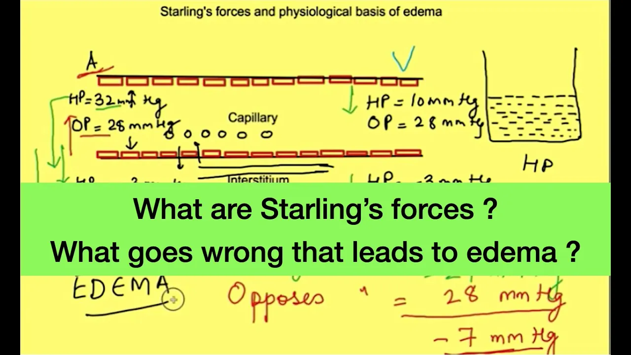












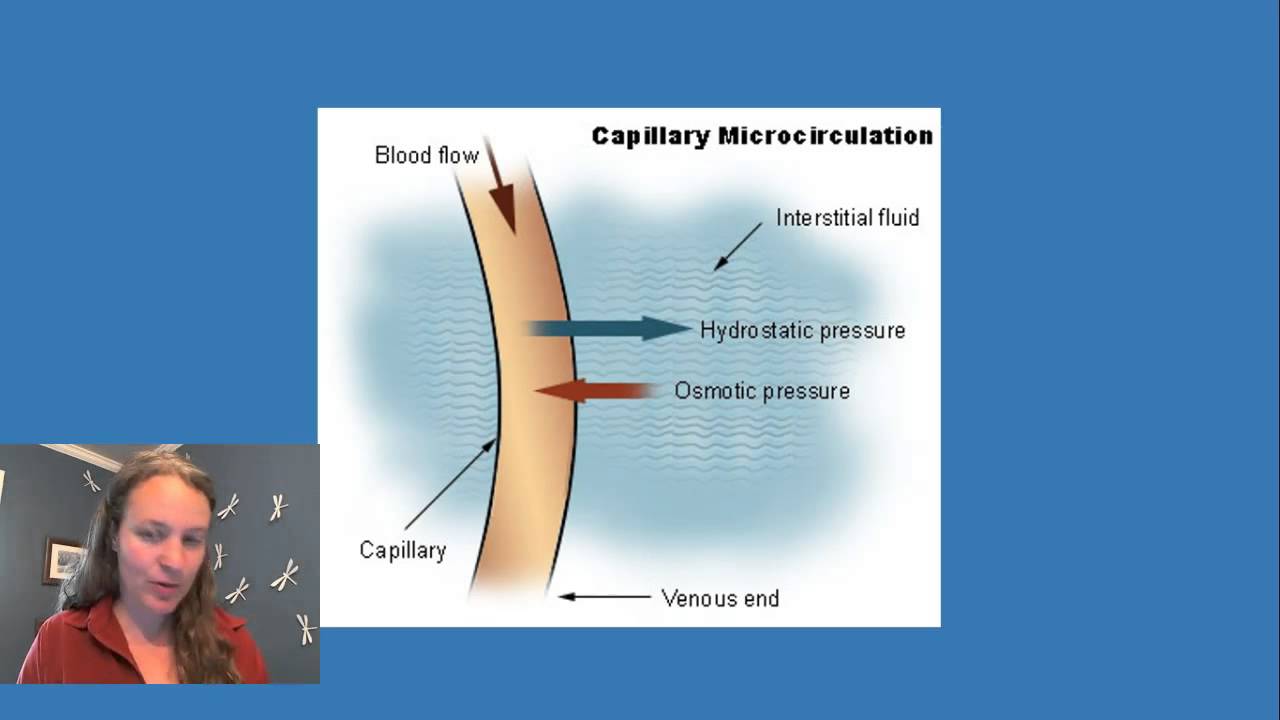
Fluid exchange between capillaries and interstitium is governed by four Starling forces: capillary hydrostatic pressure (pushes fluid out), interstitial hydrostatic pressure (pushes fluid in), capillary oncotic pressure (pulls fluid in via osmosis), and interstitial oncotic pressure (pulls fluid out); net filtration occurs when capillary pressure exceeds oncotic pressure, while reabsorption happens when oncotic pressure dominates, and imbalances in these pressures can lead to edema.
Capillary Fluid Exchange: Starling Forces Explained
Added:Basic anatomy of the cardiovascular microcirculation, specifically the structure of arterioles, capillaries, and venules.
The physical principles of diffusion, osmosis, and osmotic pressure across semipermeable membranes.
The concept of hydrostatic pressure, specifically how fluid pressure behaves inside biological vessels.
The role of major plasma proteins, particularly albumin, and their contribution to colloid osmotic (oncotic) pressure.
The clinical pathophysiology of edema, exploring how imbalances in Starling forces lead to tissue fluid accumulation in conditions like heart failure or liver disease.
The function of the lymphatic system in collecting and returning excess filtered interstitial fluid back into the venous circulation.
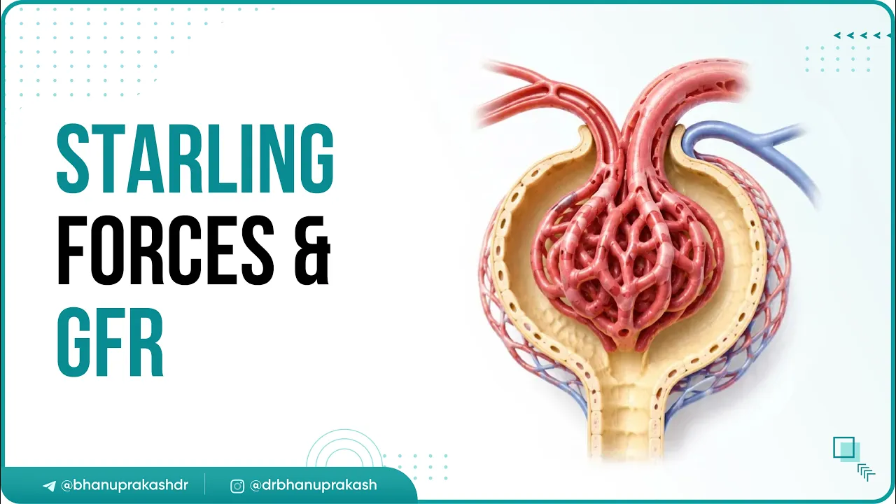
The specialized application of Starling forces in renal physiology, specifically during glomerular filtration in the kidneys.
The therapeutic administration of intravenous fluids (crystalloids vs. colloids) and how they alter capillary fluid exchange dynamics.
136.1K views3.5Klikes6:18Original Release: 2020-10-08
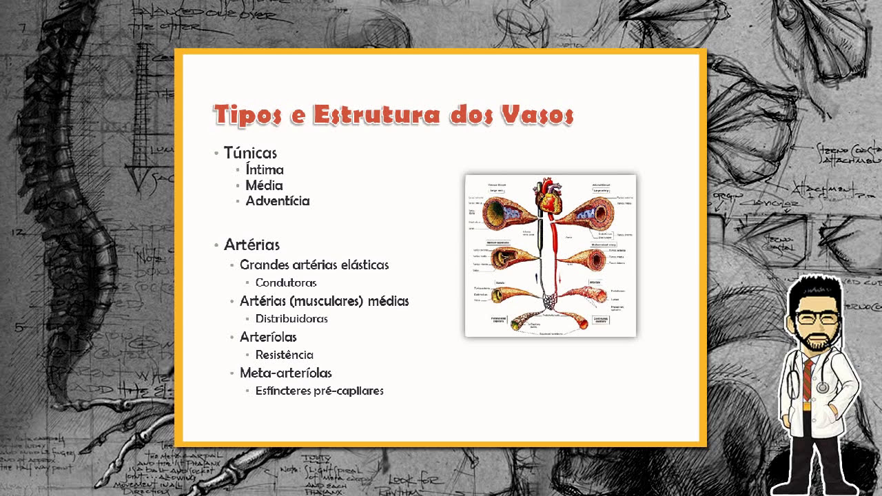
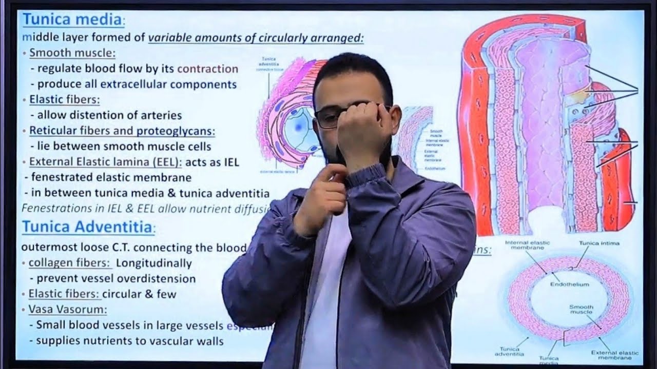
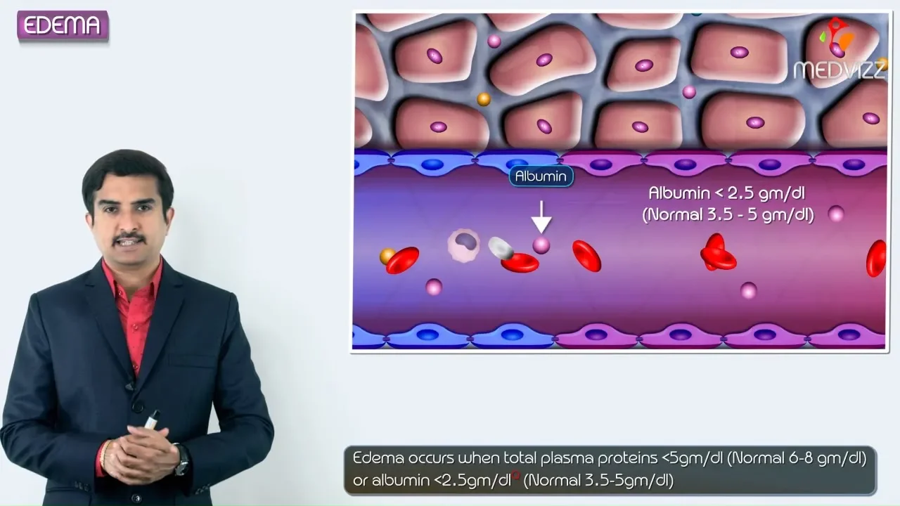
hi everyone welcome to bite size med where we talk about quick bite-sized concepts in medicine for study and rapid review this video is on the fluid exchange in capillaries each organ in the body is supplied by an artery the artery divides a bunch of times to form arterioles the arterioles then branch to form meta-arterioles and capillaries which then drain into venules and then veins at the point of origin of each capillary from the arteriole is a pre-capillary sphincter which controls the blood flow through the capillary true capillaries do not have smooth muscle in their wall versus arterioles which do what they do have is a layer of endothelial cells and a thin basement membrane this wall is just about a half micron thick there are spaces between the endothelial cells called intercellular clefts lipid-soluble substances can move across the membrane by simple diffusion like oxygen and carbon dioxide but substances that aren't lipid soluble but are water soluble and are small can go through the intercellular spaces like sodium and glucose but there's a size limitation so larger molecules like plasma proteins cannot pass through there are exceptions to this like the hepatic sinusoids where the spaces are large enough for plasma proteins to pass through the capillary is surrounded by interstitium so the two are separated by the capillary membrane the interstitium is like a gel with collagen and proteoglycans there's fluid trapped in between and that's the interstitial fluid since protein can't fit through the capillary membrane the interstitium has lesser protein the only proteins it has comes from leakage from the capillaries fluid and other solutes move between the capillary and the interstitial fluid and there are forces that either push the fluid through or oppose it there are four forces that determine which way fluids can move these are called starling forces named after ernest darling there are two hydrostatic pressures which would be the pressure exerted by the fluid itself on either side in the capillary it's pc and in the interstitial fluid it's pi there are two colloid osmotic pressures or oncotic pressures which are controlled by proteins in the capillary pi c and the interstitial fluid pi i so first let's look at pc that's the capillary hydrostatic pressure it's the pressure exerted by fluid so it sort of forces fluid to move into the interstitium encouraging filtration so let's put a plus sign next to it the pc can be affected by the changes in the arterial and the venous pressures and resistance which get transmitted to the capillary an increase in the pressure increases the pc while the interstitial fluid hydrostatic pressure pi pushes fluid back into the capillary membrane so it's negative the pi is very low because the fluid is trapped in the gel the capillary colloid osmotic pressure pi c is from plasma proteins by osmosis it would pull fluid from the interstitium into the capillary so we'll put a minus sign next to it the interstitial colloid osmotic pressure is from proteins in the interstitium in opposition it pulls fluid out of the capillary but since the capillary membrane has poured so small that proteins find it hard to get through this value is lower but it's still positive so if we put this whole thing together that gives us the net filtration pressure if this value is positive it means that there's net filtration of fluid from the capillary to the interstitium while if it's negative that means the direction of flow is towards the capillary which is reabsorption but there's one more factor the filtration coefficient kf which includes the number of capillaries the number and size of pores etc all that put together as the hydraulic conductance or the permeability of the capillary membrane so the rate of filtration will be by this starling equation kf into pc minus pi minus f pi c minus pi i physiologically the pc at the arterial end is higher than the venous so there's net filtration at the arterial end and net reabsorption at the venous end but there's a slight disequilibrium with a little higher filtration and the extra filtrate gets returned to circulation from the interstitium through the lymphatic vessels so what factors can increase filtration when pc increases filtration increases how does that happen increased arterial or venous pressure or even if the veins constrict by back pressure the pc will rise a reduction of pi though already pi is quite low a reduction of the colloid osmotic pressure in the capillary that's pi c pi c is controlled by the plasma proteins so if there's low plasma protein concentration like in something like nephrotic syndrome where there's excessive loss of proteins in the urine then pi c is low an increase in pi i so pi i is by the proteins in the interstitium which like i said is low but the lymphatics are the ones that remove proteins from the interstitium if there's lymphatic damage then pi i can increase so ultimately fluid will move out of the capillary into the interstitium expanding the fluid spaces causing edema because even the lymphatics can't handle the load and that's it that's how startling forces work for fluid exchange and capillaries i hope you found this video useful if you did give this video a thumbs up and subscribe to my channel thanks for watching and i'll see you in the next one
Up Next
12:03
Starling Forces: Oncotic and Hydrostatic Pressure Explained
@PhysiologyforHippies
255.3K views•2015-05-21

48:47
Graphic Medicine: Comics for Collaborative Healthcare Communication
@nationalpatientadvocate
189 views•2023-12-04
6:20
Lung and Chest Wall Compliance | Respiratory Physiology
@bytesizemed
239.9K views•2020-12-31

3:05
Stages of Labor and Vaginal Birth | Childbirth Animation
@nucleusmedicalmedia
52.1M views•2017-08-18
Related Study Plans & Knowledge Roadmaps
Structured learning paths in Medicine
medicine
Abnormal LFTs: Diagnostic Workup & Pathology
01Liver Anatomy and Basic Physiology
02Introduction to Liver Function Tests (LFTs)
03Hepatocellular vs. Cholestatic Patterns
+ 3 more learning modules
12h6 modules
Explore Roadmap
medicine
Analyzing Blood Smears: RBC, WBC & Platelets
01Introduction to Blood Components & Smear Preparation
02Normal Morphology of Blood Cells
03Erythrocyte Pathologies & Morphological Variants
+ 2 more learning modules
12h5 modules
Explore Roadmap
medicine
Mechanical Ventilation: Waveforms & Alarms
01Anatomy and Physiology of Breathing
02Introduction to Mechanical Ventilation & Core Modes
03Ventilator Settings and Waveform Interpretation
+ 2 more learning modules
12h5 modules
Explore Roadmap
medicine
Neuro Exam: Localization & Stroke Syndromes
01Foundations of Neuroanatomy and Vascular Supply
02The Neurological Examination: Step-by-Step
03Principles of Neurological Localization
+ 3 more learning modules
18h6 modules
Explore Roadmap